Downloaded 233 times
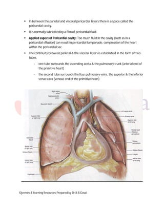


The pericardium has two layers - the fibrous pericardium and serous pericardium. The fibrous pericardium is a dense connective tissue that protects the heart. The serous pericardium contains two layers that lubricate the heart and prevent friction. It also contains the pericardial cavity filled with fluid. Too much fluid in the cavity can cause compression of the heart.


