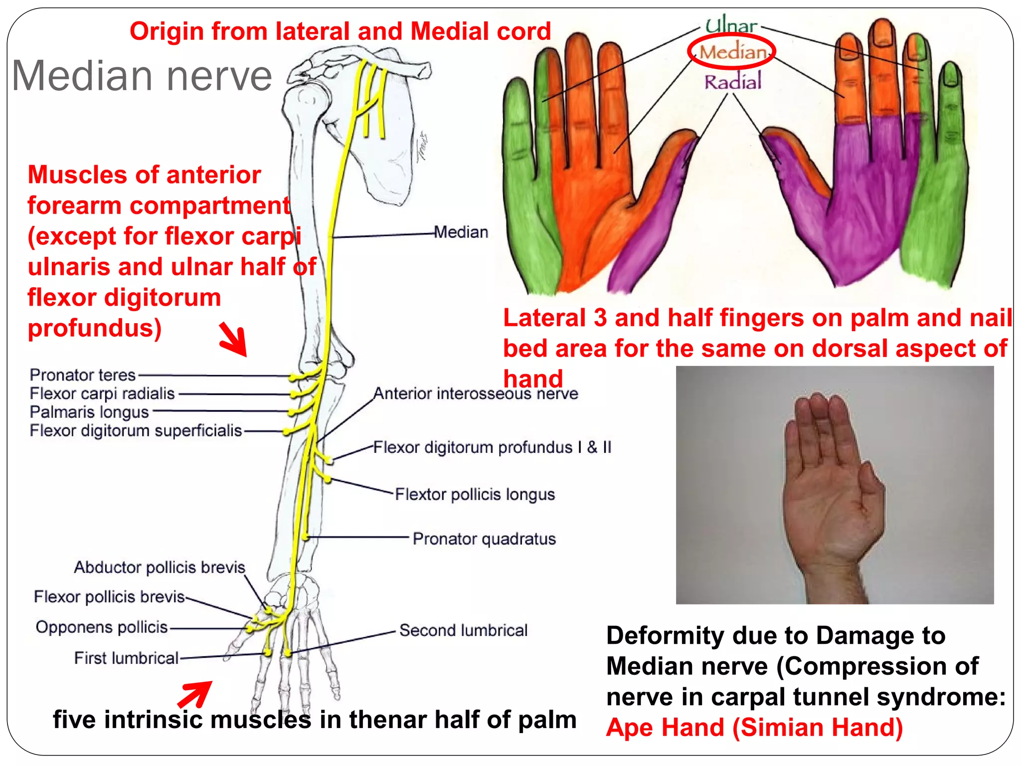
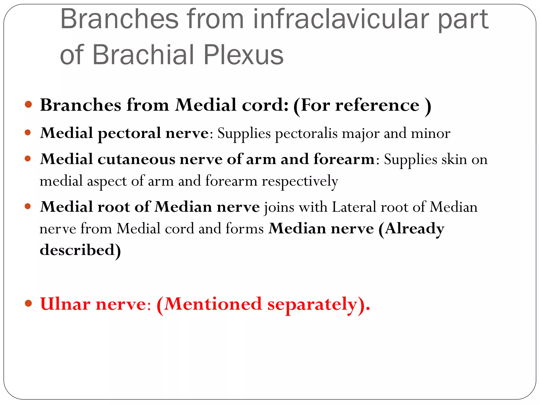
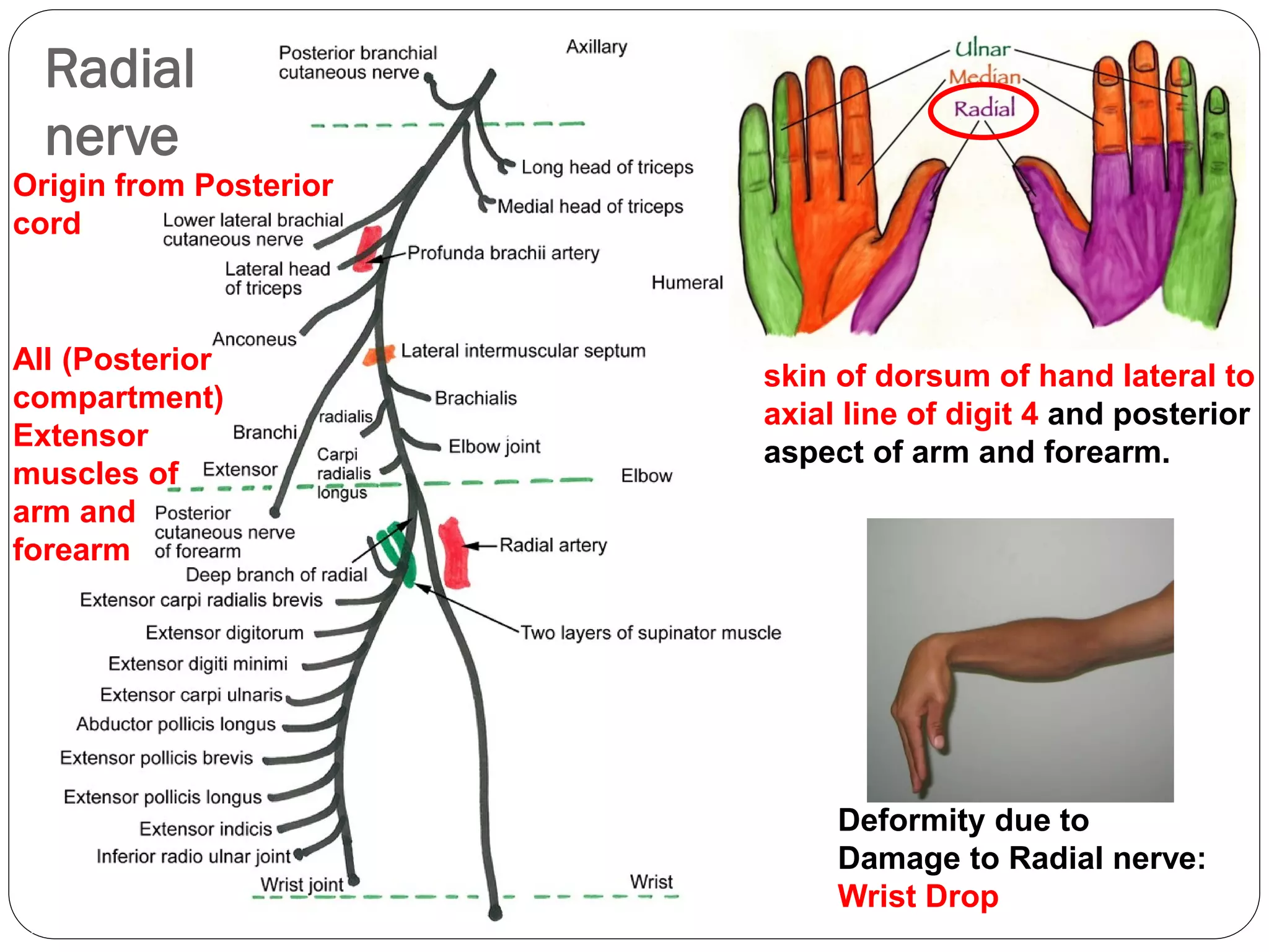
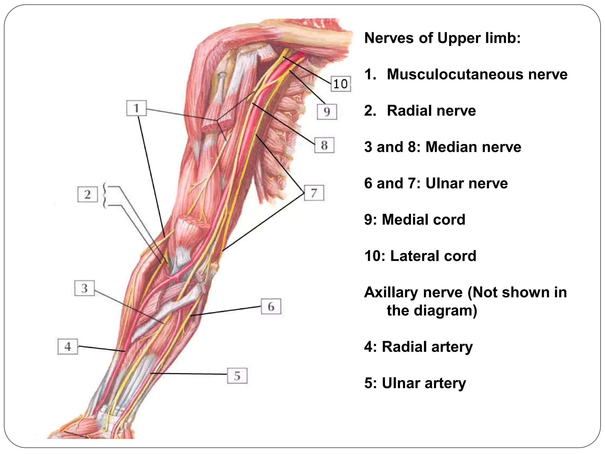
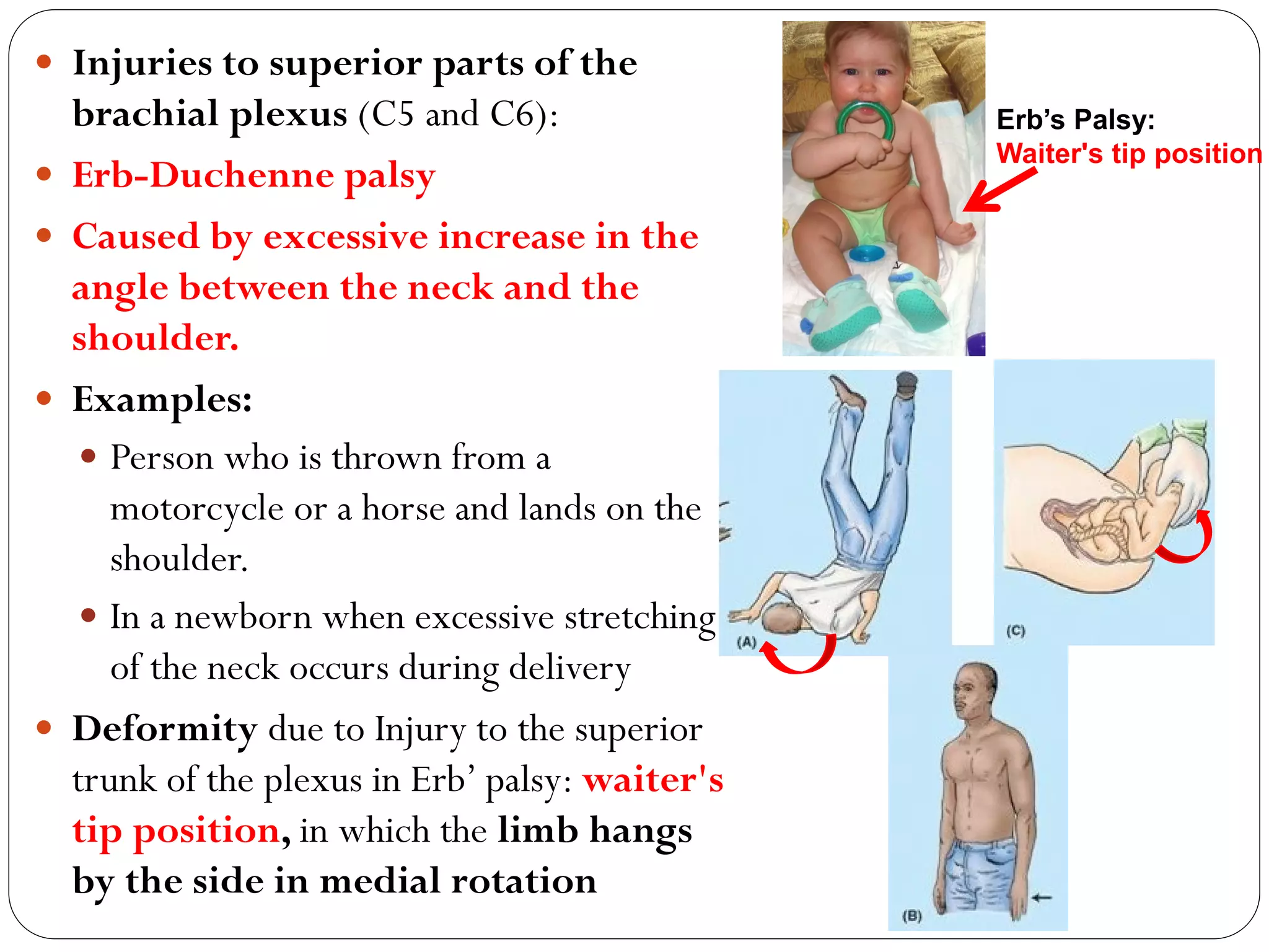
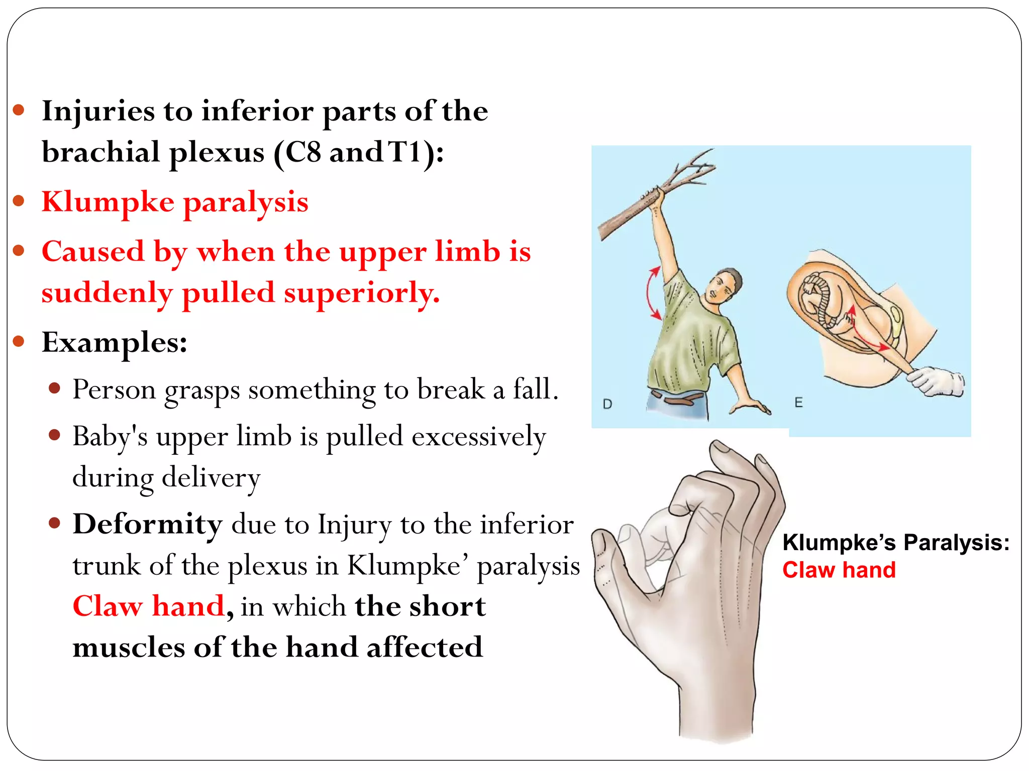
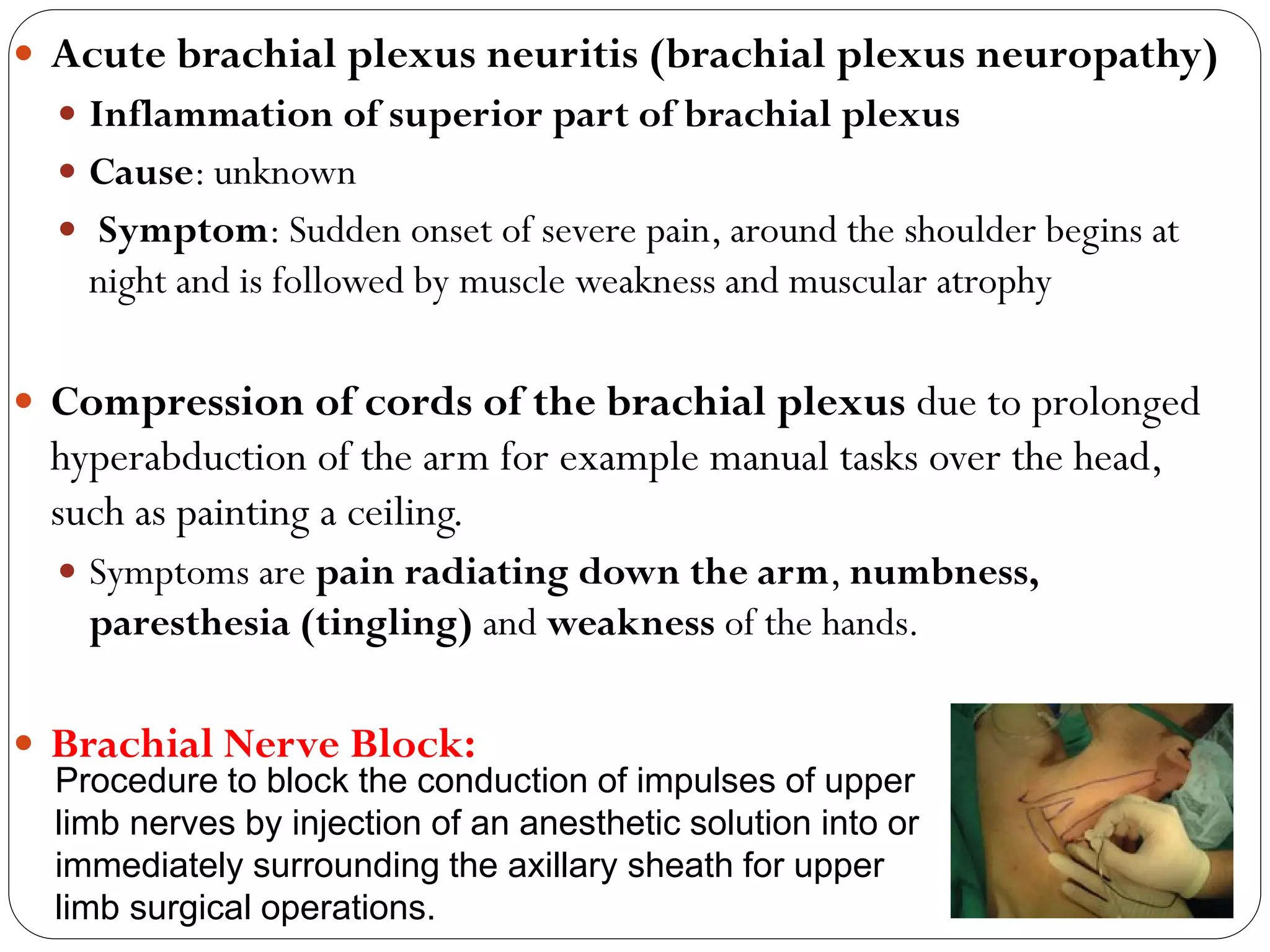
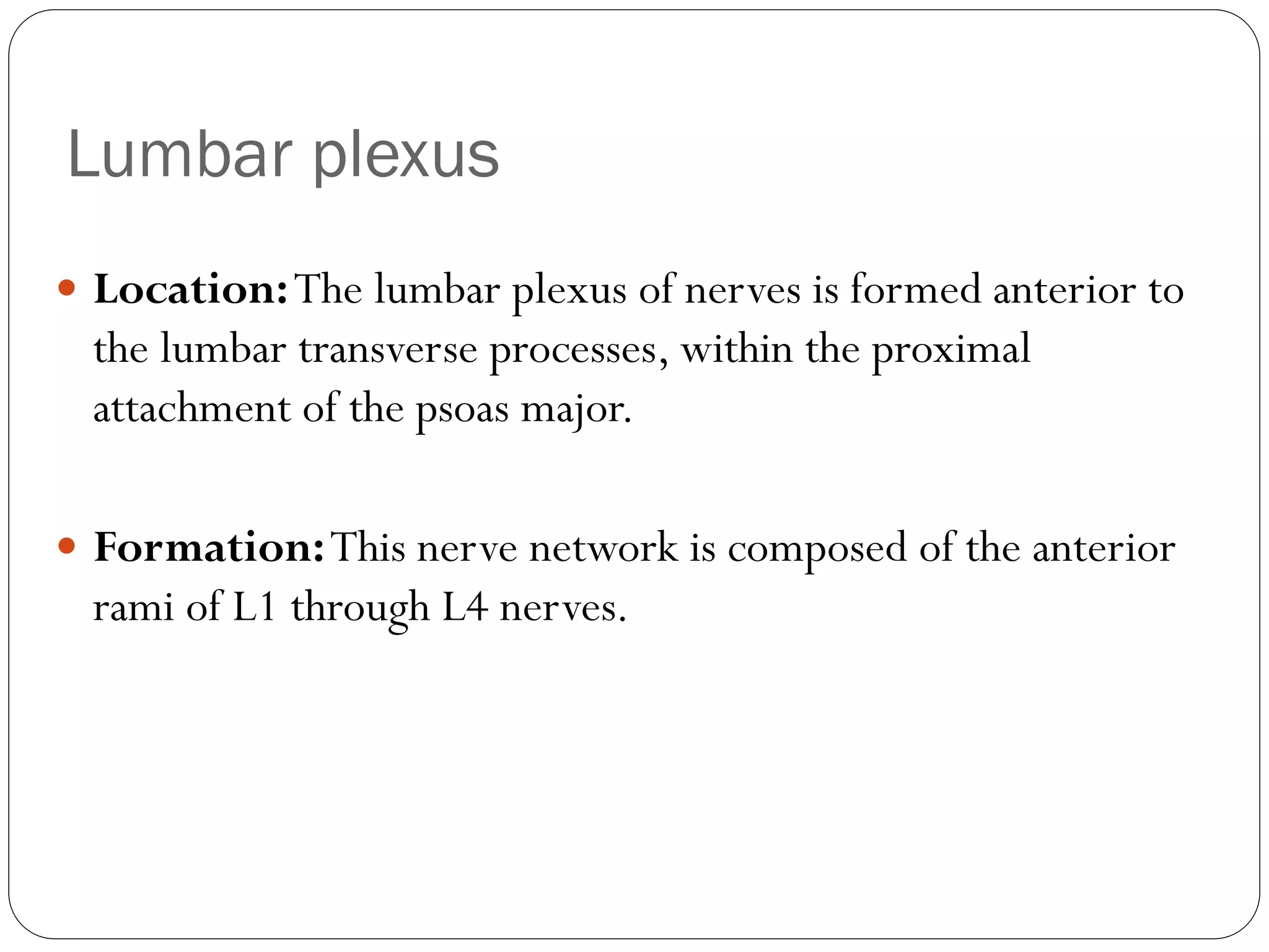
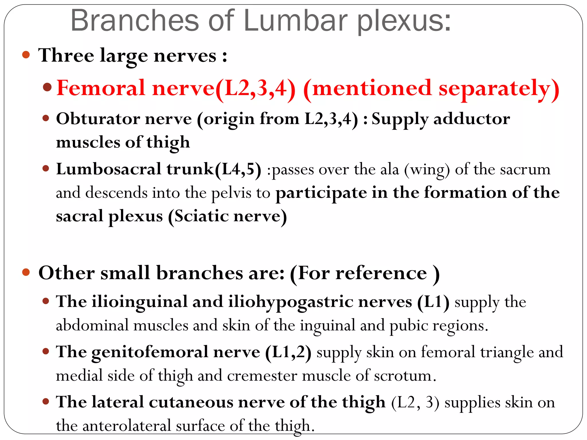
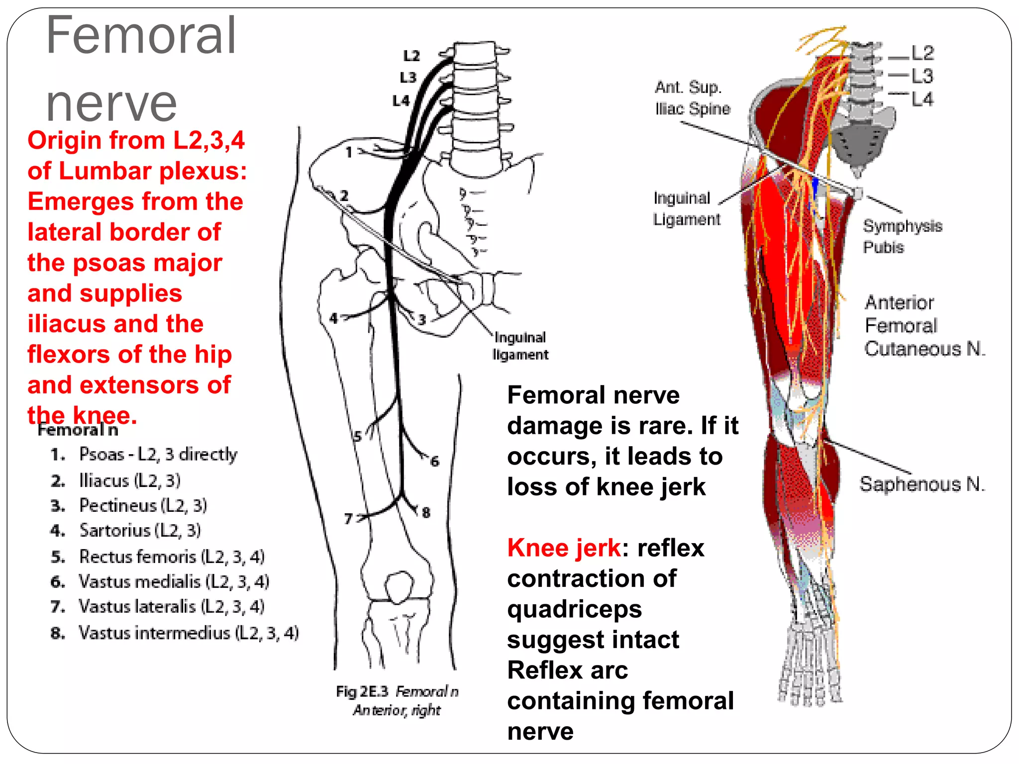
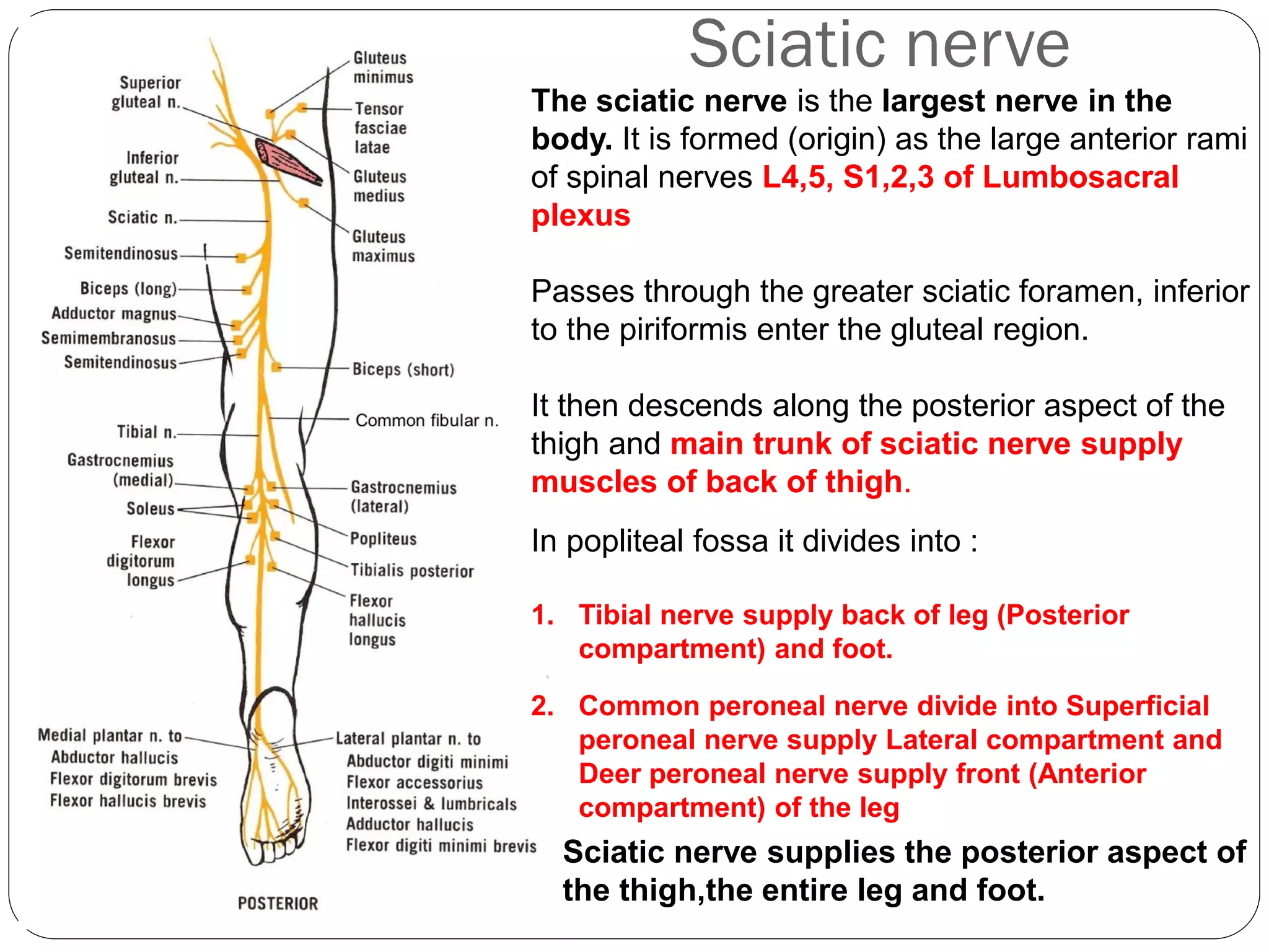
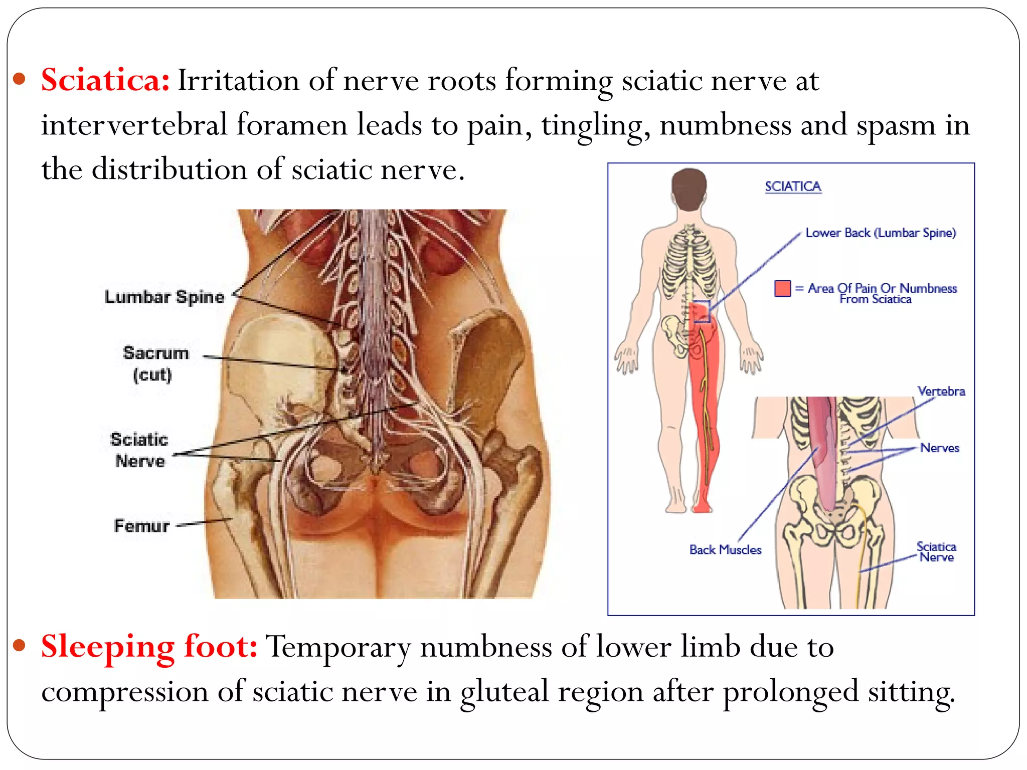
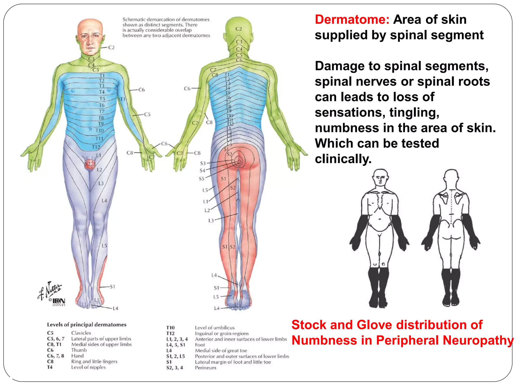
Dr.B.B.Gosai
Anatomy of Brachial and Lumbosacral Plexus
with its applied aspect
What we will learn today?
 Brachial Plexus: Formation, Branches (Specially Radial, Ulnar and
Median nerves), distribution and applied aspect (Chapter 6: PP: 721-
726, 729, 730, 743, 769, 791)
 Lumbosacral Plexus: Formation, Branches (specially Femoral and
Sciatic nerves), distribution and applied aspect
 Lumbar plexus: Chapter 2: PP: 313
 Sacral Plexus: Chapter 3: PP: 357, 359
 Femoral and Sciatic nerves: Ch 5 559, 574, 605
 References:
 Clinical Oriented Anatomy by Keith Moore :
Ch 6, Ch 2, Ch 3, Ch 5
 Atlas of Anatomy by Gilroy: Ch 24 pp. 349-359, Ch 29 pp. 449-459
 What is Plexus? Plexus means network
 Nerve Plexus is network of nerves like Cervical, Brachial,
Lumbosacral plexus.
 All these nerve plexuses are formed by Anterior (Ventral)
Rami of Spinal nerves.
Brachial Plexus
 Major nerve plexus formed in the neck and axilla and supply upper
limb.
 Formation: It is formed by ventral (Anterior) rami of Cervical
5, 6,7,8 andThoracic 1 spinal nerves (C5,6,7,8,T1).
 There are known as roots of Brachial plexus.
Note:
Roots of spinal nerves and
Roots of Brachial plexus
are not same.
Roots of spinal nerve come
from spinal cord.
Roots of brachial plexus
are from ventral rami of
spinal nerves.
C5,6,7,8,T1 form Roots of
Brachial Plexus
Location and Relations of Brachial plexus:
 In the neck: (Above clavicle)
 it is located between anterior
and middle scalene muscles
around subclavian artery.
 In the axilla: (Below clavicle)
 Nerves of brachial plexus are
arranged around axillary
artery
Formation of Brachial Plexus
 Roots of Brachial Plexus: C,5,6,7,8,T1
 Trunk of Brachial Plexus:
 Superior (Upper)Trunk: From union of C5 and C6 roots.
 MiddleTrunk: From continuation of C7 root.
 Inferior (Lower)Trunk: From union of C8 andT1 roots.
 Divisions of Brachial Plexus:
 Each trunk divides in to Anterior and Posterior divisions.
Anterior division supplyAnterior (Flexor) compartments of upper
limb and Posterior division supply Posterior (Extensor)
compartments of Upper limb.
Brachial Plexus
Formation of Brachial Plexus
 Cords of Brachial Plexus :
 Lateral cord: From union of anterior divisions of superior (upper)
and middle trunk.
 Medial cord: From continuation of anterior division of inferior
(lower) trunk.
 Posterior cord: From union of posterior divisions of all three
trunks.
 Cords named according to relation to axillary artery.
 Branches of Brachial Plexus: Divided into branches from
Supraclavicular part (Above the clavicle) and Infraclavicular part
(Below the clavicle).
THE BRACHIAL
PLEXUS
Important
nerves:
Musculocutaneo
us nerve (5)
Axillary nerve (6)
Radial nerve (8)
Ulnar nerve (14)
Median nerve
(24)
THE BRACHIAL
PLEXUS
“M” Shaped
formation by
branches of
lateral and
medial cord ie.
Musculocutaneo
us, Roots of
Median and
Ulnar nerves
Branches from supraclavicular part
of Brachial Plexus
 From Roots: (For reference )
 Dorsal scapular nerve (C4,5): Supplies Rhomboids and Levator
scapulae
 Long thoracic nerve (C5,6,7): Supplies Serratus anterior
 FromTrunk: (Superior – UpperTrunk only gives
branches)
 Suprascapular nerve (From Superior trunk): Supplies
supraspnatus and infraspinatus
 Nerve to subclavius (From Superior trunk): Supplies
Subclavius
Brachial Plexus
Branches from infraclavicular part of
Brachial Plexus
 Branches from Lateral cord:
 Lateral pectoral nerve: Supplies pectoralis
major and minor
 Musculocutaneous nerve:
 supplies biceps brachii, Brachialis and
coracobrachialis
and Skin of lateral aspect of forearm
 Lateral root of Median nerve joins with
Medial root of Median nerve from Medial cord and
forms Median nerve (Mentioned
Separately)
Median nerve
Lateral 3 and half fingers on palm and nail
bed area for the same on dorsal aspect of
hand
Muscles of anterior
forearm compartment
(except for flexor carpi
ulnaris and ulnar half of
flexor digitorum
profundus)
five intrinsic muscles in thenar half of palm
Deformity due to Damage to
Median nerve (Compression of
nerve in carpal tunnel syndrome:
Ape Hand (Simian Hand)
Origin from lateral and Medial cord
Branches from infraclavicular part
of Brachial Plexus
 Branches from Medial cord: (For reference )
 Medial pectoral nerve: Supplies pectoralis major and minor
 Medial cutaneous nerve of arm and forearm: Supplies skin on
medial aspect of arm and forearm respectively
 Medial root of Median nerve joins with Lateral root of Median
nerve from Medial cord and forms Median nerve (Already
described)
 Ulnar nerve: (Mentioned separately).
Flexor carpi ulnaris
and
ulnar half of
flexor digitorum
profundus
(forearm);
skin of hand medial to axial line of digit
4 (Medial one and half fingers of hand)
Ulnar
nerve
most (15 muscles)
intrinsic muscles of
hand (except 5
muscles supplied by
Median nerve)
Deformity due to
Damage to Ulnar nerve:
Claw Hand
Origin from Medial cord
Branches from infraclavicular part
of Brachial Plexus
 Branches from Posterior cord:
 Axillary nerve: Supplies teres minor and deltoid muscles; skin of
superolateral arm (over inferior part of deltoid)
 Radial nerve: (Mentioned Separately)
 Upper subscapular nerve: Supplies upper part of subscapularis
 Lower subscapular nerve: Supplies lower part of subscapularis
andTeres major
 Thoracodorsal nerve: Supplies Latissimus dorsi
Radial
nerve
skin of dorsum of hand lateral to
axial line of digit 4 and posterior
aspect of arm and forearm.
Deformity due to
Damage to Radial nerve:
Wrist Drop
All (Posterior
compartment)
Extensor
muscles of
arm and
forearm
Origin from Posterior
cord
Nerves of Upper limb:
1. Musculocutaneous nerve
2. Radial nerve
3 and 8: Median nerve
6 and 7: Ulnar nerve
9: Medial cord
10: Lateral cord
Axillary nerve (Not shown in
the diagram)
4: Radial artery
5: Ulnar artery
Musculocutaneous nerve
Median nerve
Ulnar nerve
Axillary nerve
Radial nerve
Quiz
 Radial nerve originate from which cord of brachial plexus?
 Most of the small muscles of hand are supplied by which
nerve?
 Compression of Median nerve in carpel tunnel leads to
which deformity?
Clinical Importance of Brachial Plexus
 Injuries to superior parts of the
brachial plexus (C5 and C6):
 Erb-Duchenne palsy
 Caused by excessive increase in the
angle between the neck and the
shoulder.
 Examples:
 Person who is thrown from a
motorcycle or a horse and lands on the
shoulder.
 In a newborn when excessive stretching
of the neck occurs during delivery
 Deformity due to Injury to the superior
trunk of the plexus in Erb’ palsy: waiter's
tip position, in which the limb hangs
by the side in medial rotation
Erb’s Palsy:
Waiter's tip position
 Injuries to inferior parts of the
brachial plexus (C8 andT1):
 Klumpke paralysis
 Caused by when the upper limb is
suddenly pulled superiorly.
 Examples:
 Person grasps something to break a fall.
 Baby's upper limb is pulled excessively
during delivery
 Deformity due to Injury to the inferior
trunk of the plexus in Klumpke’ paralysis:
Claw hand, in which the short
muscles of the hand affected
Klumpke’s Paralysis:
Claw hand
 Acute brachial plexus neuritis (brachial plexus neuropathy)
 Inflammation of superior part of brachial plexus
 Cause: unknown
 Symptom: Sudden onset of severe pain, around the shoulder begins at
night and is followed by muscle weakness and muscular atrophy
 Compression of cords of the brachial plexus due to prolonged
hyperabduction of the arm for example manual tasks over the head,
such as painting a ceiling.
 Symptoms are pain radiating down the arm, numbness,
paresthesia (tingling) and weakness of the hands.
 Brachial Nerve Block:
Procedure to block the conduction of impulses of upper
limb nerves by injection of an anesthetic solution into or
immediately surrounding the axillary sheath for upper
limb surgical operations.
Lumbosacral
Plexus
Lumbar Plexus
Sacral Plexus
Sciatic
Femoral Nerves: lateral to Psoas
Muscle (4 and 59)
Sacral plexus forming sciatic nerve
in lateral wall of pelvis (5 and 62)
Lumbar plexus
 Location:The lumbar plexus of nerves is formed anterior to
the lumbar transverse processes, within the proximal
attachment of the psoas major.
 Formation:This nerve network is composed of the anterior
rami of L1 through L4 nerves.
Branches of Lumbar plexus:
 Three large nerves :
Femoral nerve(L2,3,4) (mentioned separately)
 Obturator nerve (origin from L2,3,4) : Supply adductor
muscles of thigh
 Lumbosacral trunk(L4,5) :passes over the ala (wing) of the sacrum
and descends into the pelvis to participate in the formation of the
sacral plexus (Sciatic nerve)
 Other small branches are: (For reference )
 The ilioinguinal and iliohypogastric nerves (L1) supply the
abdominal muscles and skin of the inguinal and pubic regions.
 The genitofemoral nerve (L1,2) supply skin on femoral triangle and
medial side of thigh and cremester muscle of scrotum.
 The lateral cutaneous nerve of the thigh (L2, 3) supplies skin on
the anterolateral surface of the thigh.
Femoral
nerveOrigin from L2,3,4
of Lumbar plexus:
Emerges from the
lateral border of
the psoas major
and supplies
iliacus and the
flexors of the hip
and extensors of
the knee.
Femoral nerve
damage is rare. If it
occurs, it leads to
loss of knee jerk
Knee jerk: reflex
contraction of
quadriceps
suggest intact
Reflex arc
containing femoral
nerve
Sacral Plexus
 Location: On the posterolateral wall of the lesser pelvis.
 Formation:Ventral rami of L4,5 from lumbosacral trunk and S1,2,3
 Branches of Sacral plexus:
 The two main nerves arising from the sacral plexus:
Sciatic nerve: (Mentioned separately )
 Pudendal nerve: is the main nerve of the perineum and the chief
sensory nerve of the external genitalia.
 Other branches:
 The superior gluteal nerve supply muscles in the gluteal region-
gluteus medius and minimus.
 The inferior gluteal nerve supply gluteus maximus muscle.

Sciatic nerve
Sciatic nerve supplies the posterior aspect of
the thigh,the entire leg and foot.
The sciatic nerve is the largest nerve in the
body. It is formed (origin) as the large anterior rami
of spinal nerves L4,5, S1,2,3 of Lumbosacral
plexus
Passes through the greater sciatic foramen, inferior
to the piriformis enter the gluteal region.
It then descends along the posterior aspect of the
thigh and main trunk of sciatic nerve supply
muscles of back of thigh.
In popliteal fossa it divides into :
1. Tibial nerve supply back of leg (Posterior
compartment) and foot.
2. Common peroneal nerve divide into Superficial
peroneal nerve supply Lateral compartment and
Deer peroneal nerve supply front (Anterior
compartment) of the leg
Femoral
nerve
Common
peroneal nerve Tibial nerve
Sciatic nerve
Obturator
nerve
Gluteal nerves
Site of
intramuscular
injections to
avoid injury to
sciatic nerve
Foot drop: Damage to
sciatic nerve or
common peroneal
branch of sciatic nerve
leads to foot drop.
Applied
Anatomy of
sciatic nerve
 Sciatica: Irritation of nerve roots forming sciatic nerve at
intervertebral foramen leads to pain, tingling, numbness and spasm in
the distribution of sciatic nerve.
 Sleeping foot: Temporary numbness of lower limb due to
compression of sciatic nerve in gluteal region after prolonged sitting.
Dermatome: Area of skin
supplied by spinal segment
Damage to spinal segments,
spinal nerves or spinal roots
can leads to loss of
sensations, tingling,
numbness in the area of skin.
Which can be tested
clinically.
Stock and Glove distribution of
Numbness in Peripheral Neuropathy
Quiz
 Damage to upper trunk of brachial plexus causes which
palsy?
 Femoral nerve originate from which roots of lumbar plexus?
 Tibial nerve supply which compartment of leg?
 Damage to which nerve causes foot drop?
Summary
 Brachial plexus through its branches supply upper limb.
 Median nerve for most of the muscles of front of forearm
 Ulnar nerve for most of the muscles in hand
 Radial nerve supply back of arm and forearm
 Lumbosacral plexus through branches supply lower limb.
 Femoral nerve supply front of thigh
 Sciatic nerve (Main trunk) to back of thigh
 Tibial nerve to back of leg
 Superficial peroneal nerve to lateral compartment of leg
 Deep peroneal nerve to front of leg
Thanks for Attention
Your knowledge and skills relieves people
from pain and miseries

Brachial and lumbosacral plexus-Dr.B.B.Gosai

  • 2.
    Dr.B.B.Gosai Anatomy of Brachialand Lumbosacral Plexus with its applied aspect
  • 3.
    What we willlearn today?  Brachial Plexus: Formation, Branches (Specially Radial, Ulnar and Median nerves), distribution and applied aspect (Chapter 6: PP: 721- 726, 729, 730, 743, 769, 791)  Lumbosacral Plexus: Formation, Branches (specially Femoral and Sciatic nerves), distribution and applied aspect  Lumbar plexus: Chapter 2: PP: 313  Sacral Plexus: Chapter 3: PP: 357, 359  Femoral and Sciatic nerves: Ch 5 559, 574, 605  References:  Clinical Oriented Anatomy by Keith Moore : Ch 6, Ch 2, Ch 3, Ch 5  Atlas of Anatomy by Gilroy: Ch 24 pp. 349-359, Ch 29 pp. 449-459
  • 4.
     What isPlexus? Plexus means network  Nerve Plexus is network of nerves like Cervical, Brachial, Lumbosacral plexus.  All these nerve plexuses are formed by Anterior (Ventral) Rami of Spinal nerves.
  • 5.
    Brachial Plexus  Majornerve plexus formed in the neck and axilla and supply upper limb.  Formation: It is formed by ventral (Anterior) rami of Cervical 5, 6,7,8 andThoracic 1 spinal nerves (C5,6,7,8,T1).  There are known as roots of Brachial plexus. Note: Roots of spinal nerves and Roots of Brachial plexus are not same. Roots of spinal nerve come from spinal cord. Roots of brachial plexus are from ventral rami of spinal nerves. C5,6,7,8,T1 form Roots of Brachial Plexus
  • 6.
    Location and Relationsof Brachial plexus:  In the neck: (Above clavicle)  it is located between anterior and middle scalene muscles around subclavian artery.  In the axilla: (Below clavicle)  Nerves of brachial plexus are arranged around axillary artery
  • 8.
    Formation of BrachialPlexus  Roots of Brachial Plexus: C,5,6,7,8,T1  Trunk of Brachial Plexus:  Superior (Upper)Trunk: From union of C5 and C6 roots.  MiddleTrunk: From continuation of C7 root.  Inferior (Lower)Trunk: From union of C8 andT1 roots.  Divisions of Brachial Plexus:  Each trunk divides in to Anterior and Posterior divisions. Anterior division supplyAnterior (Flexor) compartments of upper limb and Posterior division supply Posterior (Extensor) compartments of Upper limb.
  • 9.
  • 10.
    Formation of BrachialPlexus  Cords of Brachial Plexus :  Lateral cord: From union of anterior divisions of superior (upper) and middle trunk.  Medial cord: From continuation of anterior division of inferior (lower) trunk.  Posterior cord: From union of posterior divisions of all three trunks.  Cords named according to relation to axillary artery.  Branches of Brachial Plexus: Divided into branches from Supraclavicular part (Above the clavicle) and Infraclavicular part (Below the clavicle).
  • 11.
    THE BRACHIAL PLEXUS Important nerves: Musculocutaneo us nerve(5) Axillary nerve (6) Radial nerve (8) Ulnar nerve (14) Median nerve (24)
  • 12.
    THE BRACHIAL PLEXUS “M” Shaped formationby branches of lateral and medial cord ie. Musculocutaneo us, Roots of Median and Ulnar nerves
  • 13.
    Branches from supraclavicularpart of Brachial Plexus  From Roots: (For reference )  Dorsal scapular nerve (C4,5): Supplies Rhomboids and Levator scapulae  Long thoracic nerve (C5,6,7): Supplies Serratus anterior  FromTrunk: (Superior – UpperTrunk only gives branches)  Suprascapular nerve (From Superior trunk): Supplies supraspnatus and infraspinatus  Nerve to subclavius (From Superior trunk): Supplies Subclavius
  • 14.
  • 15.
    Branches from infraclavicularpart of Brachial Plexus  Branches from Lateral cord:  Lateral pectoral nerve: Supplies pectoralis major and minor  Musculocutaneous nerve:  supplies biceps brachii, Brachialis and coracobrachialis and Skin of lateral aspect of forearm  Lateral root of Median nerve joins with Medial root of Median nerve from Medial cord and forms Median nerve (Mentioned Separately)
  • 16.
    Median nerve Lateral 3and half fingers on palm and nail bed area for the same on dorsal aspect of hand Muscles of anterior forearm compartment (except for flexor carpi ulnaris and ulnar half of flexor digitorum profundus) five intrinsic muscles in thenar half of palm Deformity due to Damage to Median nerve (Compression of nerve in carpal tunnel syndrome: Ape Hand (Simian Hand) Origin from lateral and Medial cord
  • 17.
    Branches from infraclavicularpart of Brachial Plexus  Branches from Medial cord: (For reference )  Medial pectoral nerve: Supplies pectoralis major and minor  Medial cutaneous nerve of arm and forearm: Supplies skin on medial aspect of arm and forearm respectively  Medial root of Median nerve joins with Lateral root of Median nerve from Medial cord and forms Median nerve (Already described)  Ulnar nerve: (Mentioned separately).
  • 18.
    Flexor carpi ulnaris and ulnarhalf of flexor digitorum profundus (forearm); skin of hand medial to axial line of digit 4 (Medial one and half fingers of hand) Ulnar nerve most (15 muscles) intrinsic muscles of hand (except 5 muscles supplied by Median nerve) Deformity due to Damage to Ulnar nerve: Claw Hand Origin from Medial cord
  • 19.
    Branches from infraclavicularpart of Brachial Plexus  Branches from Posterior cord:  Axillary nerve: Supplies teres minor and deltoid muscles; skin of superolateral arm (over inferior part of deltoid)  Radial nerve: (Mentioned Separately)  Upper subscapular nerve: Supplies upper part of subscapularis  Lower subscapular nerve: Supplies lower part of subscapularis andTeres major  Thoracodorsal nerve: Supplies Latissimus dorsi
  • 20.
    Radial nerve skin of dorsumof hand lateral to axial line of digit 4 and posterior aspect of arm and forearm. Deformity due to Damage to Radial nerve: Wrist Drop All (Posterior compartment) Extensor muscles of arm and forearm Origin from Posterior cord
  • 21.
    Nerves of Upperlimb: 1. Musculocutaneous nerve 2. Radial nerve 3 and 8: Median nerve 6 and 7: Ulnar nerve 9: Medial cord 10: Lateral cord Axillary nerve (Not shown in the diagram) 4: Radial artery 5: Ulnar artery
  • 22.
    Musculocutaneous nerve Median nerve Ulnarnerve Axillary nerve Radial nerve
  • 23.
    Quiz  Radial nerveoriginate from which cord of brachial plexus?  Most of the small muscles of hand are supplied by which nerve?  Compression of Median nerve in carpel tunnel leads to which deformity?
  • 24.
    Clinical Importance ofBrachial Plexus
  • 25.
     Injuries tosuperior parts of the brachial plexus (C5 and C6):  Erb-Duchenne palsy  Caused by excessive increase in the angle between the neck and the shoulder.  Examples:  Person who is thrown from a motorcycle or a horse and lands on the shoulder.  In a newborn when excessive stretching of the neck occurs during delivery  Deformity due to Injury to the superior trunk of the plexus in Erb’ palsy: waiter's tip position, in which the limb hangs by the side in medial rotation Erb’s Palsy: Waiter's tip position
  • 26.
     Injuries toinferior parts of the brachial plexus (C8 andT1):  Klumpke paralysis  Caused by when the upper limb is suddenly pulled superiorly.  Examples:  Person grasps something to break a fall.  Baby's upper limb is pulled excessively during delivery  Deformity due to Injury to the inferior trunk of the plexus in Klumpke’ paralysis: Claw hand, in which the short muscles of the hand affected Klumpke’s Paralysis: Claw hand
  • 27.
     Acute brachialplexus neuritis (brachial plexus neuropathy)  Inflammation of superior part of brachial plexus  Cause: unknown  Symptom: Sudden onset of severe pain, around the shoulder begins at night and is followed by muscle weakness and muscular atrophy  Compression of cords of the brachial plexus due to prolonged hyperabduction of the arm for example manual tasks over the head, such as painting a ceiling.  Symptoms are pain radiating down the arm, numbness, paresthesia (tingling) and weakness of the hands.  Brachial Nerve Block: Procedure to block the conduction of impulses of upper limb nerves by injection of an anesthetic solution into or immediately surrounding the axillary sheath for upper limb surgical operations.
  • 28.
  • 29.
    Femoral Nerves: lateralto Psoas Muscle (4 and 59) Sacral plexus forming sciatic nerve in lateral wall of pelvis (5 and 62)
  • 30.
    Lumbar plexus  Location:Thelumbar plexus of nerves is formed anterior to the lumbar transverse processes, within the proximal attachment of the psoas major.  Formation:This nerve network is composed of the anterior rami of L1 through L4 nerves.
  • 31.
    Branches of Lumbarplexus:  Three large nerves : Femoral nerve(L2,3,4) (mentioned separately)  Obturator nerve (origin from L2,3,4) : Supply adductor muscles of thigh  Lumbosacral trunk(L4,5) :passes over the ala (wing) of the sacrum and descends into the pelvis to participate in the formation of the sacral plexus (Sciatic nerve)  Other small branches are: (For reference )  The ilioinguinal and iliohypogastric nerves (L1) supply the abdominal muscles and skin of the inguinal and pubic regions.  The genitofemoral nerve (L1,2) supply skin on femoral triangle and medial side of thigh and cremester muscle of scrotum.  The lateral cutaneous nerve of the thigh (L2, 3) supplies skin on the anterolateral surface of the thigh.
  • 32.
    Femoral nerveOrigin from L2,3,4 ofLumbar plexus: Emerges from the lateral border of the psoas major and supplies iliacus and the flexors of the hip and extensors of the knee. Femoral nerve damage is rare. If it occurs, it leads to loss of knee jerk Knee jerk: reflex contraction of quadriceps suggest intact Reflex arc containing femoral nerve
  • 33.
    Sacral Plexus  Location:On the posterolateral wall of the lesser pelvis.  Formation:Ventral rami of L4,5 from lumbosacral trunk and S1,2,3  Branches of Sacral plexus:  The two main nerves arising from the sacral plexus: Sciatic nerve: (Mentioned separately )  Pudendal nerve: is the main nerve of the perineum and the chief sensory nerve of the external genitalia.  Other branches:  The superior gluteal nerve supply muscles in the gluteal region- gluteus medius and minimus.  The inferior gluteal nerve supply gluteus maximus muscle. 
  • 34.
    Sciatic nerve Sciatic nervesupplies the posterior aspect of the thigh,the entire leg and foot. The sciatic nerve is the largest nerve in the body. It is formed (origin) as the large anterior rami of spinal nerves L4,5, S1,2,3 of Lumbosacral plexus Passes through the greater sciatic foramen, inferior to the piriformis enter the gluteal region. It then descends along the posterior aspect of the thigh and main trunk of sciatic nerve supply muscles of back of thigh. In popliteal fossa it divides into : 1. Tibial nerve supply back of leg (Posterior compartment) and foot. 2. Common peroneal nerve divide into Superficial peroneal nerve supply Lateral compartment and Deer peroneal nerve supply front (Anterior compartment) of the leg
  • 35.
    Femoral nerve Common peroneal nerve Tibialnerve Sciatic nerve Obturator nerve Gluteal nerves
  • 36.
    Site of intramuscular injections to avoidinjury to sciatic nerve Foot drop: Damage to sciatic nerve or common peroneal branch of sciatic nerve leads to foot drop. Applied Anatomy of sciatic nerve
  • 37.
     Sciatica: Irritationof nerve roots forming sciatic nerve at intervertebral foramen leads to pain, tingling, numbness and spasm in the distribution of sciatic nerve.  Sleeping foot: Temporary numbness of lower limb due to compression of sciatic nerve in gluteal region after prolonged sitting.
  • 38.
    Dermatome: Area ofskin supplied by spinal segment Damage to spinal segments, spinal nerves or spinal roots can leads to loss of sensations, tingling, numbness in the area of skin. Which can be tested clinically. Stock and Glove distribution of Numbness in Peripheral Neuropathy
  • 39.
    Quiz  Damage toupper trunk of brachial plexus causes which palsy?  Femoral nerve originate from which roots of lumbar plexus?  Tibial nerve supply which compartment of leg?  Damage to which nerve causes foot drop?
  • 40.
    Summary  Brachial plexusthrough its branches supply upper limb.  Median nerve for most of the muscles of front of forearm  Ulnar nerve for most of the muscles in hand  Radial nerve supply back of arm and forearm  Lumbosacral plexus through branches supply lower limb.  Femoral nerve supply front of thigh  Sciatic nerve (Main trunk) to back of thigh  Tibial nerve to back of leg  Superficial peroneal nerve to lateral compartment of leg  Deep peroneal nerve to front of leg
  • 41.
    Thanks for Attention Yourknowledge and skills relieves people from pain and miseries