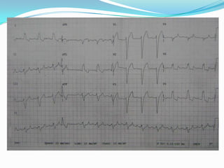
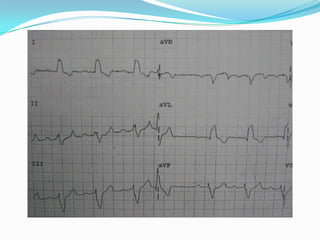
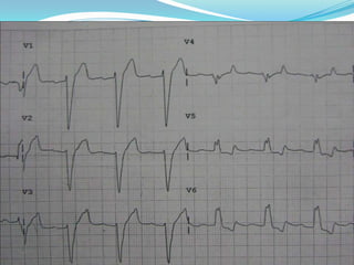
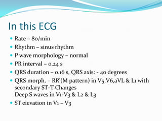
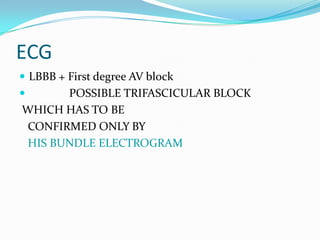
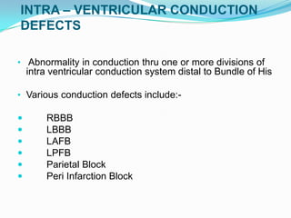
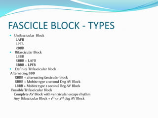
ECG OF THE WEEKProf.Dr . G.ELANGOVAN’S unitDr.M.Amudhan
73 year old male presented with c/o giddiness on & offfor past 1yr.  chest pain past 2 daysPast history;  not a k/c/o T2DM/SHT/IHD  No other significant historyO/E  pt concious ,oriented, afebrile.   no pallor/icterus/cyanosis/clubbing/LN/pedal    edemaVitals; PR 84/min  BP 150/90Cvs: s1 s2 heard. No murmurs.RS: nvbs heard, no added sounds,Other systems also normal.
In this ECGRate – 80/minRhythm – sinus rhythm P wave morphology – normal PR interval – 0.24 sQRS duration – 0.16 s, QRS axis: - 40 degrees QRS morph. – RR’(M pattern) in V5,V6,aVL & L1 with secondary ST-T Changes                                           Deep S waves in V1-V3 & L2 & L3ST eievation in V1 – V3
ECGLBBB + First degree AV block              POSSIBLE TRIFASCICULAR BLOCK WHICH HAS TO BE    CONFIRMED ONLY BYHIS BUNDLE ELECTROGRAM
                                                                              INTRA – VENTRICULAR CONDUCTION DEFECTS Abnormality in conduction thru one or more divisions of intra ventricular conduction system distal to Bundle of His
Various conduction defects include:-	RBBB	LBBB	LAFB	LPFB	Parietal BlockPeri Infarction Block
Causes Of IVCD :-Congenital ( RBBB in normal individuals )IHD ( AMI / PMI / Coronary atherosclerosis )Cardiomyopathy ( DCM / HCM )Infiltrative Lesions ( Tumors / Chagas / Hypothyroidism / Amyloidosis )Aortic stenosis ( LBBB )Infective EndocarditisHyperkalemiaCardiac InjuryMassive Pulmonary EmbolismVentricular HypertrophyMyocarditis
FASCICLE BLOCK - TYPESUnifascicular  Block       LAFB       LPFB       RBBBBifascicular Block       LBBB       RBBB + LAFB       RBBB + LPFB Definite Trifascicular Block Alternating BBB      RBBB + alternating fascicular block      RBBB + Mobitz type 2 second Deg AV Block      LBBB + Mobitz type 2 second Deg AV Block  Possible Trifascicular Block     Complete AV Block with ventricular escape rhythm     Any Bifascicular Block + 1ST or 2nd deg AV Block
FASCICULAR BLOCKSLAFBLAD (-30 to -80 )
rS in L2,3,aVf

ECG: Trifascicular Block

  • 1.
    ECG OF THEWEEKProf.Dr . G.ELANGOVAN’S unitDr.M.Amudhan
  • 2.
    73 year oldmale presented with c/o giddiness on & offfor past 1yr. chest pain past 2 daysPast history; not a k/c/o T2DM/SHT/IHD No other significant historyO/E pt concious ,oriented, afebrile. no pallor/icterus/cyanosis/clubbing/LN/pedal edemaVitals; PR 84/min BP 150/90Cvs: s1 s2 heard. No murmurs.RS: nvbs heard, no added sounds,Other systems also normal.
  • 6.
    In this ECGRate– 80/minRhythm – sinus rhythm P wave morphology – normal PR interval – 0.24 sQRS duration – 0.16 s, QRS axis: - 40 degrees QRS morph. – RR’(M pattern) in V5,V6,aVL & L1 with secondary ST-T Changes Deep S waves in V1-V3 & L2 & L3ST eievation in V1 – V3
  • 7.
    ECGLBBB + Firstdegree AV block POSSIBLE TRIFASCICULAR BLOCK WHICH HAS TO BE CONFIRMED ONLY BYHIS BUNDLE ELECTROGRAM
  • 8.
    INTRA – VENTRICULAR CONDUCTION DEFECTS Abnormality in conduction thru one or more divisions of intra ventricular conduction system distal to Bundle of His
  • 9.
    Various conduction defectsinclude:- RBBB LBBB LAFB LPFB Parietal BlockPeri Infarction Block
  • 10.
    Causes Of IVCD:-Congenital ( RBBB in normal individuals )IHD ( AMI / PMI / Coronary atherosclerosis )Cardiomyopathy ( DCM / HCM )Infiltrative Lesions ( Tumors / Chagas / Hypothyroidism / Amyloidosis )Aortic stenosis ( LBBB )Infective EndocarditisHyperkalemiaCardiac InjuryMassive Pulmonary EmbolismVentricular HypertrophyMyocarditis
  • 11.
    FASCICLE BLOCK -TYPESUnifascicular Block LAFB LPFB RBBBBifascicular Block LBBB RBBB + LAFB RBBB + LPFB Definite Trifascicular Block Alternating BBB RBBB + alternating fascicular block RBBB + Mobitz type 2 second Deg AV Block LBBB + Mobitz type 2 second Deg AV Block Possible Trifascicular Block Complete AV Block with ventricular escape rhythm Any Bifascicular Block + 1ST or 2nd deg AV Block
  • 12.
  • 13.