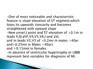
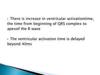
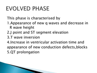
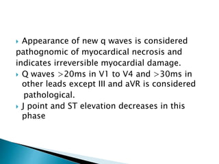
SlideShare a Scribd company logo
M.I denotes cellular damage due to prolonged
ischaemia .Sudden cardiac death is a frequent
presenting feature of MI with most of deaths
occuring within one hour due to ventricular
arrythymias.
In presence of ischaemic symptoms the diagnosisof MI
is based upon one of the following
1.Development of new pathological Q waves.
2.Presence of ST segment elevation or depression
3.Development of new LBBB
 The changes in ECG should be present in two
contiguous leads.
 Confirmation by diagnosis can be done by
demonstration by elevation in cardiac
enzymes,ECHO,or by coronary angiography.
Based on ECG, MI is further differentiated as
STEMI and NSTEMI.
Evolution of NSTEMI into STEMI is possible and
therefore both subsets should be treated as
aggresively as possible
The ECG changes evolve over a period of time
and are described as
1.HYPERACUTE PHASE(over minutes-hours)
2.EVOLVED PHASE(over hours)
3.CHRONIC STABILISED PHASE(over days-weeks)
The changes in ECG of chronic stabilised phase
persists throughout life and generally represent
changes of changes of old MI in absence of further
progession of disease.
The ECG changes in this phase are
1.Tall,symmetrical,peaked and widened T waves
2.Slope elevation of ST segment
3.Increased amplitude of R wave/changes in
Terminal QRS complex
4.Increased ventricular activation time
This phase is critical because complication of
Ventricular fibrillation is most likely to occur and
Coronary reperfusion during this phase totally
reverses changes without any residual myocardial
damage.
The Twaves are tall and wide sometimes itmay
exceed amplitude of associated R wave.
Although for individual leads,different
thresholds of calling tall T waves exist,roughly
>0.5mv in limb leads and >1mv in precordial
leads can be considerded tall.
-One of most noticeable and characteristic
feature is slope elevation of ST segment,which
loses its upwards concavity and becomes
straightened with upward slope
-New onset J point and ST elevation of >0.1m in
leads ll,III,aVF,V4,V5,V6,I and aVL
and in leads V2,V3 of >0.2mv in males >40yr
and>0.25mv in Males <40yrs
and >0.15mm in females
-in absence of ventricular hypertrophy or LBBB
represent best variables for diagnosis of MI.
The R wave becomes taller than normal.
Tall R wave,slope elevated ST segment and tall
widened T wave at times merge with each other
so that it is difficult to separate 3 deflections
and the resulting wide complex is result of
QRS-ST-T fusion.
 There is increase in ventricular activationtime,
the time from beginning of QRS complex to
apexof the R wave
 The ventricular activation time is delayed
beyond 40ms
This phase is characterised by
1.Appearance of new q waves and decrease in
R wave height
2.J point and ST segment elevation
3.T wave inversion
4.Increase in ventricular activation time and
appearance of new conduction defects,blocks
5.QT prolongation
 Appearance of new q waves is considered
pathognomic of myocardical necrosis and
indicates irreversible myocardial damage.
 Q waves >20ms in V1 to V4 and >30ms in
other leads except III and aVR is considered
pathological.
 J point and ST elevation decreases in this
phase
 Tall peaked wave of hyperacute phase start to
decrease in amplitude and becomes inverted in
the infarcted area which occurs simultaneously
along with new q waves and decrease in ST
segment
 The QRS complex becomes wider due to
increase in ventricular activation time due to
slow conduction and delayed depolarization
in affected area.
QT interval may be prolonged due to increase
in durationof depolarization and repolarization
and appearance of new conduction defects.
This phase is characterised by
1.Changes in QRS complex: the q waves evolve
maximally in QS, QR or Qr pattern and sometimes
disappear.
2.Changes in J point and ST segment: elevated
J point and ST segment returns to baseline and
isoelectric
3.Changes in T wave :the inverted T waves
regains its positivity
4.Normalization of QT interval :occurs once
ischaemia is relieved
LEFT MAIN CORONARY ARTERY :
1.Left anterior descending:upper 2/3 septum,
anterior wall, lateral wall and apex of
L.ventricle
2.Left circumflex:lateral wall and posterior/
inferior wall
RIGHT CORONARY ARTERY:
right ventricle, inferior/posterior wall and
lower 1/3 septum
1.ST elevation in lead
III>aVF>II
2.ST depression in lead
I and aVL.
3.Sum of ST depression in
lead I –III /sum of ST
elevation in lead
inferior leads <1
4.S/R ratio in lead avl >3
1.ST elevation in lead
II>aVF>III and leads V5
V6
2.No ST depression or
sometimes ST elevation
in lead I and aVL
3. 3.Sum of ST depression
in lead I –III /sum of ST
elevation in lead inferior
leads >1
4.S/R ratio in lead avl <3
New onset of LBBB suggests acute MI.
In patients with documented LBBB earlier,it is
difficult to diagnose AWMI due to masking
effect of LBBB on QRST changes.
CRITERIA USED FOR ACUTE AWMI WITH PRIOR
LBBB IS SGARBOSSA CRITERIA
1.ST elevation in atleast one lead of >1mm
concordant to positive QRS complex[5]
2.ST depression of >1mm in V1 to V3[3]
3.Discordant ST elevation >5mm in atleast one
leads with prominant negative QRS[2]
A total of >= 3 points suggests
An acute IWMI in presence of LBBB can be
diagnosed as there is no masking effect of
LBBB in inferior leads.
Characterised by ST segment depression dueto
Myocardial ischaemia of subendocardial region.
Following are ECG changes in NSTEMI:
1.ST segment depression
2.T wave inversion
There is little or no alternation of QRS
complex.
New horizontal or down sloping ST segment of
>0.05mv in two contiguous leads suggests
ongoing myocardial ischaemia
T wave inversion of >0.1mv in two contiguous
leads with prominent R wave suggests
myocardial ischaemia.
T wave inversion accompanies changes of ST
segment depression in NSTEMI
RISK OF ASSESSMENT IN NSTEMI BASED ON
ECG
1.LBBB
2.ST segment deviation of >0.05mv(1/2 small
sq)
3.T wave inversion of >0.3mv(3 small sq)
Descibed in a patient who presents with
history of chestpain.
ECG during during episode appears to be
normal, with ST segment in V2 and V3 being
either concave or straight and which is
Isoelectric or minimally elevated.
Following chest pain symmetric and deep T
Wave invwesion may develop in pain free
periods with no pathological Q waves and there
is no significant biochemical alternation.
This is due to tight proximal LAD stenosis.
The ECG dignosis reinfarction following initial
infarction may be confounded by the initial
evolutionary ECG changes.
Reinarction should be considered when ST
elevation of 0.1mv reoccurs in a patient having
lesser degree of ST elevation or new
pathognomic Q waves appear in atleast two
contiguous leads when associated with
Ischaemic symptoms for >= 20 min

More Related Content

What's hot

Constrictive pericarditis
Constrictive pericarditis Constrictive pericarditis
Constrictive pericarditis
Ramachandra Barik
 
P wave (ECG) - Dr. Akif Baig
P wave (ECG) - Dr. Akif BaigP wave (ECG) - Dr. Akif Baig
P wave (ECG) - Dr. Akif Baig
akifab93
 
AVNRT
AVNRTAVNRT
Holter monitor1
Holter  monitor1Holter  monitor1
Holter monitor1
jhundaily
 
Vpcs
VpcsVpcs
Bundle branch blocks by Dr Sujith Chadala
Bundle branch blocks by Dr Sujith ChadalaBundle branch blocks by Dr Sujith Chadala
Bundle branch blocks by Dr Sujith Chadala
Dr Sujith Chadala
 
Mitral valve stenosis
Mitral valve stenosisMitral valve stenosis
Mitral valve stenosis
Vutriloc
 
Supraventricular tachycardias
Supraventricular tachycardiasSupraventricular tachycardias
Supraventricular tachycardiasPraveen Nagula
 
HOCM Hypertrophic cardiomyopathy
HOCM Hypertrophic cardiomyopathyHOCM Hypertrophic cardiomyopathy
HOCM Hypertrophic cardiomyopathy
Please hit like if you really liked my PPTs
 
Ecg in acs
Ecg in acsEcg in acs
Cardiac Arrhythmias
Cardiac ArrhythmiasCardiac Arrhythmias
Cardiac Arrhythmias
Kathiri Venkat
 
Sick sinus syndrome
Sick sinus syndrome Sick sinus syndrome
Sick sinus syndrome
Rawalpindi Medical College
 
Bradyarrhythmia Management
Bradyarrhythmia ManagementBradyarrhythmia Management
Bradyarrhythmia Management
SCGH ED CME
 
Aortic stenosis
Aortic stenosisAortic stenosis
Aortic stenosis
Silah Aysha
 
Chronotropic Incompetence
Chronotropic Incompetence Chronotropic Incompetence
Chronotropic Incompetence
Frank Meissner
 
Complications of Myocardial Infarction (MI)
Complications of Myocardial Infarction (MI)Complications of Myocardial Infarction (MI)
Complications of Myocardial Infarction (MI)
Eneutron
 
Ventricular fibrillation
Ventricular fibrillationVentricular fibrillation
Ventricular fibrillation
robert robert
 
Atrial fibrillation & Atrial flutter
Atrial fibrillation & Atrial flutterAtrial fibrillation & Atrial flutter
Atrial fibrillation & Atrial flutter
Kobee Jai
 

What's hot (20)

Constrictive pericarditis
Constrictive pericarditis Constrictive pericarditis
Constrictive pericarditis
 
P wave (ECG) - Dr. Akif Baig
P wave (ECG) - Dr. Akif BaigP wave (ECG) - Dr. Akif Baig
P wave (ECG) - Dr. Akif Baig
 
AVNRT
AVNRTAVNRT
AVNRT
 
Holter monitor1
Holter  monitor1Holter  monitor1
Holter monitor1
 
Vpcs
VpcsVpcs
Vpcs
 
Bundle branch blocks by Dr Sujith Chadala
Bundle branch blocks by Dr Sujith ChadalaBundle branch blocks by Dr Sujith Chadala
Bundle branch blocks by Dr Sujith Chadala
 
Mitral valve stenosis
Mitral valve stenosisMitral valve stenosis
Mitral valve stenosis
 
Supraventricular tachycardias
Supraventricular tachycardiasSupraventricular tachycardias
Supraventricular tachycardias
 
HOCM Hypertrophic cardiomyopathy
HOCM Hypertrophic cardiomyopathyHOCM Hypertrophic cardiomyopathy
HOCM Hypertrophic cardiomyopathy
 
Ecg in acs
Ecg in acsEcg in acs
Ecg in acs
 
Cardiac Arrhythmias
Cardiac ArrhythmiasCardiac Arrhythmias
Cardiac Arrhythmias
 
Sick sinus syndrome
Sick sinus syndrome Sick sinus syndrome
Sick sinus syndrome
 
Ecg & arrhythmias
Ecg & arrhythmiasEcg & arrhythmias
Ecg & arrhythmias
 
Bradyarrhythmia Management
Bradyarrhythmia ManagementBradyarrhythmia Management
Bradyarrhythmia Management
 
11.atrial flutter for basic ep.final
11.atrial flutter for basic ep.final11.atrial flutter for basic ep.final
11.atrial flutter for basic ep.final
 
Aortic stenosis
Aortic stenosisAortic stenosis
Aortic stenosis
 
Chronotropic Incompetence
Chronotropic Incompetence Chronotropic Incompetence
Chronotropic Incompetence
 
Complications of Myocardial Infarction (MI)
Complications of Myocardial Infarction (MI)Complications of Myocardial Infarction (MI)
Complications of Myocardial Infarction (MI)
 
Ventricular fibrillation
Ventricular fibrillationVentricular fibrillation
Ventricular fibrillation
 
Atrial fibrillation & Atrial flutter
Atrial fibrillation & Atrial flutterAtrial fibrillation & Atrial flutter
Atrial fibrillation & Atrial flutter
 

Similar to Ecg changes in mi

Mimicks Masks of Myocardial Infarction.pptx
Mimicks Masks of Myocardial Infarction.pptxMimicks Masks of Myocardial Infarction.pptx
Mimicks Masks of Myocardial Infarction.pptx
RajeshPonnada3
 
Mimicks n Masks of MI.pptx
Mimicks n Masks of MI.pptxMimicks n Masks of MI.pptx
Mimicks n Masks of MI.pptx
rajeshkumar276599
 
CARDIAC ARRTHYMIAs PART 2 BY DR. QAZI IMTIAZ RASOOL
CARDIAC ARRTHYMIAs   PART 2  BY DR. QAZI IMTIAZ RASOOLCARDIAC ARRTHYMIAs   PART 2  BY DR. QAZI IMTIAZ RASOOL
CARDIAC ARRTHYMIAs PART 2 BY DR. QAZI IMTIAZ RASOOL
Dr Qazi Imtiaz RASOOL
 
Ecg ecg abnormalities
Ecg ecg abnormalitiesEcg ecg abnormalities
Ecg ecg abnormalities
jhundaily
 
Myocardial infarction (MI) ecg localisation
Myocardial infarction (MI) ecg localisationMyocardial infarction (MI) ecg localisation
Myocardial infarction (MI) ecg localisation
Malleswara rao Dangeti
 
Myocardial Ischemia and Infarction
Myocardial Ischemia and InfarctionMyocardial Ischemia and Infarction
Myocardial Ischemia and Infarctionmssa_500
 
7th part ECG Basics: ECG changes in IHD Dr Salah Mabrouk
7th part ECG Basics: ECG changes in IHD Dr Salah Mabrouk7th part ECG Basics: ECG changes in IHD Dr Salah Mabrouk
7th part ECG Basics: ECG changes in IHD Dr Salah Mabrouk
Dr Salah Mabrouk Khallaf
 
Ecg basics
Ecg basicsEcg basics
Ecg basics
Krishna Kishore
 
ECG Changes in Myocardial ischemia, myocardial infarction.pptx
ECG Changes in Myocardial ischemia, myocardial infarction.pptxECG Changes in Myocardial ischemia, myocardial infarction.pptx
ECG Changes in Myocardial ischemia, myocardial infarction.pptx
sandeepsharma637519
 
Basic ECG &rhythm interpretation
Basic ECG &rhythm interpretationBasic ECG &rhythm interpretation
Basic ECG &rhythm interpretation
Dr Abd Elaal Elbahnasy
 
ECG life in the fastlane.pptx
ECG life in the fastlane.pptxECG life in the fastlane.pptx
ECG life in the fastlane.pptx
abdogamala2
 
The ecg in chmaber enlargement approach
The ecg in chmaber enlargement approachThe ecg in chmaber enlargement approach
The ecg in chmaber enlargement approach
Dheeraj kumar
 
STEMI EQUIVALENT 1.pptx
STEMI EQUIVALENT 1.pptxSTEMI EQUIVALENT 1.pptx
STEMI EQUIVALENT 1.pptx
HadinurJufri
 
St segment elevations
St  segment elevationsSt  segment elevations
St segment elevations
Ramesh Babu
 
ECG Interpretation
ECG InterpretationECG Interpretation
ECG Interpretation
Alireza Bahmani
 
ECG-T wave inversion , Dr. Malala Rajapaksha ,Cardiology unit,General Hospit...
 ECG-T wave inversion , Dr. Malala Rajapaksha ,Cardiology unit,General Hospit... ECG-T wave inversion , Dr. Malala Rajapaksha ,Cardiology unit,General Hospit...
ECG-T wave inversion , Dr. Malala Rajapaksha ,Cardiology unit,General Hospit...
malala720
 
Beginners Guide for ECG Interpretation
Beginners Guide for ECG InterpretationBeginners Guide for ECG Interpretation
Beginners Guide for ECG Interpretationmeducationdotnet
 

Similar to Ecg changes in mi (20)

Mimicks Masks of Myocardial Infarction.pptx
Mimicks Masks of Myocardial Infarction.pptxMimicks Masks of Myocardial Infarction.pptx
Mimicks Masks of Myocardial Infarction.pptx
 
Mimicks n Masks of MI.pptx
Mimicks n Masks of MI.pptxMimicks n Masks of MI.pptx
Mimicks n Masks of MI.pptx
 
CARDIAC ARRTHYMIAs PART 2 BY DR. QAZI IMTIAZ RASOOL
CARDIAC ARRTHYMIAs   PART 2  BY DR. QAZI IMTIAZ RASOOLCARDIAC ARRTHYMIAs   PART 2  BY DR. QAZI IMTIAZ RASOOL
CARDIAC ARRTHYMIAs PART 2 BY DR. QAZI IMTIAZ RASOOL
 
Ecg ecg abnormalities
Ecg ecg abnormalitiesEcg ecg abnormalities
Ecg ecg abnormalities
 
Myocardial infarction (MI) ecg localisation
Myocardial infarction (MI) ecg localisationMyocardial infarction (MI) ecg localisation
Myocardial infarction (MI) ecg localisation
 
Myocardial Ischemia and Infarction
Myocardial Ischemia and InfarctionMyocardial Ischemia and Infarction
Myocardial Ischemia and Infarction
 
7th part ECG Basics: ECG changes in IHD Dr Salah Mabrouk
7th part ECG Basics: ECG changes in IHD Dr Salah Mabrouk7th part ECG Basics: ECG changes in IHD Dr Salah Mabrouk
7th part ECG Basics: ECG changes in IHD Dr Salah Mabrouk
 
Ecg
EcgEcg
Ecg
 
Ecg basics
Ecg basicsEcg basics
Ecg basics
 
ECG Changes in Myocardial ischemia, myocardial infarction.pptx
ECG Changes in Myocardial ischemia, myocardial infarction.pptxECG Changes in Myocardial ischemia, myocardial infarction.pptx
ECG Changes in Myocardial ischemia, myocardial infarction.pptx
 
Basic ECG &rhythm interpretation
Basic ECG &rhythm interpretationBasic ECG &rhythm interpretation
Basic ECG &rhythm interpretation
 
ECG life in the fastlane.pptx
ECG life in the fastlane.pptxECG life in the fastlane.pptx
ECG life in the fastlane.pptx
 
ECG-REVISION.pptx
ECG-REVISION.pptxECG-REVISION.pptx
ECG-REVISION.pptx
 
Case 004
Case 004Case 004
Case 004
 
The ecg in chmaber enlargement approach
The ecg in chmaber enlargement approachThe ecg in chmaber enlargement approach
The ecg in chmaber enlargement approach
 
STEMI EQUIVALENT 1.pptx
STEMI EQUIVALENT 1.pptxSTEMI EQUIVALENT 1.pptx
STEMI EQUIVALENT 1.pptx
 
St segment elevations
St  segment elevationsSt  segment elevations
St segment elevations
 
ECG Interpretation
ECG InterpretationECG Interpretation
ECG Interpretation
 
ECG-T wave inversion , Dr. Malala Rajapaksha ,Cardiology unit,General Hospit...
 ECG-T wave inversion , Dr. Malala Rajapaksha ,Cardiology unit,General Hospit... ECG-T wave inversion , Dr. Malala Rajapaksha ,Cardiology unit,General Hospit...
ECG-T wave inversion , Dr. Malala Rajapaksha ,Cardiology unit,General Hospit...
 
Beginners Guide for ECG Interpretation
Beginners Guide for ECG InterpretationBeginners Guide for ECG Interpretation
Beginners Guide for ECG Interpretation
 

More from Indhu Reddy

Timing of SURGERY IN CHD
Timing of SURGERY IN CHDTiming of SURGERY IN CHD
Timing of SURGERY IN CHD
Indhu Reddy
 
PEDIATRIC Cardiac ct
PEDIATRIC Cardiac ctPEDIATRIC Cardiac ct
PEDIATRIC Cardiac ct
Indhu Reddy
 
TETRALOGY OF FALLOT
TETRALOGY OF FALLOTTETRALOGY OF FALLOT
TETRALOGY OF FALLOT
Indhu Reddy
 
acute pericarditis
 acute pericarditis acute pericarditis
acute pericarditis
Indhu Reddy
 
Non hodgkins lymphoma nandhu
Non hodgkins lymphoma nandhuNon hodgkins lymphoma nandhu
Non hodgkins lymphoma nandhu
Indhu Reddy
 
Megaloblastic anaemia
Megaloblastic anaemiaMegaloblastic anaemia
Megaloblastic anaemia
Indhu Reddy
 
Noncompressive myelopathy
Noncompressive myelopathyNoncompressive myelopathy
Noncompressive myelopathy
Indhu Reddy
 
Spinalshock 151204153237-lva1-app6891
Spinalshock 151204153237-lva1-app6891Spinalshock 151204153237-lva1-app6891
Spinalshock 151204153237-lva1-app6891
Indhu Reddy
 
Update on diabetes treatment strategies 2017
Update on diabetes treatment strategies 2017Update on diabetes treatment strategies 2017
Update on diabetes treatment strategies 2017
Indhu Reddy
 
Medical emergency on scorpion sting new 4
Medical emergency on scorpion sting new 4Medical emergency on scorpion sting new 4
Medical emergency on scorpion sting new 4
Indhu Reddy
 
Medical emergency on paracetamol poisoning
Medical emergency on paracetamol poisoningMedical emergency on paracetamol poisoning
Medical emergency on paracetamol poisoning
Indhu Reddy
 
Lupusnephritis vamsivihari
Lupusnephritis  vamsivihariLupusnephritis  vamsivihari
Lupusnephritis vamsivihari
Indhu Reddy
 
Imaging in stroke
Imaging in strokeImaging in stroke
Imaging in stroke
Indhu Reddy
 
Hocm
HocmHocm
Supervasmol
SupervasmolSupervasmol
Supervasmol
Indhu Reddy
 
Facialnerve 160502100010
Facialnerve 160502100010Facialnerve 160502100010
Facialnerve 160502100010
Indhu Reddy
 
Evans syndrome
Evans syndromeEvans syndrome
Evans syndrome
Indhu Reddy
 
Fluid therapy in medical disorders
Fluid therapy in medical disordersFluid therapy in medical disorders
Fluid therapy in medical disorders
Indhu Reddy
 
Chronic pancreatitis seminar
Chronic pancreatitis seminarChronic pancreatitis seminar
Chronic pancreatitis seminar
Indhu Reddy
 
Cardiac chanellopathies
Cardiac chanellopathiesCardiac chanellopathies
Cardiac chanellopathies
Indhu Reddy
 

More from Indhu Reddy (20)

Timing of SURGERY IN CHD
Timing of SURGERY IN CHDTiming of SURGERY IN CHD
Timing of SURGERY IN CHD
 
PEDIATRIC Cardiac ct
PEDIATRIC Cardiac ctPEDIATRIC Cardiac ct
PEDIATRIC Cardiac ct
 
TETRALOGY OF FALLOT
TETRALOGY OF FALLOTTETRALOGY OF FALLOT
TETRALOGY OF FALLOT
 
acute pericarditis
 acute pericarditis acute pericarditis
acute pericarditis
 
Non hodgkins lymphoma nandhu
Non hodgkins lymphoma nandhuNon hodgkins lymphoma nandhu
Non hodgkins lymphoma nandhu
 
Megaloblastic anaemia
Megaloblastic anaemiaMegaloblastic anaemia
Megaloblastic anaemia
 
Noncompressive myelopathy
Noncompressive myelopathyNoncompressive myelopathy
Noncompressive myelopathy
 
Spinalshock 151204153237-lva1-app6891
Spinalshock 151204153237-lva1-app6891Spinalshock 151204153237-lva1-app6891
Spinalshock 151204153237-lva1-app6891
 
Update on diabetes treatment strategies 2017
Update on diabetes treatment strategies 2017Update on diabetes treatment strategies 2017
Update on diabetes treatment strategies 2017
 
Medical emergency on scorpion sting new 4
Medical emergency on scorpion sting new 4Medical emergency on scorpion sting new 4
Medical emergency on scorpion sting new 4
 
Medical emergency on paracetamol poisoning
Medical emergency on paracetamol poisoningMedical emergency on paracetamol poisoning
Medical emergency on paracetamol poisoning
 
Lupusnephritis vamsivihari
Lupusnephritis  vamsivihariLupusnephritis  vamsivihari
Lupusnephritis vamsivihari
 
Imaging in stroke
Imaging in strokeImaging in stroke
Imaging in stroke
 
Hocm
HocmHocm
Hocm
 
Supervasmol
SupervasmolSupervasmol
Supervasmol
 
Facialnerve 160502100010
Facialnerve 160502100010Facialnerve 160502100010
Facialnerve 160502100010
 
Evans syndrome
Evans syndromeEvans syndrome
Evans syndrome
 
Fluid therapy in medical disorders
Fluid therapy in medical disordersFluid therapy in medical disorders
Fluid therapy in medical disorders
 
Chronic pancreatitis seminar
Chronic pancreatitis seminarChronic pancreatitis seminar
Chronic pancreatitis seminar
 
Cardiac chanellopathies
Cardiac chanellopathiesCardiac chanellopathies
Cardiac chanellopathies
 

Recently uploaded

263778731218 Abortion Clinic /Pills In Harare ,
263778731218 Abortion Clinic /Pills In Harare ,263778731218 Abortion Clinic /Pills In Harare ,
263778731218 Abortion Clinic /Pills In Harare ,
sisternakatoto
 
Pulmonary Thromboembolism - etilogy, types, medical- Surgical and nursing man...
Pulmonary Thromboembolism - etilogy, types, medical- Surgical and nursing man...Pulmonary Thromboembolism - etilogy, types, medical- Surgical and nursing man...
Pulmonary Thromboembolism - etilogy, types, medical- Surgical and nursing man...
VarunMahajani
 
Triangles of Neck and Clinical Correlation by Dr. RIG.pptx
Triangles of Neck and Clinical Correlation by Dr. RIG.pptxTriangles of Neck and Clinical Correlation by Dr. RIG.pptx
Triangles of Neck and Clinical Correlation by Dr. RIG.pptx
Dr. Rabia Inam Gandapore
 
ACUTE SCROTUM.....pdf. ACUTE SCROTAL CONDITIOND
ACUTE SCROTUM.....pdf. ACUTE SCROTAL CONDITIONDACUTE SCROTUM.....pdf. ACUTE SCROTAL CONDITIOND
ACUTE SCROTUM.....pdf. ACUTE SCROTAL CONDITIOND
DR SETH JOTHAM
 
Maxilla, Mandible & Hyoid Bone & Clinical Correlations by Dr. RIG.pptx
Maxilla, Mandible & Hyoid Bone & Clinical Correlations by Dr. RIG.pptxMaxilla, Mandible & Hyoid Bone & Clinical Correlations by Dr. RIG.pptx
Maxilla, Mandible & Hyoid Bone & Clinical Correlations by Dr. RIG.pptx
Dr. Rabia Inam Gandapore
 
How STIs Influence the Development of Pelvic Inflammatory Disease.pptx
How STIs Influence the Development of Pelvic Inflammatory Disease.pptxHow STIs Influence the Development of Pelvic Inflammatory Disease.pptx
How STIs Influence the Development of Pelvic Inflammatory Disease.pptx
FFragrant
 
basicmodesofventilation2022-220313203758.pdf
basicmodesofventilation2022-220313203758.pdfbasicmodesofventilation2022-220313203758.pdf
basicmodesofventilation2022-220313203758.pdf
aljamhori teaching hospital
 
Novas diretrizes da OMS para os cuidados perinatais de mais qualidade
Novas diretrizes da OMS para os cuidados perinatais de mais qualidadeNovas diretrizes da OMS para os cuidados perinatais de mais qualidade
Novas diretrizes da OMS para os cuidados perinatais de mais qualidade
Prof. Marcus Renato de Carvalho
 
Charaka Samhita Sutra sthana Chapter 15 Upakalpaniyaadhyaya
Charaka Samhita Sutra sthana Chapter 15 UpakalpaniyaadhyayaCharaka Samhita Sutra sthana Chapter 15 Upakalpaniyaadhyaya
Charaka Samhita Sutra sthana Chapter 15 Upakalpaniyaadhyaya
Dr KHALID B.M
 
New Directions in Targeted Therapeutic Approaches for Older Adults With Mantl...
New Directions in Targeted Therapeutic Approaches for Older Adults With Mantl...New Directions in Targeted Therapeutic Approaches for Older Adults With Mantl...
New Directions in Targeted Therapeutic Approaches for Older Adults With Mantl...
i3 Health
 
ARTHROLOGY PPT NCISM SYLLABUS AYURVEDA STUDENTS
ARTHROLOGY PPT NCISM SYLLABUS AYURVEDA STUDENTSARTHROLOGY PPT NCISM SYLLABUS AYURVEDA STUDENTS
ARTHROLOGY PPT NCISM SYLLABUS AYURVEDA STUDENTS
Dr. Vinay Pareek
 
KDIGO 2024 guidelines for diabetologists
KDIGO 2024 guidelines for diabetologistsKDIGO 2024 guidelines for diabetologists
KDIGO 2024 guidelines for diabetologists
د.محمود نجيب
 
Ophthalmology Clinical Tests for OSCE exam
Ophthalmology Clinical Tests for OSCE examOphthalmology Clinical Tests for OSCE exam
Ophthalmology Clinical Tests for OSCE exam
KafrELShiekh University
 
Flu Vaccine Alert in Bangalore Karnataka
Flu Vaccine Alert in Bangalore KarnatakaFlu Vaccine Alert in Bangalore Karnataka
Flu Vaccine Alert in Bangalore Karnataka
addon Scans
 
ANATOMY AND PHYSIOLOGY OF URINARY SYSTEM.pptx
ANATOMY AND PHYSIOLOGY OF URINARY SYSTEM.pptxANATOMY AND PHYSIOLOGY OF URINARY SYSTEM.pptx
ANATOMY AND PHYSIOLOGY OF URINARY SYSTEM.pptx
Swetaba Besh
 
New Drug Discovery and Development .....
New Drug Discovery and Development .....New Drug Discovery and Development .....
New Drug Discovery and Development .....
NEHA GUPTA
 
TEST BANK for Operations Management, 14th Edition by William J. Stevenson, Ve...
TEST BANK for Operations Management, 14th Edition by William J. Stevenson, Ve...TEST BANK for Operations Management, 14th Edition by William J. Stevenson, Ve...
TEST BANK for Operations Management, 14th Edition by William J. Stevenson, Ve...
kevinkariuki227
 
Alcohol_Dr. Jeenal Mistry MD Pharmacology.pdf
Alcohol_Dr. Jeenal Mistry MD Pharmacology.pdfAlcohol_Dr. Jeenal Mistry MD Pharmacology.pdf
Alcohol_Dr. Jeenal Mistry MD Pharmacology.pdf
Dr Jeenal Mistry
 
POST OPERATIVE OLIGURIA and its management
POST OPERATIVE OLIGURIA and its managementPOST OPERATIVE OLIGURIA and its management
POST OPERATIVE OLIGURIA and its management
touseefaziz1
 
Knee anatomy and clinical tests 2024.pdf
Knee anatomy and clinical tests 2024.pdfKnee anatomy and clinical tests 2024.pdf
Knee anatomy and clinical tests 2024.pdf
vimalpl1234
 

Recently uploaded (20)

263778731218 Abortion Clinic /Pills In Harare ,
263778731218 Abortion Clinic /Pills In Harare ,263778731218 Abortion Clinic /Pills In Harare ,
263778731218 Abortion Clinic /Pills In Harare ,
 
Pulmonary Thromboembolism - etilogy, types, medical- Surgical and nursing man...
Pulmonary Thromboembolism - etilogy, types, medical- Surgical and nursing man...Pulmonary Thromboembolism - etilogy, types, medical- Surgical and nursing man...
Pulmonary Thromboembolism - etilogy, types, medical- Surgical and nursing man...
 
Triangles of Neck and Clinical Correlation by Dr. RIG.pptx
Triangles of Neck and Clinical Correlation by Dr. RIG.pptxTriangles of Neck and Clinical Correlation by Dr. RIG.pptx
Triangles of Neck and Clinical Correlation by Dr. RIG.pptx
 
ACUTE SCROTUM.....pdf. ACUTE SCROTAL CONDITIOND
ACUTE SCROTUM.....pdf. ACUTE SCROTAL CONDITIONDACUTE SCROTUM.....pdf. ACUTE SCROTAL CONDITIOND
ACUTE SCROTUM.....pdf. ACUTE SCROTAL CONDITIOND
 
Maxilla, Mandible & Hyoid Bone & Clinical Correlations by Dr. RIG.pptx
Maxilla, Mandible & Hyoid Bone & Clinical Correlations by Dr. RIG.pptxMaxilla, Mandible & Hyoid Bone & Clinical Correlations by Dr. RIG.pptx
Maxilla, Mandible & Hyoid Bone & Clinical Correlations by Dr. RIG.pptx
 
How STIs Influence the Development of Pelvic Inflammatory Disease.pptx
How STIs Influence the Development of Pelvic Inflammatory Disease.pptxHow STIs Influence the Development of Pelvic Inflammatory Disease.pptx
How STIs Influence the Development of Pelvic Inflammatory Disease.pptx
 
basicmodesofventilation2022-220313203758.pdf
basicmodesofventilation2022-220313203758.pdfbasicmodesofventilation2022-220313203758.pdf
basicmodesofventilation2022-220313203758.pdf
 
Novas diretrizes da OMS para os cuidados perinatais de mais qualidade
Novas diretrizes da OMS para os cuidados perinatais de mais qualidadeNovas diretrizes da OMS para os cuidados perinatais de mais qualidade
Novas diretrizes da OMS para os cuidados perinatais de mais qualidade
 
Charaka Samhita Sutra sthana Chapter 15 Upakalpaniyaadhyaya
Charaka Samhita Sutra sthana Chapter 15 UpakalpaniyaadhyayaCharaka Samhita Sutra sthana Chapter 15 Upakalpaniyaadhyaya
Charaka Samhita Sutra sthana Chapter 15 Upakalpaniyaadhyaya
 
New Directions in Targeted Therapeutic Approaches for Older Adults With Mantl...
New Directions in Targeted Therapeutic Approaches for Older Adults With Mantl...New Directions in Targeted Therapeutic Approaches for Older Adults With Mantl...
New Directions in Targeted Therapeutic Approaches for Older Adults With Mantl...
 
ARTHROLOGY PPT NCISM SYLLABUS AYURVEDA STUDENTS
ARTHROLOGY PPT NCISM SYLLABUS AYURVEDA STUDENTSARTHROLOGY PPT NCISM SYLLABUS AYURVEDA STUDENTS
ARTHROLOGY PPT NCISM SYLLABUS AYURVEDA STUDENTS
 
KDIGO 2024 guidelines for diabetologists
KDIGO 2024 guidelines for diabetologistsKDIGO 2024 guidelines for diabetologists
KDIGO 2024 guidelines for diabetologists
 
Ophthalmology Clinical Tests for OSCE exam
Ophthalmology Clinical Tests for OSCE examOphthalmology Clinical Tests for OSCE exam
Ophthalmology Clinical Tests for OSCE exam
 
Flu Vaccine Alert in Bangalore Karnataka
Flu Vaccine Alert in Bangalore KarnatakaFlu Vaccine Alert in Bangalore Karnataka
Flu Vaccine Alert in Bangalore Karnataka
 
ANATOMY AND PHYSIOLOGY OF URINARY SYSTEM.pptx
ANATOMY AND PHYSIOLOGY OF URINARY SYSTEM.pptxANATOMY AND PHYSIOLOGY OF URINARY SYSTEM.pptx
ANATOMY AND PHYSIOLOGY OF URINARY SYSTEM.pptx
 
New Drug Discovery and Development .....
New Drug Discovery and Development .....New Drug Discovery and Development .....
New Drug Discovery and Development .....
 
TEST BANK for Operations Management, 14th Edition by William J. Stevenson, Ve...
TEST BANK for Operations Management, 14th Edition by William J. Stevenson, Ve...TEST BANK for Operations Management, 14th Edition by William J. Stevenson, Ve...
TEST BANK for Operations Management, 14th Edition by William J. Stevenson, Ve...
 
Alcohol_Dr. Jeenal Mistry MD Pharmacology.pdf
Alcohol_Dr. Jeenal Mistry MD Pharmacology.pdfAlcohol_Dr. Jeenal Mistry MD Pharmacology.pdf
Alcohol_Dr. Jeenal Mistry MD Pharmacology.pdf
 
POST OPERATIVE OLIGURIA and its management
POST OPERATIVE OLIGURIA and its managementPOST OPERATIVE OLIGURIA and its management
POST OPERATIVE OLIGURIA and its management
 
Knee anatomy and clinical tests 2024.pdf
Knee anatomy and clinical tests 2024.pdfKnee anatomy and clinical tests 2024.pdf
Knee anatomy and clinical tests 2024.pdf
 

Ecg changes in mi

  • 1.
  • 2. M.I denotes cellular damage due to prolonged ischaemia .Sudden cardiac death is a frequent presenting feature of MI with most of deaths occuring within one hour due to ventricular arrythymias. In presence of ischaemic symptoms the diagnosisof MI is based upon one of the following 1.Development of new pathological Q waves. 2.Presence of ST segment elevation or depression 3.Development of new LBBB
  • 3.  The changes in ECG should be present in two contiguous leads.  Confirmation by diagnosis can be done by demonstration by elevation in cardiac enzymes,ECHO,or by coronary angiography.
  • 4. Based on ECG, MI is further differentiated as STEMI and NSTEMI. Evolution of NSTEMI into STEMI is possible and therefore both subsets should be treated as aggresively as possible
  • 5. The ECG changes evolve over a period of time and are described as 1.HYPERACUTE PHASE(over minutes-hours) 2.EVOLVED PHASE(over hours) 3.CHRONIC STABILISED PHASE(over days-weeks) The changes in ECG of chronic stabilised phase persists throughout life and generally represent changes of changes of old MI in absence of further progession of disease.
  • 6. The ECG changes in this phase are 1.Tall,symmetrical,peaked and widened T waves 2.Slope elevation of ST segment 3.Increased amplitude of R wave/changes in Terminal QRS complex 4.Increased ventricular activation time This phase is critical because complication of Ventricular fibrillation is most likely to occur and Coronary reperfusion during this phase totally reverses changes without any residual myocardial damage.
  • 7. The Twaves are tall and wide sometimes itmay exceed amplitude of associated R wave. Although for individual leads,different thresholds of calling tall T waves exist,roughly >0.5mv in limb leads and >1mv in precordial leads can be considerded tall.
  • 8. -One of most noticeable and characteristic feature is slope elevation of ST segment,which loses its upwards concavity and becomes straightened with upward slope -New onset J point and ST elevation of >0.1m in leads ll,III,aVF,V4,V5,V6,I and aVL and in leads V2,V3 of >0.2mv in males >40yr and>0.25mv in Males <40yrs and >0.15mm in females -in absence of ventricular hypertrophy or LBBB represent best variables for diagnosis of MI.
  • 9. The R wave becomes taller than normal. Tall R wave,slope elevated ST segment and tall widened T wave at times merge with each other so that it is difficult to separate 3 deflections and the resulting wide complex is result of QRS-ST-T fusion.
  • 10.  There is increase in ventricular activationtime, the time from beginning of QRS complex to apexof the R wave  The ventricular activation time is delayed beyond 40ms
  • 11. This phase is characterised by 1.Appearance of new q waves and decrease in R wave height 2.J point and ST segment elevation 3.T wave inversion 4.Increase in ventricular activation time and appearance of new conduction defects,blocks 5.QT prolongation
  • 12.  Appearance of new q waves is considered pathognomic of myocardical necrosis and indicates irreversible myocardial damage.  Q waves >20ms in V1 to V4 and >30ms in other leads except III and aVR is considered pathological.  J point and ST elevation decreases in this phase
  • 13.  Tall peaked wave of hyperacute phase start to decrease in amplitude and becomes inverted in the infarcted area which occurs simultaneously along with new q waves and decrease in ST segment  The QRS complex becomes wider due to increase in ventricular activation time due to slow conduction and delayed depolarization in affected area.
  • 14. QT interval may be prolonged due to increase in durationof depolarization and repolarization and appearance of new conduction defects.
  • 15. This phase is characterised by 1.Changes in QRS complex: the q waves evolve maximally in QS, QR or Qr pattern and sometimes disappear. 2.Changes in J point and ST segment: elevated J point and ST segment returns to baseline and isoelectric 3.Changes in T wave :the inverted T waves regains its positivity 4.Normalization of QT interval :occurs once ischaemia is relieved
  • 16.
  • 17.
  • 18. LEFT MAIN CORONARY ARTERY : 1.Left anterior descending:upper 2/3 septum, anterior wall, lateral wall and apex of L.ventricle 2.Left circumflex:lateral wall and posterior/ inferior wall RIGHT CORONARY ARTERY: right ventricle, inferior/posterior wall and lower 1/3 septum
  • 19.
  • 20.
  • 21.
  • 22.
  • 23. 1.ST elevation in lead III>aVF>II 2.ST depression in lead I and aVL. 3.Sum of ST depression in lead I –III /sum of ST elevation in lead inferior leads <1 4.S/R ratio in lead avl >3 1.ST elevation in lead II>aVF>III and leads V5 V6 2.No ST depression or sometimes ST elevation in lead I and aVL 3. 3.Sum of ST depression in lead I –III /sum of ST elevation in lead inferior leads >1 4.S/R ratio in lead avl <3
  • 24.
  • 25.
  • 26. New onset of LBBB suggests acute MI. In patients with documented LBBB earlier,it is difficult to diagnose AWMI due to masking effect of LBBB on QRST changes. CRITERIA USED FOR ACUTE AWMI WITH PRIOR LBBB IS SGARBOSSA CRITERIA 1.ST elevation in atleast one lead of >1mm concordant to positive QRS complex[5] 2.ST depression of >1mm in V1 to V3[3] 3.Discordant ST elevation >5mm in atleast one leads with prominant negative QRS[2] A total of >= 3 points suggests
  • 27. An acute IWMI in presence of LBBB can be diagnosed as there is no masking effect of LBBB in inferior leads.
  • 28. Characterised by ST segment depression dueto Myocardial ischaemia of subendocardial region. Following are ECG changes in NSTEMI: 1.ST segment depression 2.T wave inversion There is little or no alternation of QRS complex.
  • 29. New horizontal or down sloping ST segment of >0.05mv in two contiguous leads suggests ongoing myocardial ischaemia
  • 30. T wave inversion of >0.1mv in two contiguous leads with prominent R wave suggests myocardial ischaemia. T wave inversion accompanies changes of ST segment depression in NSTEMI
  • 31. RISK OF ASSESSMENT IN NSTEMI BASED ON ECG 1.LBBB 2.ST segment deviation of >0.05mv(1/2 small sq) 3.T wave inversion of >0.3mv(3 small sq)
  • 32. Descibed in a patient who presents with history of chestpain. ECG during during episode appears to be normal, with ST segment in V2 and V3 being either concave or straight and which is Isoelectric or minimally elevated. Following chest pain symmetric and deep T Wave invwesion may develop in pain free periods with no pathological Q waves and there is no significant biochemical alternation. This is due to tight proximal LAD stenosis.
  • 33. The ECG dignosis reinfarction following initial infarction may be confounded by the initial evolutionary ECG changes. Reinarction should be considered when ST elevation of 0.1mv reoccurs in a patient having lesser degree of ST elevation or new pathognomic Q waves appear in atleast two contiguous leads when associated with Ischaemic symptoms for >= 20 min