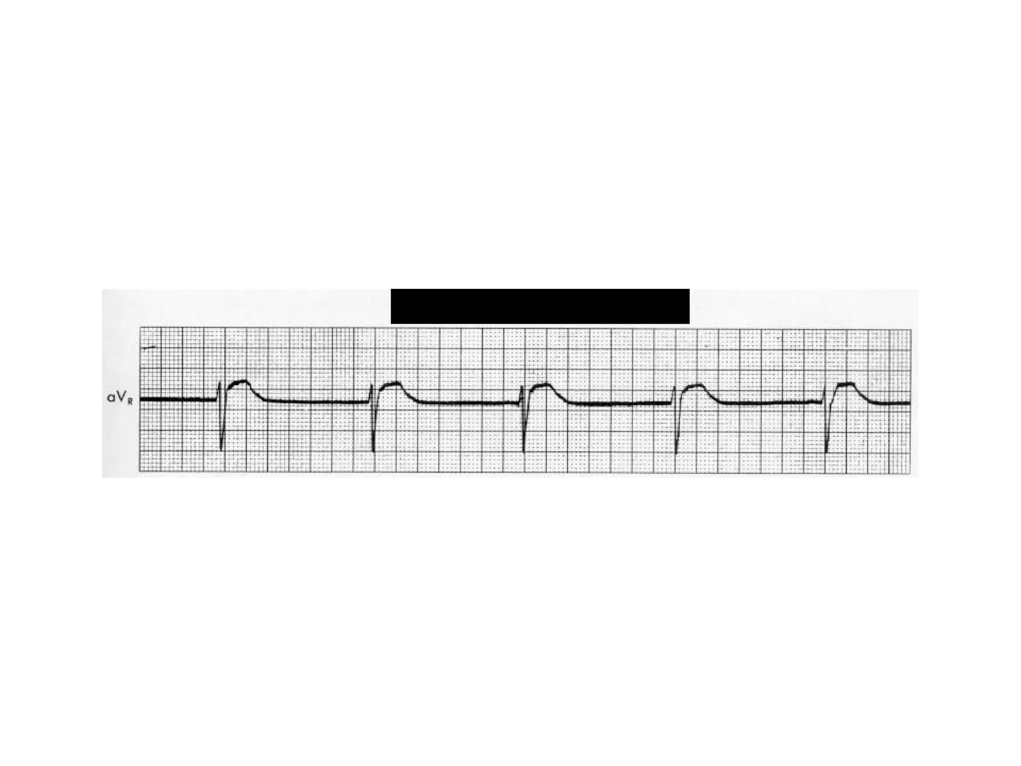
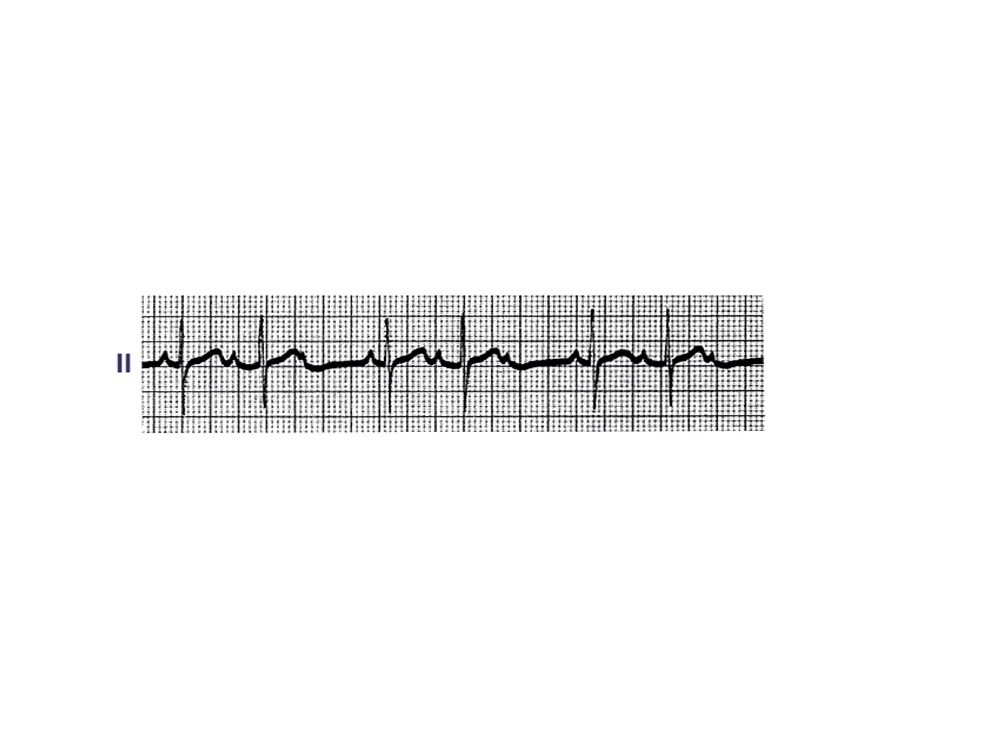
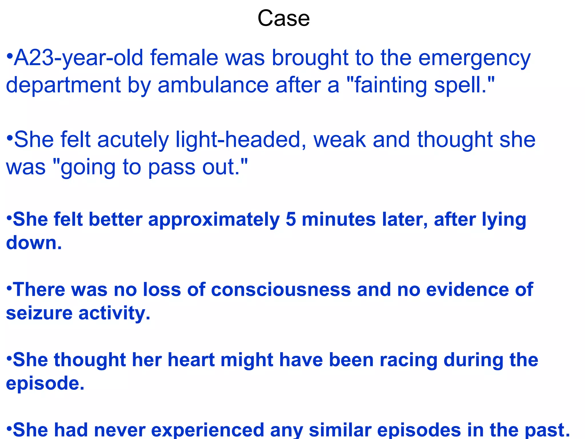
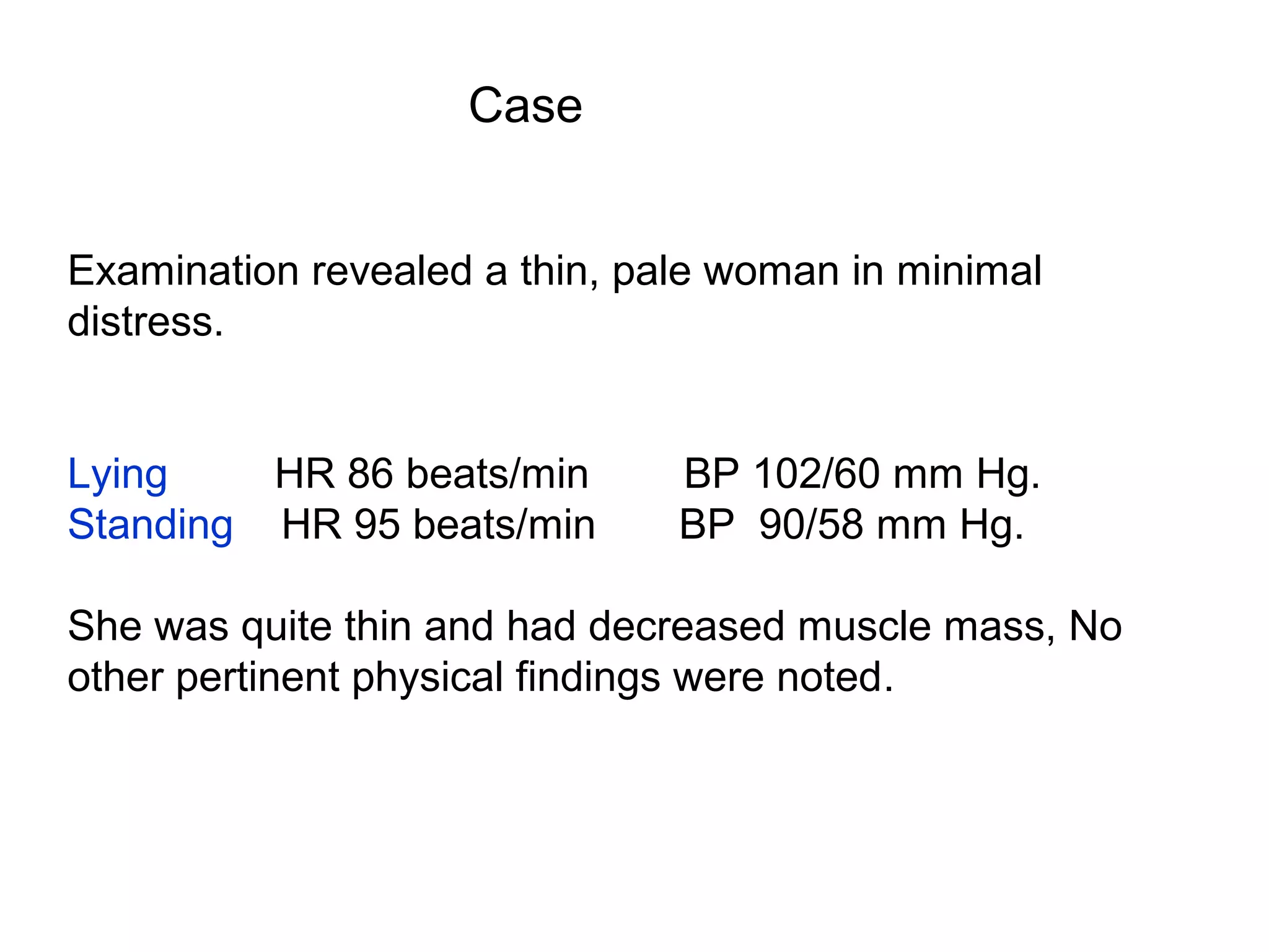
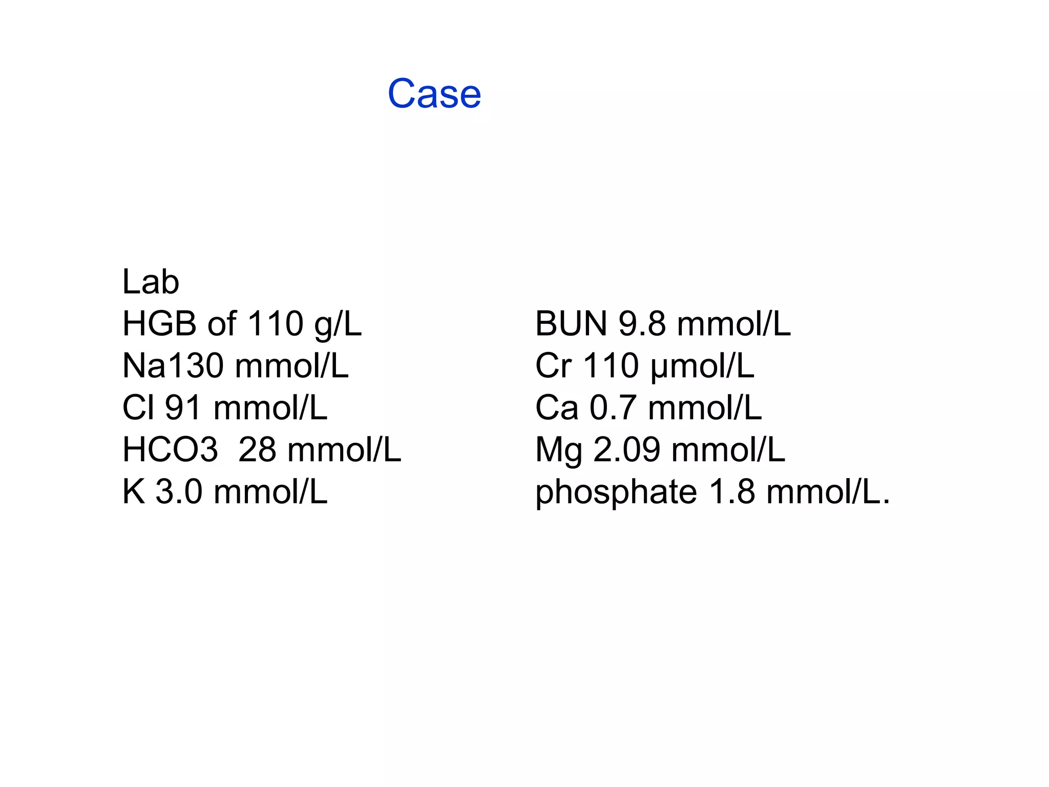
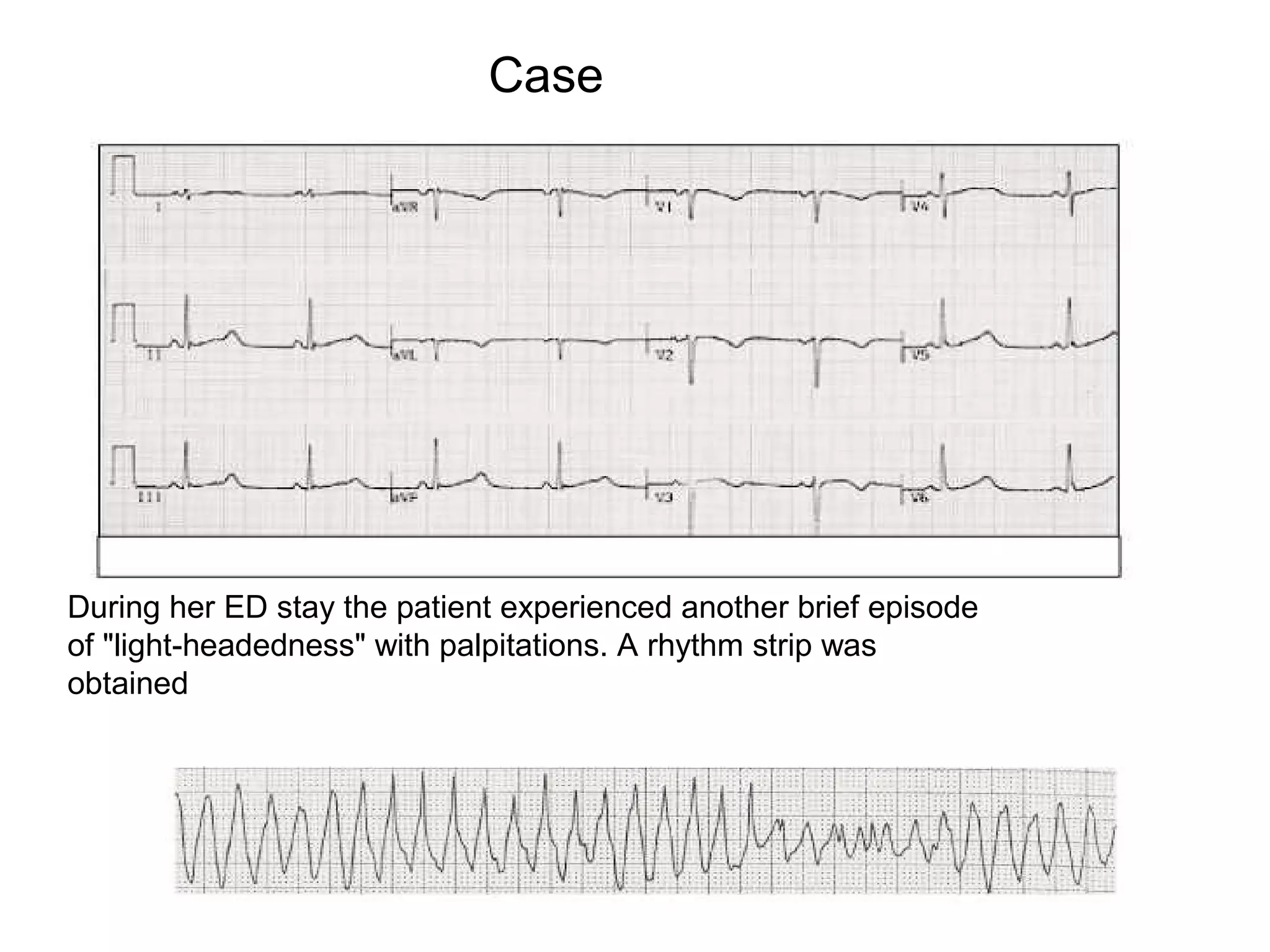
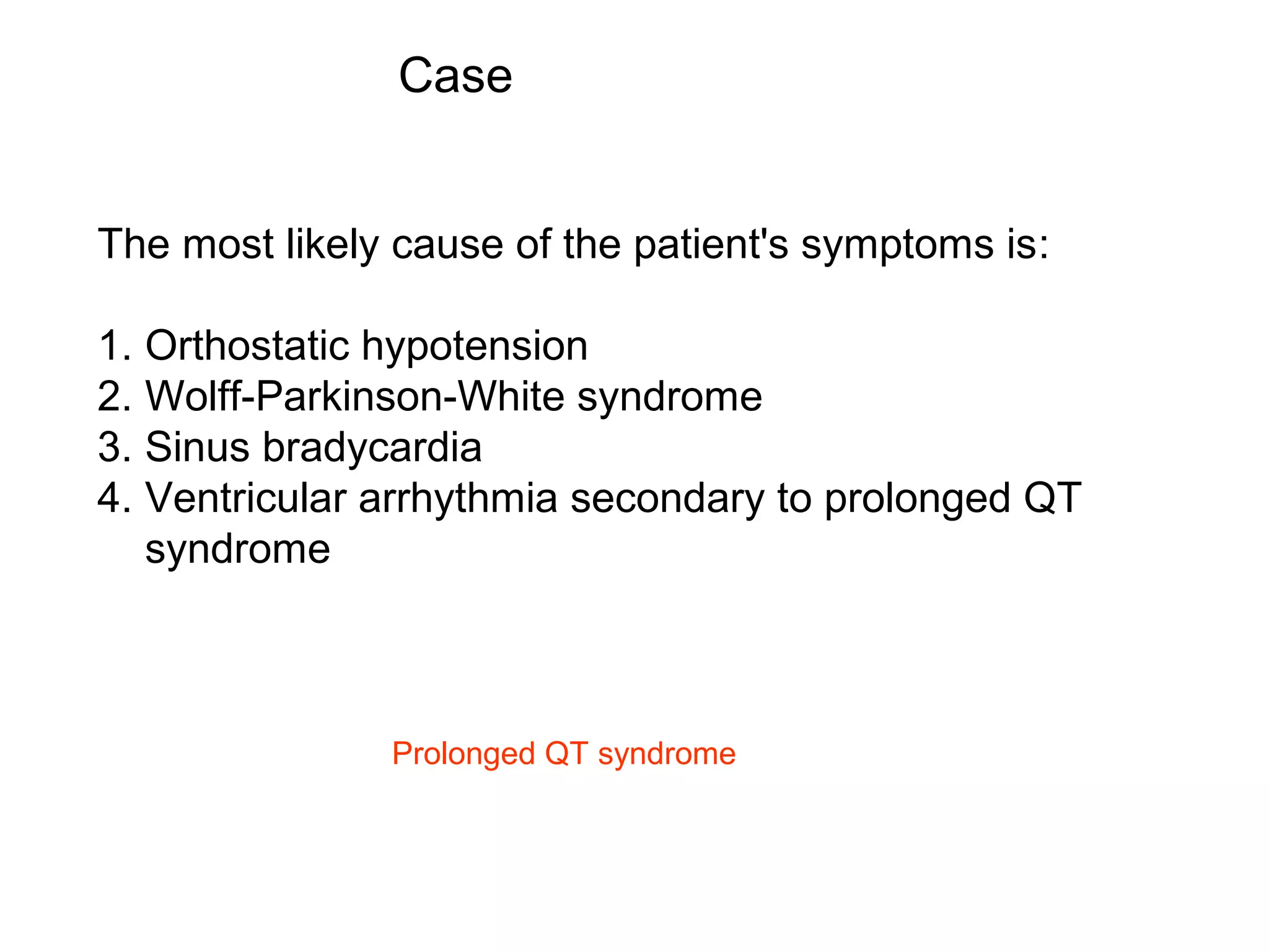
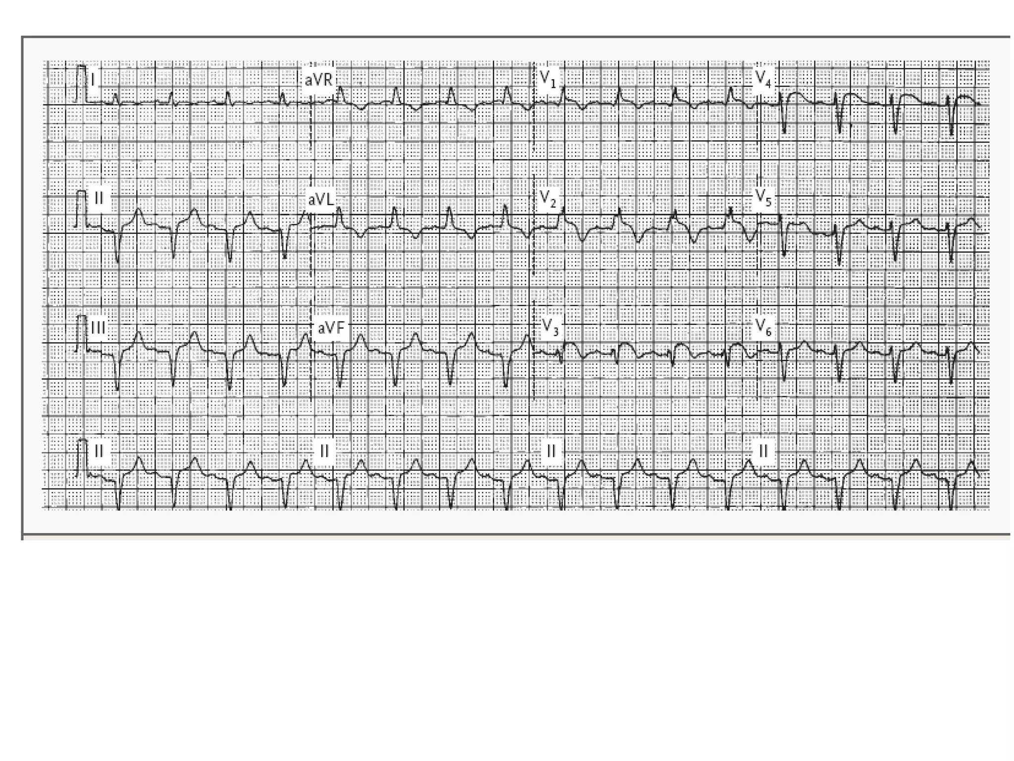
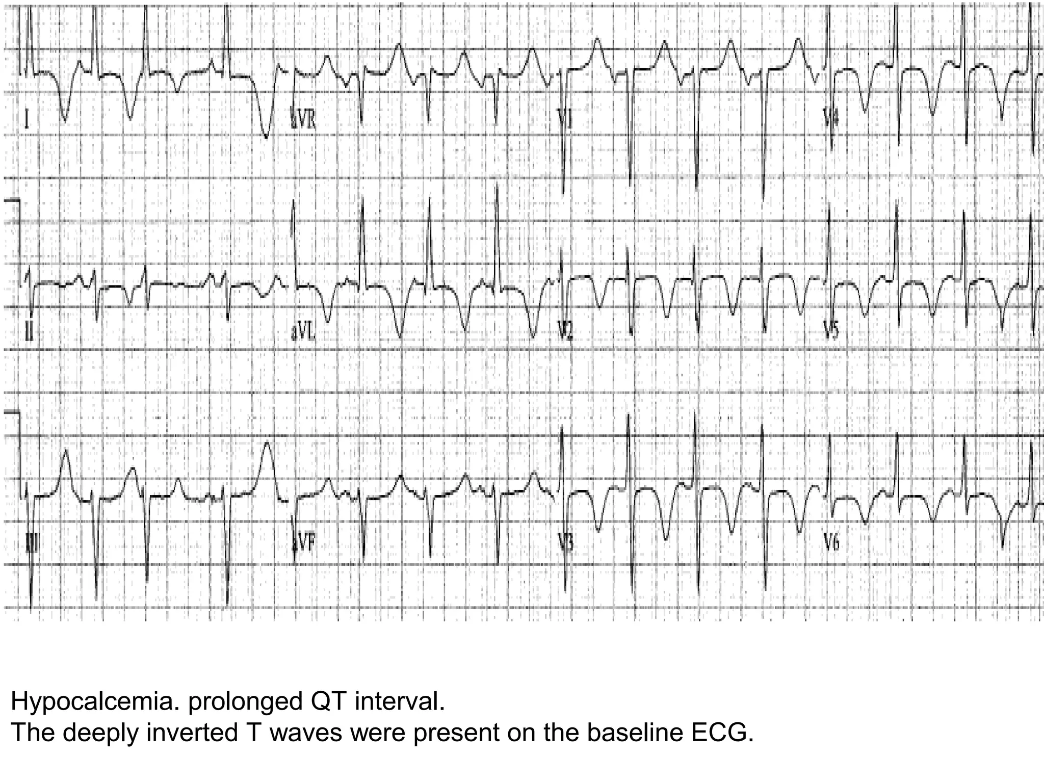
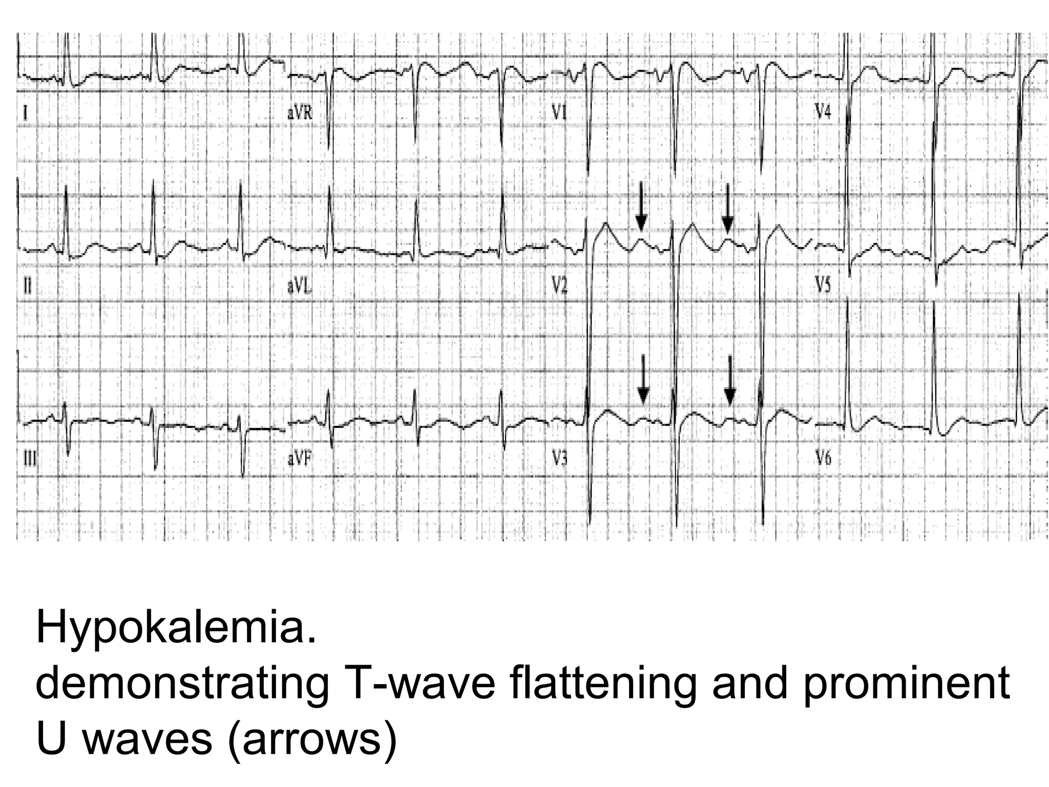
ECG Workshop

Dr. Zohair Alaseri, MD
FRCPc, Emergency Medicine
FRCPc, Critical Care Medicine
Assistant Professor
Chairman, Department of Emergency Medicine
King Saud University Hospitals, Riyadh, KSA
Y. O. F. presented with sever palpitation and waeakness 60
Arrested in 3 minutes from arrival to your department
Arrest ECG

Severe hyperkalemia
prolonged PR interval and
widened QRS complex
year old with sever crushing, heavy chest pain started 45 m. ago 60
with BP of 85/45
Family want to know what is going on & what do you want to do
for their relative??
Elderly man with this rhythm and with previously normal ECG??
State your intervention please??
????Spot DX please
D/D???

Electrical Alternans
????Explain what do you see

Wolff-Parkinson-White pattern
The short PR interval is due to a bypass track, also known as the Kent pathway. By
. bypassing the AV node the PR shortens
The delta wave represents early activation of the ventricles from the bypass tract. The
fusion QRS is the result of two activation sequences, one from the bypass tract and one
. from the AV node
. The ST-T changes are secondary to changes in the ventricular activation sequence
year old lady history of hyperthyroidism presented with 54
palpitation and sweating
Med inderal and digoxin & carbimazol
,y.o. m. with sever submental pain 50
redness and tenderness ass with chest pain
50 y.o. m. with sever submental pain,
redness and tenderness ass with chest pain
Peritonsillar, Parapharyngeal
(contains carotids, jug vein,
cerv symp chain and CN IX XII)
Retropharyngeal (skull base
to mediastinum – T2)
“danger”
(post to retropharyngeal
space from skull base to
diagphragm)
Prevertebral spaces (skull ,coccyx)
Elderly bed ridden man with increased weakness & fatigabulity with
BP of 90/50
What do you want to give??

Prolonged QT
y o m sever chest typical ischemic pain for 30 minutes 70

The deep S waves in the inferior leads and a left axis deviation
. indicates Left Anterior hemiblock+ poor R wave progression
year old lady with suden onset of Palpitation 60

The rhythm is atrial flutter sawtoothed pattern.
The conduction ratio is variable between 12:1 and
6:1
year old with sudden onset of sever dizziness for 15 minutes 18

PR interval is short at about 100ms.
QRS is broad even running into the P wave in lead II.
Initial slurring of the QRS is evident.
This is a case of WPW , and it seems to be intermittent with a
recovery to normal in the last 3 beats of the rhythm strip.
69 y o f with sever pleuritc chest pain and diaphoresis for 30
minutes
PMH DM IHD HTN

Anteroseptal Infarction
The elevated S-T segements in leads V1,V2,V3
65 year old male with sever chest pain radiating to his back
similar to his pain when he had MI 1 year ago
His bp is 90/30

acute anterolateral myocardial infarctio & left anterior
hemiblock
45 year o. f. head of emergency department
with sever headache, nausea & vomiting

Complete heart block third degree AV block & PVC
QS complexes are commonly found in leads V1, and V2 as
a normal variant.
y. o. 2 hours after streptokinase for anterior wall MI 80

Mobitz type II AV block
The ventricular rate is 75. The atrial rate is 150
SVT with a 2:1 block.
P waves are inverted in the inferior limb leads
This points towards a low atrial location as the site of the ectopy.
This is most likely a paroxysmal atrial tachycardia with block
Right bundle branch block & left anterior hemiblock
77 y o m with palpitation history of MI 3 years ago

Wide QRS complex tachycardia.
ventricular tachycardia
The QRS complex is wider than 140 ms.
The R:S ratio in lead V6 is less than 1.
Nodal idiojunctional
year old male sudden loc 30
No PMH
His brother died at age of 39

down-sloping Coved type or less

commonly saddle-back type ST
segment elevation followed by
inverted T waves

Brugada
Second-degree AV block
Mobitz type I AV block
Ventricular fibrillation
y. o. m. heavy smoker with worsening SOB 45

Multifocal atrial tachycardia
different P wave morphologies
Atrial flutter with 2:1 AV conduction.

Atrial flutter with 4:1 AV conduction.
year old with sudden palpitation 17

Atrial fibrillation with ventricular preexcitation

.S. rhythm with ventricular preexcitation
Sinus arrhythmia.
year old with diarrhea and BP of 50/29 7

MAT
Multifocal atrial tachycardia.
The presence of at least three distinct P-wave
morphologies and varying P-P, R-R, and PR
intervals
YEAR OLD WITH SYNCOPE AND HYPOTENSION 65
year old with on/off palpitation 19

WPW Type Preexcitation
There are ectopic P wave before them; therefore
these are PAC's with RBBB aberration
LBBB and 2nd degree AV Block, Mobitz Type II
RBBB + LAFB = Bifascicular block-KH
The LAFB is recognized by the marked left axis deviation (-75 degrees) in the
frontal plane, rS complexes in II, III, aVF, and the tiny q-wave in aVL.
RBBB plus Mobitz II 2nd Degree AV Block
In this rhythm strip of sinus arrhythmia, the faster rates have a LBBB
morphology. In some patients with a diseased left bundle branch, the
onset of LBBB usually occurs initially as a rate-dependent block; i.e., the
left bundle fails to conduct at the faster rate because of prolonged
refractoriness
year old with pulmonary edema 50

Advanced Hyperkalemia
Marked widenening of the QRS duration combined with tall, peaked T
waves are suggestive of advanced hyperkalemia. Note the absence of P
waves, suggesting a junctional rhythm, but in hyperkalemia the atrial
muscle may be paralyzed while still in sinus rhythm. The sinus impulse
conducts to the AV node through internodal tracts without activating
the atrial muscle
Lateral Myocardial Infarction
Case
•A23-year-old female was brought to the emergency
department by ambulance after a "fainting spell."
•She felt acutely light-headed, weak and thought she
was "going to pass out."
•She felt better approximately 5 minutes later, after lying
down.
•There was no loss of consciousness and no evidence of
seizure activity.
•She thought her heart might have been racing during the
episode.
•She had never experienced any similar episodes in the past.
Case

PMH revealed a 3-year history of anorexia nervosa.
Over the past 4 weeks she had significantly reduced
her oral intake and had lost approximately 20 lbs.
Her present weight was 90 lbs.
She smoked half a pack of cigarettes a day and
denied recent alcohol intake.
Case
Examination revealed a thin, pale woman in minimal
distress.
Lying
HR 86 beats/min
Standing HR 95 beats/min

BP 102/60 mm Hg.
BP 90/58 mm Hg.

She was quite thin and had decreased muscle mass, No
other pertinent physical findings were noted.
Case

Lab
HGB of 110 g/L
Na130 mmol/L
Cl 91 mmol/L
HCO3 28 mmol/L
K 3.0 mmol/L

BUN 9.8 mmol/L
Cr 110 µmol/L
Ca 0.7 mmol/L
Mg 2.09 mmol/L
phosphate 1.8 mmol/L.
Case

During her ED stay the patient experienced another brief episode
of "light-headedness" with palpitations. A rhythm strip was
obtained
Case
The most likely cause of the patient's symptoms is:
1. Orthostatic hypotension
2. Wolff-Parkinson-White syndrome
3. Sinus bradycardia
4. Ventricular arrhythmia secondary to prolonged QT
syndrome

Prolonged QT syndrome
60 year old female presented with sever
dizziness and sweating started 2 hours ago
20 minute later C/O C_P
65 y. o. m. p. with typical CP.
(TCA ingestion
sinus tachycardia
widened QRS complex, deep S wave in lead I,
and prominent R wave in lead aVR.
Ventricular paced rhythm with underlying
complete heart block
A 68-year-old man presented with chest pain radiating to the
. left arm of 3 hours’ duration
.He was diaphoretic and pale

ST-segment depression, prominent R wave, and upright T waves in leads V1 to
V3 in addition, ST-segment depression was seen in the inferior and lateral leads
Posterior Leads

V7, posterior axillary
line;
V8, posterior
scapular line;
V9, left border of
spine.

All in the same
horizontal plane of V4
to V6
ST-segment elevation in leads V8 and V9
minimal ST-segment depression with large R waves in the right
. precordial leads (V1 to V3), confirming posterior wall AMI
Inferior & RV MI

•Inferior STE  &  reciprocal STD 
•STE in lead V1 is typical for RV AMI 
 
• widespread STE
V1R is same as standard V2, 
V2R is the same as standard V1,
V3R is halfway between V2R 
V4R, V4R is fifth intercostals 
space at right midclavicular line,
 
V5R is same level as V4R in right 
anterior axillary line
V6R is same level in right 
midaxillary line
70-yr-old man presents with severe chest pain, nausea, vomiting and
diaphoresis. Right-sided chest leads are shown (i.e., V1R to V6R).

• A. fib with subtle signs of acute inferior (MI) with ST elevations 
intermittently apparent in leads II, II and aVF. 
• The right-sided chest leads show evidence of concomitant (RV) 
MI with slight ST elevations in leads V4R to V6R. 
SVT
Multifocal atrial tachycardia.
Wide complex tachycardia: rate-related BBB.

After adenosine became
VT
Ventricular Tachycardia.
Ventricular tachycardia with AV 
dissociation. 
(arrows denote regular P waves, although 
Wide complex tachycardia. A fusion beat 
(arrow) is seen in the rhythm strip;
its presence confirms VT.
Accelerated idioventricular rhythm. 
Wolff Parkinson White syndrome and atrial 
fibrillation. 
(A) Hypercalcemia shortened QT interval.prominent U waves (arrows) 
(B) Normocalcemia. This is baseline ECG from the same patient as in (A) when her 
serum calcium level was normalized (7.8 mg/dL). 
Hypocalcemia. prolonged QT interval. 
The deeply inverted T waves were present on the baseline ECG.
(A) Hyperkalemia. B) Resolving hyperkalemia..
Sine wave rhythm of hyperkalemia. 
The patient was administered IV calcium 
gluconate, causing an immediate narrowing 
of the QRS complex.
Hyperkalemia. 
minimal narrowed peaking of the T waves. 
Hypokalemia. 
demonstrating T-wave flattening and prominent 
U waves (arrows)
Thank You
Ecg workshop

Ecg workshop

Editor's Notes

  • #85 Note the small pacemaker spikes before the QRS complexes in many of the leads. In addition, the QRS complex in V1 exhibits ventricular ectopic morphology; i.e., there is a slur or notch at the beginning of the S wave, and >60ms delay from onset to QRS to nadir of S wave. This rules against a supraventricular rhythm with LBBB