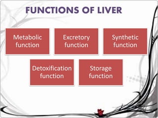
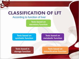
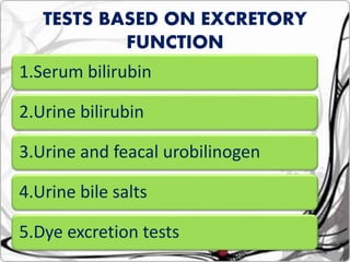
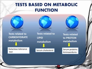
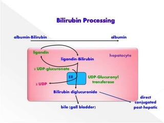
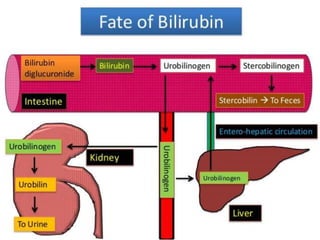
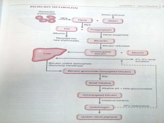
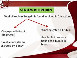
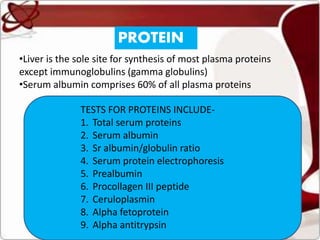
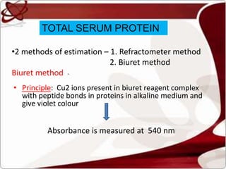
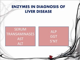
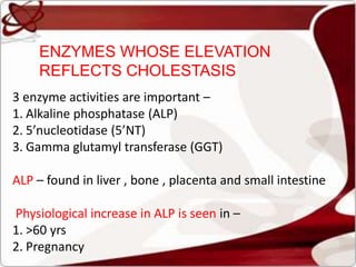
Liver function tests can detect, distinguish, and monitor various types of liver disease. Tests are grouped based on the liver's excretory, metabolic, detoxification, storage and synthetic functions. Enzyme tests like ALT and AST indicate hepatocyte damage, while elevated ALP and GGT suggest cholestasis. Protein tests such as albumin, PT and AFP evaluate synthetic function. Bilirubin, bile salts and dye excretion tests examine excretory function. Together these tests provide insight into liver health and disease categories.
The liver is the largest organ in the body
It is located below the diaphragm in the right upper quadrant of the abdominal cavity and extended approximately from the right 5th rib to the lower border of the rib cage.
The liver is the largest organ in the body
It is located below the diaphragm in the right upper quadrant of the abdominal cavity and extended approximately from the right 5th rib to the lower border of the rib cage.
Liver function tests and their clinical applicationsrohini sane
A comprehensive presentation on Liver Function Tests and their clinical applications for MBBS, BDS, B Pharm & Biotechnology students to facilitate self- study.
Renal function tests are very useful for effective clinical evaluation of renal failure for effective management. So it is useful for medical and allied professional students and clinical practitioners.
Serum Protein and Albumin-Globulin RatioASHIKH SEETHY
For MBBS Biochemistry Practical. Explains various methods of protein estimation and estimation of AG ratio, conditions leading to alterations in AG ratio etc.
LIVER FUNCTION TESTS BY DR. PREMJEET KAUR, ASSISTANT PROFESSOR BIOCHEMISTRY Premjeet Kaur
BY THE END OF THIS PRESENTATION YOU WILL BE ABLE TO ANSWER WHAT, WHY, WHICH ABOUT LIVER FUNCTION TESTS , WHAT IS JAUNDICE , METABOLISM OF HEME , FORMATION OF BILE PIGMENTS FROM HEME , TRASFER OF LILIRUBIN FROM BLOOD TO BILE , DETERMINATION OF SERUM BILIRUBIN, RETENTION JAUNDICE , REGURGITATION JAUNDICE ,DETERMINATION OF AMMONIA IN BLOOD ,ANTIPYRINE TEST, SERUM ENZYMES IN LIVER DISEASE, ASSESING EXTENT OF LIVER DAMAGE , DIAGNOSIS OF SUBCLINICAL JAUNDICE , BCG TEST , PLASMA PROTEINS , DETOXIFICATION FUNCTION OF LIVER
Disorders of liver and kidney, Nitrogen metabolism.pdfshinycthomas
Disorders of liver and kidney – Jaundice, fatty liver, normal and abnormal functions of liver and kidney. Inulin and urea clearance.
Abnormalities of nitrogen metabolism
The prostate is an exocrine gland of the male mammalian reproductive system
It is a walnut-sized gland that forms part of the male reproductive system and is located in front of the rectum and just below the urinary bladder
Function is to store and secrete a clear, slightly alkaline fluid that constitutes 10-30% of the volume of the seminal fluid that along with the spermatozoa, constitutes semen
A healthy human prostate measures (4cm-vertical, by 3cm-horizontal, 2cm ant-post ).
It surrounds the urethra just below the urinary bladder. It has anterior, median, posterior and two lateral lobes
It’s work is regulated by androgens which are responsible for male sex characteristics
Generalised disease of the prostate due to hormonal derangement which leads to non malignant enlargement of the gland (increase in the number of epithelial cells and stromal tissue)to cause compression of the urethra leading to symptoms (LUTS
These lecture slides, by Dr Sidra Arshad, offer a quick overview of physiological basis of a normal electrocardiogram.
Learning objectives:
1. Define an electrocardiogram (ECG) and electrocardiography
2. Describe how dipoles generated by the heart produce the waveforms of the ECG
3. Describe the components of a normal electrocardiogram of a typical bipolar leads (limb II)
4. Differentiate between intervals and segments
5. Enlist some common indications for obtaining an ECG
Study Resources:
1. Chapter 11, Guyton and Hall Textbook of Medical Physiology, 14th edition
2. Chapter 9, Human Physiology - From Cells to Systems, Lauralee Sherwood, 9th edition
3. Chapter 29, Ganong’s Review of Medical Physiology, 26th edition
4. Electrocardiogram, StatPearls - https://www.ncbi.nlm.nih.gov/books/NBK549803/
5. ECG in Medical Practice by ABM Abdullah, 4th edition
6. ECG Basics, http://www.nataliescasebook.com/tag/e-c-g-basics
Prix Galien International 2024 Forum ProgramLevi Shapiro
June 20, 2024, Prix Galien International and Jerusalem Ethics Forum in ROME. Detailed agenda including panels:
- ADVANCES IN CARDIOLOGY: A NEW PARADIGM IS COMING
- WOMEN’S HEALTH: FERTILITY PRESERVATION
- WHAT’S NEW IN THE TREATMENT OF INFECTIOUS,
ONCOLOGICAL AND INFLAMMATORY SKIN DISEASES?
- ARTIFICIAL INTELLIGENCE AND ETHICS
- GENE THERAPY
- BEYOND BORDERS: GLOBAL INITIATIVES FOR DEMOCRATIZING LIFE SCIENCE TECHNOLOGIES AND PROMOTING ACCESS TO HEALTHCARE
- ETHICAL CHALLENGES IN LIFE SCIENCES
- Prix Galien International Awards Ceremony
Factory Supply Best Quality Pmk Oil CAS 28578–16–7 PMK Powder in Stockrebeccabio
Factory Supply Best Quality Pmk Oil CAS 28578–16–7 PMK Powder in Stock
Telegram: bmksupplier
signal: +85264872720
threema: TUD4A6YC
You can contact me on Telegram or Threema
Communicate promptly and reply
Free of customs clearance, Double Clearance 100% pass delivery to USA, Canada, Spain, Germany, Netherland, Poland, Italy, Sweden, UK, Czech Republic, Australia, Mexico, Russia, Ukraine, Kazakhstan.Door to door service
Hot Selling Organic intermediates
Pulmonary Thromboembolism - etilogy, types, medical- Surgical and nursing man...VarunMahajani
Disruption of blood supply to lung alveoli due to blockage of one or more pulmonary blood vessels is called as Pulmonary thromboembolism. In this presentation we will discuss its causes, types and its management in depth.
Tom Selleck Health: A Comprehensive Look at the Iconic Actor’s Wellness Journeygreendigital
Tom Selleck, an enduring figure in Hollywood. has captivated audiences for decades with his rugged charm, iconic moustache. and memorable roles in television and film. From his breakout role as Thomas Magnum in Magnum P.I. to his current portrayal of Frank Reagan in Blue Bloods. Selleck's career has spanned over 50 years. But beyond his professional achievements. fans have often been curious about Tom Selleck Health. especially as he has aged in the public eye.
Follow us on: Pinterest
Introduction
Many have been interested in Tom Selleck health. not only because of his enduring presence on screen but also because of the challenges. and lifestyle choices he has faced and made over the years. This article delves into the various aspects of Tom Selleck health. exploring his fitness regimen, diet, mental health. and the challenges he has encountered as he ages. We'll look at how he maintains his well-being. the health issues he has faced, and his approach to ageing .
Early Life and Career
Childhood and Athletic Beginnings
Tom Selleck was born on January 29, 1945, in Detroit, Michigan, and grew up in Sherman Oaks, California. From an early age, he was involved in sports, particularly basketball. which played a significant role in his physical development. His athletic pursuits continued into college. where he attended the University of Southern California (USC) on a basketball scholarship. This early involvement in sports laid a strong foundation for his physical health and disciplined lifestyle.
Transition to Acting
Selleck's transition from an athlete to an actor came with its physical demands. His first significant role in "Magnum P.I." required him to perform various stunts and maintain a fit appearance. This role, which he played from 1980 to 1988. necessitated a rigorous fitness routine to meet the show's demands. setting the stage for his long-term commitment to health and wellness.
Fitness Regimen
Workout Routine
Tom Selleck health and fitness regimen has evolved. adapting to his changing roles and age. During his "Magnum, P.I." days. Selleck's workouts were intense and focused on building and maintaining muscle mass. His routine included weightlifting, cardiovascular exercises. and specific training for the stunts he performed on the show.
Selleck adjusted his fitness routine as he aged to suit his body's needs. Today, his workouts focus on maintaining flexibility, strength, and cardiovascular health. He incorporates low-impact exercises such as swimming, walking, and light weightlifting. This balanced approach helps him stay fit without putting undue strain on his joints and muscles.
Importance of Flexibility and Mobility
In recent years, Selleck has emphasized the importance of flexibility and mobility in his fitness regimen. Understanding the natural decline in muscle mass and joint flexibility with age. he includes stretching and yoga in his routine. These practices help prevent injuries, improve posture, and maintain mobilit
micro teaching on communication m.sc nursing.pdfAnurag Sharma
Microteaching is a unique model of practice teaching. It is a viable instrument for the. desired change in the teaching behavior or the behavior potential which, in specified types of real. classroom situations, tends to facilitate the achievement of specified types of objectives.
Title: Sense of Smell
Presenter: Dr. Faiza, Assistant Professor of Physiology
Qualifications:
MBBS (Best Graduate, AIMC Lahore)
FCPS Physiology
ICMT, CHPE, DHPE (STMU)
MPH (GC University, Faisalabad)
MBA (Virtual University of Pakistan)
Learning Objectives:
Describe the primary categories of smells and the concept of odor blindness.
Explain the structure and location of the olfactory membrane and mucosa, including the types and roles of cells involved in olfaction.
Describe the pathway and mechanisms of olfactory signal transmission from the olfactory receptors to the brain.
Illustrate the biochemical cascade triggered by odorant binding to olfactory receptors, including the role of G-proteins and second messengers in generating an action potential.
Identify different types of olfactory disorders such as anosmia, hyposmia, hyperosmia, and dysosmia, including their potential causes.
Key Topics:
Olfactory Genes:
3% of the human genome accounts for olfactory genes.
400 genes for odorant receptors.
Olfactory Membrane:
Located in the superior part of the nasal cavity.
Medially: Folds downward along the superior septum.
Laterally: Folds over the superior turbinate and upper surface of the middle turbinate.
Total surface area: 5-10 square centimeters.
Olfactory Mucosa:
Olfactory Cells: Bipolar nerve cells derived from the CNS (100 million), with 4-25 olfactory cilia per cell.
Sustentacular Cells: Produce mucus and maintain ionic and molecular environment.
Basal Cells: Replace worn-out olfactory cells with an average lifespan of 1-2 months.
Bowman’s Gland: Secretes mucus.
Stimulation of Olfactory Cells:
Odorant dissolves in mucus and attaches to receptors on olfactory cilia.
Involves a cascade effect through G-proteins and second messengers, leading to depolarization and action potential generation in the olfactory nerve.
Quality of a Good Odorant:
Small (3-20 Carbon atoms), volatile, water-soluble, and lipid-soluble.
Facilitated by odorant-binding proteins in mucus.
Membrane Potential and Action Potential:
Resting membrane potential: -55mV.
Action potential frequency in the olfactory nerve increases with odorant strength.
Adaptation Towards the Sense of Smell:
Rapid adaptation within the first second, with further slow adaptation.
Psychological adaptation greater than receptor adaptation, involving feedback inhibition from the central nervous system.
Primary Sensations of Smell:
Camphoraceous, Musky, Floral, Pepperminty, Ethereal, Pungent, Putrid.
Odor Detection Threshold:
Examples: Hydrogen sulfide (0.0005 ppm), Methyl-mercaptan (0.002 ppm).
Some toxic substances are odorless at lethal concentrations.
Characteristics of Smell:
Odor blindness for single substances due to lack of appropriate receptor protein.
Behavioral and emotional influences of smell.
Transmission of Olfactory Signals:
From olfactory cells to glomeruli in the olfactory bulb, involving lateral inhibition.
Primitive, less old, and new olfactory systems with different path
MANAGEMENT OF ATRIOVENTRICULAR CONDUCTION BLOCK.pdfJim Jacob Roy
Cardiac conduction defects can occur due to various causes.
Atrioventricular conduction blocks ( AV blocks ) are classified into 3 types.
This document describes the acute management of AV block.
Ozempic: Preoperative Management of Patients on GLP-1 Receptor Agonists Saeid Safari
Preoperative Management of Patients on GLP-1 Receptor Agonists like Ozempic and Semiglutide
ASA GUIDELINE
NYSORA Guideline
2 Case Reports of Gastric Ultrasound
2. SEVERAL BIOCHEMICAL TESTS
CAN BE USED –
•To detect presence of liver disease
•Distinguish among different types of liver disorders
•Gauge the extent of known liver disease
•Follow the response to treatment
3. •Liver tests rarely suggest a specific diagnosis
•They suggest a general category of liver disease,
such as hepatocellular or cholestatic
•This further directs the evaluation
5. CLASSIFICATION OF LFT
According to function of liver
Tests based on
excretory function
Tests based on
metabolic function
Tests based on
detoxification function
Tests based in
storage function
Tests based on
synthetic function
9. Tests related to
CARBOHYDRATE
metabolism
Tests related to
LIPID
metabolism
Tests related
to PROTEIN
metabolism
Galactose tolerance
test
Serum cholesterol Serum proteins
Aminoaciduria
TESTS BASED ON METABOLIC
FUNCTION
18. SERUM BILIRUBIN
Total bilirubin (<1mg/dl) is found in blood in 2 fractions
•Conjugated bilirubin
(<0.3mg/dl)
•Soluble in water so
excreted by kidney
•Unconjugated bilirubin
•Insoluble in water so
bound to albumin in
blood
19. Significance of hyperbilirubinemia
•Daily production of bilirubin <500mg
•But normal liver can conjugate upto 1500mg/day
•So plasma bilirubin concentration is an insensitive test
for liver disease - since it begins to rise only after
significant liver damage has occured
20. CAUSES OF HYPERBILIRUBINEMIA
Isolated increase in unconjugated bilirubin is due to –
1. Hemolytic disease
2. Genetic disorders – crigler najjar and gilbert’s syndrome
3. Neonatal jaundice/physiological jaundice
Isolated increase in conjugated bilirubin is due to –
1. Cholestasis
2. Genetic disorders – Dubin johnson syndrome and rotor’s
syndrome
Increase in both conjugated and unconjugated bilirubin is due
to –
1. Intrahepatic /liver disorders
21.
22.
23. Pigment deposition in
hepatocytes in DJ syndrome
• Coarsely- granular brown pigment
• Diffuse but more heavily concentrated in the
perivenular zone
• grey to black colour grossly
• pigment shares some of its physiochemical
properties with lipofuscin and melanin
• Oil Red O-positive (in frozen sections), stains
black with the Fontana stain
• autofluorescent when examined by ultraviolet
microscopy
24. DELTA BILIRUBIN / BILIPROTEIN
•When diseases/cholestasis prevents excretion of
conjugated bilirubin into bile
it enters the plasma
Filtered by the kidneys
Excreted in urine
•Some monoconjugated bilrubin can become
covalently bound to albumin
25. •This protein bound conjugated bilirubin is
known as - biliprotein or delta – bilirubin
•Normally present in very small amount
•Increases in cholestasis
•Half life is longer – 20 days (like albumin)
•Normally half life of conjugated bilirubin is 24
hrs
27. DIAZO METHOD
•Sample – Serum or plasma EDTA or heparin
•Note – protect from light
•Exposure to direct sunlight can decrease bilirubin
in samples by 50% within one hour
28. Van Den Bergh reaction
Serum bilirubin Diazotized sulphanilic acid
(Ehrlich diazo reagent)
Azobilirubin( red)
Direct bilirubin – reading is taken at one minute
Add activator /accelerator
2nd reading at 30 minutes – Total bilirubin
Measure absorbance at 600nm
29. Test Principle Comments
Evelyn Malloy method Diazo method – product is
azobilirubin
Activator – ethanol
Readings- at one minute and 30
minutes
Sensitive to pH changes and
serum protein changes
Overestimates dB
Jendrassik-Groff Diazo method
Product- azobilirubin
Activator – caffeine sodium
benzoate
Insensitive to pH changes
And serum protein changes
Direct
Spectrophotometric
Method
Absorbance of bilirubin in serum at
455 nm
Abs Hb at 455 nm is corrected by
subtracting the
abs at 575 nm.
Insensitive to hemolysis
Affected by the presence of
lipochromes and turbidity
Only used in new born infants
HPLC Alkaline methanolysis method
Conjugated forms are methylated
Unconjugated forms remain
unchanged
Extracted into chloroform
30. URINE BILIRUBIN
•Any bilirubin found in urine is conjugated bilirubin
•Presence of bilirubinuria is s/o liver disease
•Can be tested by dipstick test
•When dipstick test show presence of urine bilirubin
– confirmatory test to be done
•Ictotest tablet – based on diazotization reaction –
coupling of solid diazonium salt with bilirubin in
an acidic medium gives a
blue/purple color
31. •Ictotest can be used to rule out presence of any
interfering substance that give false positive reaction
with dipstick test
• While recovering from jaundice urine bilirubin clears
before serum bilirubin
•Presence of bilirubin in urine impart it dark brown
color
• Also fouchet’s test done – if bilirubin is
present a green color develops
due to formation of biliverdin
32. Colour Cause
Orange Conc. Urine , urobilin , drugs
Orange-reddish
brown
Drugs , rhubarb ingestion
Dark brown Altered blood , myoglobin, porphyrins , phenolic drugs
Red Blood, beetroot or blackberry ingestion , food dyes
Purple-red Phenolphthalein laxative
Brown black Altered blood, melanin, homogentisic acid
33. URINE UROBILINOGEN
• Increase in urine is sensitive indicator of
hepatocellular disease
• It is markedly increased in hemolysis
• In cholestatic jaundice urobilinogen disappears
from urine
• Urine strips are available
• Fresh urine should be used
• Ehrlich’s test – gives
pink-red color
34. BILE SALTS
•Products of cholesterol metabolism
•Facilitate absorption of fat from intestine
•Constitute a substantial amount of bile in bilirubin
excretion and can be used in diagnosing cholestasis
•Primary bile salts – cholate and
chenodeoxycholate are produced in liver
Metabolised by bacteria in intestine
35. Produces secondary bile salts – lithocholate,
deoxycholate and ursodeoxycholate
•In cirrhosis – reduced ratio of primary to secondary
bile salts
•In cholestasis – as secondary bile salts are not
formed – so increased ratio of primary to secondary
bile salts
36. •In normal condition – renal excretion of bile salts is
negligible
•In cholestasis – increased renal excretion of bile salts
•
•For measuremnet – chromatography (HPLC)
•Hay’s test – bile salts when present lower the surface
tension of urine
• When sulphur powder is added to the urine, sulphur
particles sink to the bottom of the tube
• In case of normal urine,
it will float on the surface
37. DYE EXCRETION
TEST
3 synthetic dyes are commonly employed to test liver
function
1. Bromosulphthalein (BSP)
2. Indocyanine green
3. Rose bengal
Bromosulphthalein excretion test –
5ml/kg weight i.v is given
BSP is taken up by hepatocytes, conjugated and
excreted in bile
38. •A blood specimen is taken after 45 mins and 2 hrs
•After 45 mins – if >50% dye is retained in blood –
abnormal Liver function is present
• This test is useful in differentiating dubin johnson
from rotor syndrome
•In DJS – At 45 mins – normal blood levels of BSP
• At 2 hrs – higher level of BSP
•In rotor syndrome – at 45 mins – higher levels
• at 2 hrs – lower levels
40. BLOOD AMMONIA
•Produced in body by normal protein metabolism
and by intestinal bacteria
•For detoxification of ammonia
In liver
converted to urea
Excreted by kidneys
In striated muscles
Combines with glutamic acid
Forms glutamine
42. PROTEIN
•Liver is the sole site for synthesis of most plasma proteins
except immunoglobulins (gamma globulins)
•Serum albumin comprises 60% of all plasma proteins
TESTS FOR PROTEINS INCLUDE-
1. Total serum proteins
2. Serum albumin
3. Sr albumin/globulin ratio
4. Serum protein electrophoresis
5. Prealbumin
6. Procollagen III peptide
7. Ceruloplasmin
8. Alpha fetoprotein
9. Alpha antitrypsin
43. TOTAL SERUM PROTEIN
•2 methods of estimation – 1. Refractometer method
2. Biuret method
Biuret method -
• Principle: Cu2 ions present in biuret reagent complex
with peptide bonds in proteins in alkaline medium and
give violet colour
Absorbance is measured at 540 nm
44. ALBUMIN
•Synthesized exclusively by liver
•Its half life is 18-20 days
•Due to its slow turn over – not a good indicator of
acute or mild hepatic dysfunction
• In hepatitis - <3g/dl of albumin – possibility of
chronic liver disease
•Non hepatic causes of Hypoalbuminemia -
• Protein losing enteropathy
• Nephrotic syndrome
46. BROMOCRESYL GREEN METHOD
•Most common method
•The complex becomes blue in color
•Absorbance at 632 nm is directly proportional to
the concentration of albumin
47. GLOBULINS
•Made up of – alpha, beta and gamma globulins
•Gamma globulin is produced by plasma cells
• alpha and beta globulins are synthesized in liver
• In cirrhosis – gamma globulin is increased
•Cirrhotic liver fails to clear bacterial antigens that
normally reach the liver – Abs are formed against such Ags
– so , increased gamma globulin
• Polyclonal gamma globulin if increases by 100% -
autoimmune hepatitis
•Increased IgM – primary biliary cirrhosis
•Increased IgA – alcoholic liver disease
48. PREALBUMIN /TRANSTHYRETIN
• Levels fall in liver disease
• Half life – 2 days
• Sensitive indicator of any changes affecting its
synthesis and catabolism
• PAB is a negative acute phase reactant
• Particularly useful in drug-induced
hepatotoxicity
52. PROCOLLAGEN III PEPTIDE
• Cleavage product of the type III procollagen
molecule
• Radioimmunoassay
• Elevated Conc. Of PIIIP- the transformation of
viable hepatic tissue into connective
tissue/fibrosis
53. • AFP is a gp and MW – 70,000 daltons
• Normally present in fetus
• Liver, yolk sac and small intestine
• AFP- ELISA
HCC
Non seminomatous testicular cancer
Ataxia telangiectasia
Hereditary tyrosinemia
Neonatal hyperbilirubinemia
Chronic active hepatitis
α- FETO PROTEIN
54. COAGULATION FACTORS
•With the exception of F-VIII , all other factors are
synthesized in liver
•Half life ranges from 6hrs for F-VII to 5 days for fibrinogen
• So their measurement is the single best measure of
hepatic synthetic function
• Tests – Serum prothrombin time
•Marked increase in PT >5secs above the control and not
corrected by Vit K administration – is a poor prognostic
sign in acute viral hepatitis and other acute and
chronic liver disease
57. 1.Enzymes whose elevation reflects damage to
hepatocytes
2. Enzymes whose elevation reflects cholestasis
3. Enzyme test that do not fit into either pattern
• ENZYMES WHOSE ELEVATION REFLECTS DAMAGE TO
HEPATOCYTES
• AMINOTRANSFERASES (transaminases) –
They include AST and ALT
SERUM EMZYME TESTS ARE
GROUPED IN 3 CATEGORIES
58. • AST(SGOT) – found in liver> cardiac muscle > skeletal
muscle> kidneys >brain
• ALT(SGPT) – found primarily in liver
• Normally present in serum in low concentration (0-40
IU/L)
• When there is damage to liver cell membrane –
increased permeability and so increased serum
concentration
• Liver cell necrosis is not required for release of these
enzymes
59. •Levels of >1000 IU/L occurs in –
• Acute viral hepatitis
• Toxin and drug induced hepatitis
• Ischaemic liver injury
• In most acute hepatocellular disorders ALT is higher or
equal to AST
60. • Normal ratio is 0.7 to 1.4
• Useful in Wilson disease, chronic liver disease
and alcoholic liver disease
• AST/ALT ratio of > 2:1 is suggestive of and >3:1
is highly suggestive of ALCOHOLIC liver disease
• AST in Alcoholic live disease is rarely >300 IU/L
AST/ALT RATIO
61. • ALT is usually normal in alcoholic liver disease ; can
be sometimes low due to an alcohol induced
deficiency of pyridoxal phosphate
• AST/ALT <1 is seen in NASH and viral hepatitis
62. • 2 forms of AST are known-
1. Cytosolic
2. Mitochondrial (mAST) – it is synthesized in
precursor form (pre-mAST)
converted to mature mAST
• Accounts for 80% of total AST activity within liver
cells
63. • mAST/total AST ratio – marker of chronic alcohol
consumption
• This distinguishes those who consume excess alcohol
from normal subjects irrespective of the presence or
absence of liver disease
64. • Isolated rise of ALT is seen in
1. Chronic HepC infection
2. Fatty liver
• Isolated AST elevation
1. Alcohol -related
2. Drug -induced liver injury,
3. Hemolysis
4. Myopathic processes
65. •Determination of these enzymes are helpful in
distinguishing hepatocellular from cholestatic
jaundice
•Increase in AST and ALT is much more ( >500 IU/L)in
hepatocellular jaundice than in cholestatic jaundice
(>200 IU/L)
•Persistence of elevated ALT and AST beyond 6
months in a case of hepatitis indicates development
of chronic hepatitis
66. •This AST procedure utilizes a modification of the
methodology recommended by the IFCC(International
Federation of Clinical Chemistry)
• In this method, aspartate aminotransferase (AST)
catalyzes the transamination of aspartate and α-
oxoglutarate, forming L-glutamate and oxalacetate
• The oxalacetate is then reduced to L-malate by malate
dehydrogenase, while NADH is simultaneously
converted to NAD+
67. •The decrease in absorbance due to the consumption
of NADH is measured at 340 nm and is proportional to
the AST activity in the sample
• L-Aspartate + α-Oxoglutarate ---------AST----------------
L-Glutamate + Oxalacetate
• Oxalacetate + NADH + H+ ------------MDH---------------
L-Malate + NAD+
68. • ALT procedure is based on a modification of the
methodology recommended by the International
Federation of Clinical Chemistry (IFCC)
• ALT transfers the amino group from alanine to α-
oxoglutarate to form pyruvate and glutamate
•The pyruvate enters a lactate dehydrogenase (LD)
catalyzed reaction with NADH to produce lactate and
NAD+
69. The decrease in absorbance due to the consumption of
NADH is measured at 340nm and is proportional to the
ALT activity in the sample
70.
71. 3 enzyme activities are important –
1. Alkaline phosphatase (ALP)
2. 5’nucleotidase (5’NT)
3. Gamma glutamyl transferase (GGT)
ALP – found in liver , bone , placenta and small intestine
Physiological increase in ALP is seen in –
1. >60 yrs
2. Pregnancy
ENZYMES WHOSE ELEVATION
REFLECTS CHOLESTASIS
72. 3. Blood groups O and B – after fatty meal influx of
intestinal ALP into blood
4. In children and adolescent during rapid bone
growth
ALP >4 times the Normal is seen in –
1. Cholestatic liver disease
2. Infiltrative liver disease such as cancer and
amyloidosis
3. Paget’s disease of bone
73. • If an isolated increase in ALP is seen , identification
of the source of elevated isoenzyme is helpful-
1. Fractionation of ALP by electrophoresis
2. Different isoenzymes have diiferent susceptibility
to inactivation by heat
• If increased heat stable fraction is found – MC
from placenta
• Most sensitive to heat inactivation is – bone ALP
3. Measure serum levels of GGT and 5’NT – they are
elevated in only liver disease
74.
75. • Serum GGT is increased in –
1. Alcoholism- Is a helpful clue in suspected cases
of alcoholism ( even in absence of alcoholic liver
disease)
2. Cholestasis
• GGT and 5’NT is especially used to assess the
nature of ALP
76. • Normal range: 10-47 IU/L
Serum γ-Glutamyl Transferase
• Normal range: 2-17 IU/L
Serum 5’-Nucleotidase
• Normal range: 39-117 IU/L
Serum alkaline phosphatase
77. LFT IN ANTI TB TREATMENT
•LFT should be performed before starting anti TB
treatment
•With use of rifampicin and isoniazid , onset of liver
damage may be as soon as 10days after commencing
therapy
•Onset of liver injury may be upto 1 year after starting
therapy – so there is no point at which it is
safe to stop performing routine LFT
78. High risk groups are –
1. Known case of liver disease – such as
alcoholic liver disease or hepatitis B or C
infection
2. Malnourished
3. Children and elderly
79.
80. NON INVASIVE BIOMARKERS(NIBM) FOR
ASSESSING LIVER FIBROSIS
•For assessing live fibrosis – liver biopsy is a
preferred method
•Utilization of NIBM for liver histology can reduce
but not replace the requirement for liver biopsy
•Classification of NIBMs
Class 1 fibrosis
markers/ direct
biomarkers
Class 2 fibrosis
markers/indirect
biomarkers
81. DIRECT BIOMARKERS –
•These are parts of liver matrix produced by
stellate cells during fibrosis process
•These include
Procollagen type 1 and 2
Type IV collagen
Hyaluronic acid
Laminin
MMP-1 (collagenases)
MMP-2 (gelatinase A)
MMP-9 (gelatinase B)
TIMPs (tissue inhibitor of matrix metalloproteinase)
82. INDIRECT MARKERS –
• These are molecules released into blood due to liver
inflammation
• These include
1. Serum ALT
2. Serum AST/ALT ratio (AAR) - >1 predictive of
cirrhosis
BARD score – AAR + BMI + diabetes
3. AST/platelet ratio (APRI)
4. Forn’s index – age + 3 lab tests (platelet count +
cholesterol level + GGT)
83. 5. PGA index – a marker to differentiate alcoholic
liver disease from cirrhosis
Includes – GTT + prothrombin index +
apolipoprotein A
Modified – PGAA – additional alpha2
macroglobulin
6. Fibrotest and fibrosure
7. Fibro index
8. FIB – 4 score
9. Fibro Q test
10. Steato test
84. COMBINED DIRECT AND INDIRECT
MARKERS –
Fibrometer test
Fibrospect II test
SHASTA index – used in case of HIV/HCV coinfected patients
Hepascore model
European liver fibrosis panel test (ELF)
85. For assessing fibrosis in HIV/HBV coinfected
patients –
1. Fibrometer
2. Hepascore
3. Zeng’s score
86.
87.
88. REFERENCES
1.Harrison’s – text book of medicine
2.Kawthalkar clinical pathology
3.Henry’s textbook of clinical
pathology