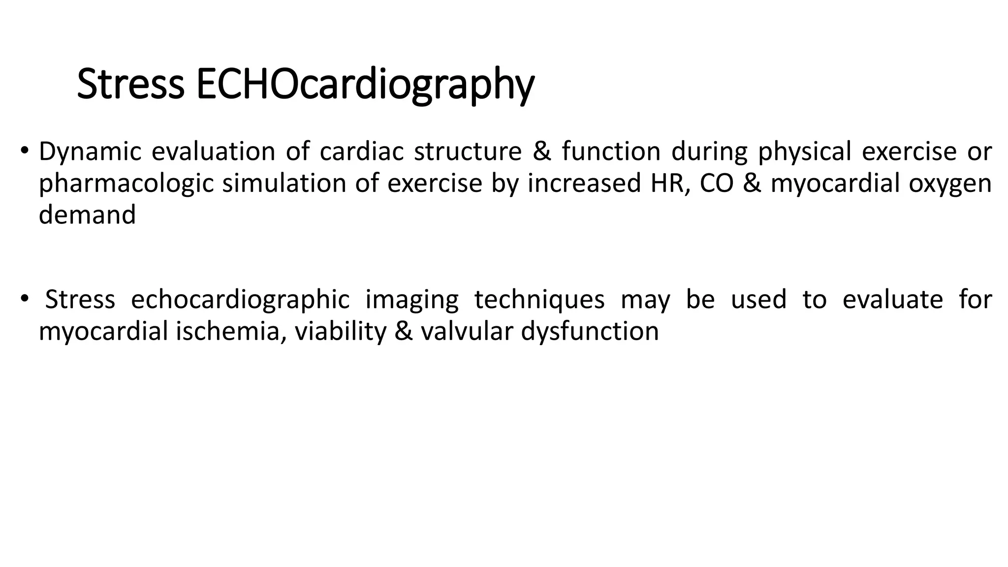
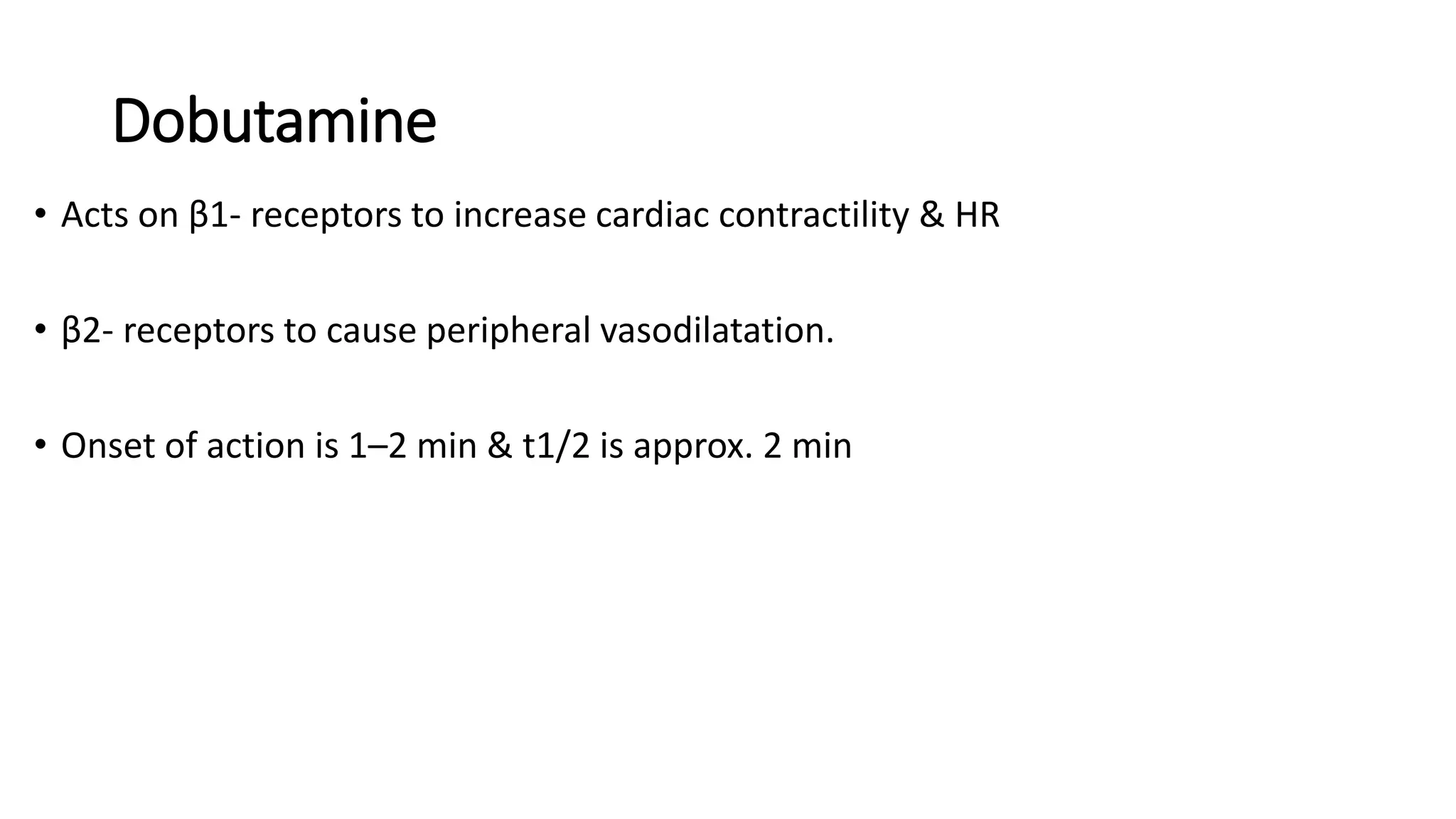
This document summarizes dobutamine stress echocardiography (DSE). Key points include:
- DSE uses the drug dobutamine to simulate exercise and increase heart rate, contractility, and myocardial oxygen demand to detect ischemia.
- It is useful for evaluating ischemia, viability, and valvular dysfunction in patients unable to exercise.
- The document reviews the DSE protocol, interpretation of wall motion abnormalities, indications, side effects, and applications for assessing ischemic heart disease, viability, valvular stenosis like mitral and aortic stenosis, and pulmonary hypertension.
2. Stress ECHOcardiography
ā¢ Dynamic evaluation of cardiac structure & function during physical exercise or
pharmacologic simulation of exercise by increased HR, CO & myocardial oxygen
demand
ā¢ Stress echocardiographic imaging techniques may be used to evaluate for
myocardial ischemia, viability & valvular dysfunction
4. Principles of pharmacologically induced stress
ā¢ Exercise-induced physiologic stress - preferred as
ā¢ Assessment of patientās functional capacity &
ā¢ symptoms reproducibility
ā¢ All major guidelines recommend exercise over pharmacological stress ECHO
ā¢ Patients unable to exercise, pharmacologic stress should be pursued.
ā¢ Commonly used pharmacologic stressors include vasodilators such as adenosine &
dipyridamole, or inotropes, such as dobutamine.
5. Treadmill stress echo
ā¢ Difficult to image while walking
ā¢ Able to get image only post exercise
ā¢ Peak exercise image is not obtainable
ā¢ Should acquire image with in 1 to 1.5 min of exercise
Exercise capacity
Blood pressure response
Arrythmia assesment
6. Do a bicycle ergometry
ā¢ Done in supine position & able to image throughout exercise
ā¢ Image is obtainable at peak exercise
ā¢ Ischemia can induce at lower HR because of increased venous return & preload
in supine position can cause a greater BP response
Difficult to attain the adequate stress
11. In the absence of a flow-limiting coronary stenosis, physiologic stress results in
Increase in
ļ Heart rate
ļ Contractility
ļ Systolic wall thickening
ļ Endocardial excursion
ļ Global contractility
ļ Ejection fraction
Decrease in end-systolic volume
12.
13. severity of the wall motion abnormality (hypokinesis versus dyskinesis) will depend on
ļ¶Magnitude of the blood flow change
ļ¶ Spatial extent of the defect
ļ¶ The presence of collateral blood flow
ļ¶ Duration of ischemia.
14. Dobutamine
ā¢ Acts on Ī²1- receptors to increase cardiac contractility & HR
ā¢ Ī²2- receptors to cause peripheral vasodilatation.
ā¢ Onset of action is 1ā2 min & t1/2 is approx. 2 min
15. ā¢ At low doses - recruits potentially contractile myocardium, & increases
myocardial contractility
ā¢ At high dose - increases myocardial oxygen demand
ā¢ So if flow limiting stenosis present - myocardial ischaemia & deterioration of
regional function d/t demand/supply mismatch.
16. Goal of DSE
ā¢ To detect myocardial ischemia in early stages of CAD by identifying RWMAs under
conditions of pharmacologic stress.
ā¢ Coronary flow limitation initially causes diastolic dysfunction followed by
manifestations of ischemia, including the visualization of systolic dysfunction.
18. ā¢ Adenosine ā direct coronary vasodilator
ā¢ Dipyridamol ā inhibit adenosine uptake
ā¢ As pharmacological stressor both are less poplular
ā¢ Redistibution can occur without RWMA
ā¢ Better suited for nucler imaging that relay on redistirbution
20. Testing Pre-requisites
ā¢ The protocol was defined by ASE(2007) & ESE(2008)
ā¢ Machine should have the ability to trigger image acquisition based on ECG
ā¢ Also machine should have split screen & quadruple screen so that simultaneous comparison
of image is possible
ā¢ Contrast enhancement should be considered to augment endocardial definition when ā„ 2
contiguous endocardial segments not visualized at rest.
21. Testing protocol
ā¢ Typically fasting for 4 hrs
ā¢ The sn of DSE for detection of ischemia may be reduced by Ī²-Bās, CCBās, and nitrates
ā¢ Stop all negative chronotropic agents and nitrates 8 to 12 hr before procedure ā unless,
goal is to assess efficacy of anti-ischemic regimen
ā¢ Graded dobutamine infusion given typically @ starting dose of 5 Ī¼g/kg/min
22. ā¢ Dobutamine dose increased every 3ā5 minutes to doses of 10, 20, 30 & finally to 40
Ī¼g/kg/min
ā¢ The goal is to achieve heart rate 85% of MPHR for patientās age
ā¢ Low-dose dobutamine stage is optimal for detection of ischemia & assessment of viability by
searching for ābiphasic responseā
ā¢ Therefore, if viability assessment is the principal aim, the initial dose is often lowered to 2.5
Ī¼g/kg/min.
23. ā¢ If THR is not achieved with dobutamine alone, atropine (max. dose 2 mg, divided into four
0.5 mg doses) can be added
ā¢ Some also consider trial of increased dose of dobutamine (50 Ī¼g/kg/min) if patient is close
to achieving 85% MPHR
ā¢ Images at each echocardiographic window are obtained during rest, low-dose, intermediate
dose, peak-dose & post-stress
24.
25. End points of the DSE protocol
ā¢ Achievement of the THR
ā¢ Detection of moderate wall motion abnormalities in ā„ 2 territories
ā¢ Symptomatic or sustained arrhythmias,
ā¢ Hypotension or severe hypertension (typically SBP 220ā240 mmHg or DBP 120 mmHg), or
ā¢ Patientās inability to tolerate the test
ā¢ Significant ST-T changes
ā¢ In some cases, reversal of RWMA is assessed with addition of post-stress beta-blockers (typically
1ā5 mg of iv metoprolol).
26. Testing interpretation
ā¢ Stress report must mention
ā¢ the adequacy of stress
o achievement of THR or
o estimated workload,
ā¢ symptoms,
ā¢ blood pressure response,
ā¢ any electrocardiographic (ischemic or arrhythmic) &
ā¢ echocardiographic changes.
ā¢ The typical stress echocardiography report should comment both on global LV
function and regional LV function, along with summative comment on the
significance of the findings
27. ā¢ Hyperdynamic ventricular response
ā¢ Increase in endocardial excursion ā„ 5 mm
ā¢ Uniform thickening during systole
ā¢ Increase in LVEF with decrease in end-sysolic volume
NORMAL RESPONSE?
28. CATEGORIZATION OF WALL MOTION
HYPOKINESIA
AKINESIA
DYSKINESIA
TARDOKINESIA
LESS THAN 30% WALL THICKENING OR <5 MM ENDOCARDIAL
EXCURSION
LESS THAN 10 % WALL THICKENING
SYSTOLIC THINNING AND OUTWARD BULGING
DELAYED INWARD MOTION AND THICKENING
Ischemic response is generally defined as decreased wall thickening in ā„1 segment
29. Pseudodyskinesia (Raisinghani sign)
ā¢ Diastolic flattening of dyskinetic motion of inferoseptal & inferobasilar
segments d/t diaphragmatic compression f/b systolic bulging
ā¢ Causes:
ā¢ Liver failure with severe ascites
ā¢ Hepatomegaly
ā¢ Pregnancy 3rd trimester
ā¢ Where a myocardial territory augments its contraction at low dose but becomes hypokinetic/akinetic at
higher doses
Biphasic response
35. Indications - dobutamine stress echocardiography
Indication Assessment
Risk stratification of IHD Presence & extent of ischemic myocardium as well as viable and/or hibernating
myocardium
Mitral stenosis(MS) 1. Indicated in asymptomatic patients with severe MS,
2. Patients having symptoms discordant with rest echocardiographic measures of
stenosis,
3. Assessment of transvalvular gradients at stress
Aortic stenosis(AS) 1. Differentiation of low gradient AS versus pseudostenosis, and
2. Prognosis in low gradient AS (eg, assessment of contractile reserve)
Pulmonary hypertension Assessment of pulmonary artery systolic pressure at stress when patient is unable to
exercise
36. Ischemic Heart disease
ā¢ Stress echocardiography has highest sp (88%) among ischemic tests including
exercise ECG testing, thallium perfusion imaging, SPECT & PET, hence high NPV
ā¢ More recent data suggest that in patients with LVH, stress echocardiography
may be the test of choice in an ischemic evaluation
ā¢ Also, heterogenous uptake of radionuclide due to LVH may generate a high
false positive rate of perfusion imaging, which is not applicable to stress
echocardiography
40. ā¢ Normal WMSI-1 at baseline and stress
ā¢ Any score>1-abnormal
ā¢ Good prognostic value
ā¢ Help to approach in a standardized and systematic way
41. False negative
ā¢ Submaximal stress
ā¢ Late image acquisition
ā¢ Single vessel disease especially LCx territory
ā¢ Marked LVH and small LV cavity
ā¢ Beta blocker and antianginal
ā¢ Elderly women
42. False positive
ā¢ Hypertensive response to stress
ā¢ Coronary spasm due to dobutamine
ā¢ Paradoxical septal motion in LBBB
ā¢ Myocardial infarction(old)
ā¢ Cardiomyopathy
ā¢ Myocarditis
ā¢ Hibernating myocardium
ā¢ Stunned myocardium
ā¢ Postoperative state
ā¢ Right ventricular volume/pressure overload
45. Types of response
ā¢ Biphasic response- most specific sign of viability
ā¢ Sustained improvement-viable stunned, remodeled or myopathic
ā¢ Deterioration-critical low flow
ā¢ No response- akinetic remain akinetic(non viable scarred)
46.
47.
48. ā¢ Four or more segments out of a total of 16 displaying a biphasic response
exhibited a specificity of 81% for predicting a ā„5% increase in post-
revascularization EF at a median 14-month follow-up of ICMP, with sn of
83% if the EF was >35% & of 92% if the EF was ā¤35%.
ā¢ DSE one week after MI with either an ischemic or biphasic response has a
sensitivity of 82% and a specificity of 80% for detecting a residual stenosis
subtending an area of hibernating myocardium.
ā¢ Myocardial viability identified with stress echocardiography is associated
with improved survival after revascularization
49. Its been shown that the combined nitroglycerin and dobutamine stress
echocardiogram had the highest specificity (83%) but rest-redistribution
thallium single proton emission tomography had the highest sensitivity
(95%) for detecting viable myocardium.
51. ā¢ Stress echocardiography has the ability to comprehensively assess valvular
function, both at rest & during stress & indirectly evaluate hemodynamics in real
time.
ā¢ Stenosis & regurgitation of all valves can be evaluated with stress
echocardiography
ā¢ MC stress echocardiogram applications are in left-sided stenotic lesions. Exercise
echocardiography is the general technique for assessment of valvular disease
except in aortic stenosis with left ventricular dysfunction.
ā¢ There are limited data for assessment of AR & MR by DSE, as there is concern that
the afterload-reducing properties of dobutamine will reduce the degree of valvular
regurgitation.
52. Mitral stenosis
ā¢ The 2007 ASE guidelines recommend stress echocardiography for
evaluation of asymptomatic patients with echocardiographically severe
mitral stenosis and patients with symptoms disproportionate to their
echocardiographic disease
ā¢ The ability to evaluate both valvular function and dynamic transvalvular
gradients is particularly helpful in patients whose symptoms are more
severe than their resting echocardiographic evaluation would suggest.
53. ā¢ A multivariate analysis of rheumatic MS defined a transmitral mean gradient ā„18
mmHg as āhigh riskā & best predictor of future events (eg, heart failure related
to mitral stenosis, surgical or balloon intervention, hemodynamically significant
arrhythmia, or death) with sn - 90% & sp - 87%.
ā¢ These high-risk patients likely warrant more aggressive clinical care and
consideration for earlier more aggressive interventions
54. Whenever there is an Increse
in flow rate across aortic valve
Associated increase in valve area
If flow rate is low valve opening get inhibited lead to
underestimation of valve area
This phenomina is a practical challenge for the doctor because when LV dysfunction is present
quantitative assessment of AS is difficult
Aortic stenosis
55. ā¢ Patients with a low LVEF (<35%ā40%) and a low mean transvalvular gradient (<30ā40
mmHg), called ālow-gradient low output aortic stenosisā, pose a diagnostic dilemma.
ā¢ This is one of the clinical situations where DSE is uniquely helpful.
ā¢ It is very difficult to determine if they have true stenosis & should be considered for valve
replacement, or instead have āpseudostenosisā, namely LV dysfunction that makes aortic
valve appear stenotic, but carries a high risk of dying with valve replacement.
56. ā¢ DSE can be uniquely useful in differentiating these patients and determining who
might benefit from surgical intervention
ā¢ Specifically, patients with aortic valve pseudostenosis are able to increase their aortic
valve area & decrease the transaortic gradient in response to dobutamine on
ā¢ In contrast, patients with true aortic stenosis are unable to increase their valve area in
response to an increased cardiac output, and instead the transvalvular gradient
increases
57. ā¢ Another use of DSE in AS is to assess contractile reserve.
ā¢ Contractile reserve is defined as a 20% increase in stroke volume (as assessed
by the left ventricular outflow tract velocity time integral) or a 20% relative
increase in ejection fraction
ā¢ For patients with low gradient aortic stenosis, surgery is most beneficial for
those with documented left ventricular contractile reserve
ā¢ The postoperative outcomes for patients with low gradient aortic stenosis
and without contractile reserve is poor
58. Cut Offs for Diagnosis
Contractile Reserve ā 20% of stroke volume
Valve area improvement to differentiate true from Pseudostenosis ā 0.2%
Asymptomatic Sev AS, mean gradient rise on exercise - > 20 mmHg
59. Pulmonary hypertension
ā¢ Doppler evaluation of the TR velocity is used to estimate RVSP with a derivation of the
Bernoulli equation (RVSP & PASP will be equal except in RVOTO & PS).
ā¢ Doppler evaluation is feasible both at rest and under stress,
ā¢ It may be helpful in quantifying the severity of pulmonary hypertension as well as
detecting occult or exercise-induced pulmonary hypertension.
60. ā¢ A TR velocity ā¤2.5 m/sec at rest corresponds to PASP of ā¤35 mmHg (assuming RAP of 10
mmHg) & defines ULN at rest.
ā¢ DSE may be used for detecting pulmonary hypertension secondary to left-sided valvular
disease such as MR & AS, although exercise is preferable to DSE if patient can exercise, &
also for regurgitant lesions.
ā¢ Detection of elevated PASP during stress predicts poorer outcomes in these patients &
should prompt consideration of more aggressive valvular intervention
61. PRETRANSPLANT EVALUATION
ā¢ Patients with ESRD are at increased risk of CAD, and CVD is the mc cause of
death after renal transplant.
ā¢ So, accurate assessment of extent & severity of CAD prior to renal
transplant is essential.
ā¢ The accuracy of DSE for detecting CAD in ESRD patients is variable (sn 37%ā
95% & sp 71%ā95%)
ā¢ Recent Cochrane analysis of pretransplant cardiac imaging modalities found
DSE to be superior to myocardial perfusion imagng for CAD detection in
pre-renal transplant patients.
62. ā¢ DSE is also often performed as part of the preoperative evaluation for liver
transplantation
ā¢ its overall sensitivity for CAD is poor, but the test has a reasonable NPV in this
population, making it useful to exclude CAD and perioperative events.
ā¢ DSE as an echocardiographic modality also gives the option of estimating
RVSP may be important given the possibility of portopulmonary hypertension
63. DSE IN WOMEN
ā¢ Women often present with symptoms considered atypical of CAD & are more
underdiagnosed and undertreated.
ā¢ Stress echocardiography provides prognostic information, even after
controlling for clinical and laboratory data, in women with known or suspected
CAD.
ā¢ When exercise ECG was directly compared with exercise echocardiography in
women, exercise or stress echocardiography had a higher sensitivity (81%
versus 77%) and specificity (80% versus 56%) for the detection of CAD.
64. ā¢ Exercise echocardiography also provided the ābest balanceā between
accuracy and cost for the diagnosis of CAD in women.
ā¢ When echocardiography was compared directly with stress thallium-201
SPECT, there was no difference in diagnostic accuracy.
ā¢ These benefits of exercise echocardiography are believed to be shared by
DSE, but exercise would remain the preferred stress modality if the patient is
able to exercise.
ā¢ For young and middle-aged women, stress echocardiography avoids the
long-term risk of radiation-induced carcinogenesis in other analogous
radionuclide-based cardiac imaging modalities.