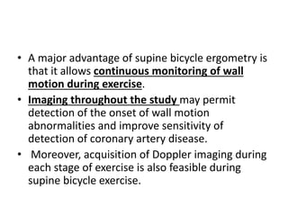
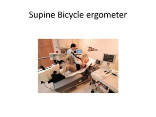
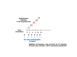
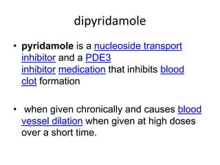
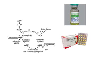
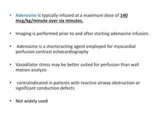
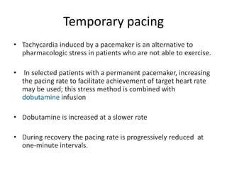
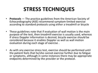
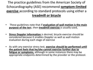
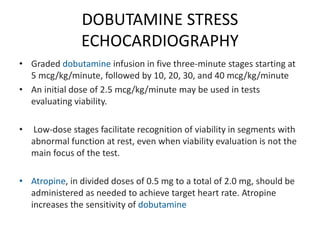
Stress echocardiography enables evaluation of cardiac function at rest and during exercise or pharmacologic stress. It can detect wall motion abnormalities indicative of ischemia and assess valvular function, left ventricular outflow tract gradients, and pulmonary pressures. Exercise or pharmacologic agents like dobutamine are used to induce stress. Indications include evaluating known or suspected coronary artery disease, viability, and valvular diseases. The test is contraindicated in acute coronary syndromes or hemodynamically significant valvular stenosis. Imaging is performed at rest and peak stress to detect new or worsening wall motion abnormalities. Doppler can also evaluate hemodynamic changes with stress.
rotablation is procedure used in complex pci with heavily calcified lesion for adequate expansion of stent.if used in indicated case and well aware of contraindication is necessary for achieving good results.
Speckle tracking echocardiography (STE) is an echocardiographic imaging technique that analyzes the motion of tissues in the heart by using the naturally occurring speckle pattern in the myocardium or blood when imaged by ultrasound.
Stress echocardiography usually done to detect viable myocardium in patients with left ventricular dysfunction who had myocardial infarction. So it helps in deciding the revascularization process.
Exercise Testing in Cardiology : Dr. Akif Baigakifab93
The testing modality and protocol should be selected in accordance with the patient’s estimated functional capacity based on age, estimated physical fitness from the patient’s history, and underlying disease
Several exercise test protocols are available for both treadmill and stationary cycle ergometers
Patients who have low estimated fitness levels or are deemed to be at higher risk because of underlying disease (e.g., recent MI, heart failure) should be tested with a less aggressive exercise protocol
Treadmill and cycle ergometers may use stepped or continuous ramp protocols
Work rate increments (stages) during stepped protocols can vary from 1 to 2.5 METs
Ramp protocols are designed with stages that are no longer than 1 minute and for the patient to attain peak effort within 8 to 12 minutes
This presentation will help physiotherapy students for their theory as well as practical purpose for measuring the exercise tolerance level of the individual.
This presentation includes maximal and sub maximal exercise testing with it's VO2 max formula
This presentation gives brief description of the treadmill test, am-strand cycle ergo-meter test, 6MWT, symptom limited testing, shuttle walk test
- Video recording of this lecture in English language: https://youtu.be/lK81BzxMqdo
- Video recording of this lecture in Arabic language: https://youtu.be/Ve4P0COk9OI
- Link to download the book free: https://nephrotube.blogspot.com/p/nephrotube-nephrology-books.html
- Link to NephroTube website: www.NephroTube.com
- Link to NephroTube social media accounts: https://nephrotube.blogspot.com/p/join-nephrotube-on-social-media.html
Explore natural remedies for syphilis treatment in Singapore. Discover alternative therapies, herbal remedies, and lifestyle changes that may complement conventional treatments. Learn about holistic approaches to managing syphilis symptoms and supporting overall health.
TEST BANK for Operations Management, 14th Edition by William J. Stevenson, Ve...kevinkariuki227
TEST BANK for Operations Management, 14th Edition by William J. Stevenson, Verified Chapters 1 - 19, Complete Newest Version.pdf
Ethanol (CH3CH2OH), or beverage alcohol, is a two-carbon alcohol
that is rapidly distributed in the body and brain. Ethanol alters many
neurochemical systems and has rewarding and addictive properties. It
is the oldest recreational drug and likely contributes to more morbidity,
mortality, and public health costs than all illicit drugs combined. The
5th edition of the Diagnostic and Statistical Manual of Mental Disorders
(DSM-5) integrates alcohol abuse and alcohol dependence into a single
disorder called alcohol use disorder (AUD), with mild, moderate,
and severe subclassifications (American Psychiatric Association, 2013).
In the DSM-5, all types of substance abuse and dependence have been
combined into a single substance use disorder (SUD) on a continuum
from mild to severe. A diagnosis of AUD requires that at least two of
the 11 DSM-5 behaviors be present within a 12-month period (mild
AUD: 2–3 criteria; moderate AUD: 4–5 criteria; severe AUD: 6–11 criteria).
The four main behavioral effects of AUD are impaired control over
drinking, negative social consequences, risky use, and altered physiological
effects (tolerance, withdrawal). This chapter presents an overview
of the prevalence and harmful consequences of AUD in the U.S.,
the systemic nature of the disease, neurocircuitry and stages of AUD,
comorbidities, fetal alcohol spectrum disorders, genetic risk factors, and
pharmacotherapies for AUD.
Lung Cancer: Artificial Intelligence, Synergetics, Complex System Analysis, S...Oleg Kshivets
RESULTS: Overall life span (LS) was 2252.1±1742.5 days and cumulative 5-year survival (5YS) reached 73.2%, 10 years – 64.8%, 20 years – 42.5%. 513 LCP lived more than 5 years (LS=3124.6±1525.6 days), 148 LCP – more than 10 years (LS=5054.4±1504.1 days).199 LCP died because of LC (LS=562.7±374.5 days). 5YS of LCP after bi/lobectomies was significantly superior in comparison with LCP after pneumonectomies (78.1% vs.63.7%, P=0.00001 by log-rank test). AT significantly improved 5YS (66.3% vs. 34.8%) (P=0.00000 by log-rank test) only for LCP with N1-2. Cox modeling displayed that 5YS of LCP significantly depended on: phase transition (PT) early-invasive LC in terms of synergetics, PT N0—N12, cell ratio factors (ratio between cancer cells- CC and blood cells subpopulations), G1-3, histology, glucose, AT, blood cell circuit, prothrombin index, heparin tolerance, recalcification time (P=0.000-0.038). Neural networks, genetic algorithm selection and bootstrap simulation revealed relationships between 5YS and PT early-invasive LC (rank=1), PT N0—N12 (rank=2), thrombocytes/CC (3), erythrocytes/CC (4), eosinophils/CC (5), healthy cells/CC (6), lymphocytes/CC (7), segmented neutrophils/CC (8), stick neutrophils/CC (9), monocytes/CC (10); leucocytes/CC (11). Correct prediction of 5YS was 100% by neural networks computing (area under ROC curve=1.0; error=0.0).
CONCLUSIONS: 5YS of LCP after radical procedures significantly depended on: 1) PT early-invasive cancer; 2) PT N0--N12; 3) cell ratio factors; 4) blood cell circuit; 5) biochemical factors; 6) hemostasis system; 7) AT; 8) LC characteristics; 9) LC cell dynamics; 10) surgery type: lobectomy/pneumonectomy; 11) anthropometric data. Optimal diagnosis and treatment strategies for LC are: 1) screening and early detection of LC; 2) availability of experienced thoracic surgeons because of complexity of radical procedures; 3) aggressive en block surgery and adequate lymph node dissection for completeness; 4) precise prediction; 5) adjuvant chemoimmunoradiotherapy for LCP with unfavorable prognosis.
Report Back from SGO 2024: What’s the Latest in Cervical Cancer?bkling
Are you curious about what’s new in cervical cancer research or unsure what the findings mean? Join Dr. Emily Ko, a gynecologic oncologist at Penn Medicine, to learn about the latest updates from the Society of Gynecologic Oncology (SGO) 2024 Annual Meeting on Women’s Cancer. Dr. Ko will discuss what the research presented at the conference means for you and answer your questions about the new developments.
The prostate is an exocrine gland of the male mammalian reproductive system
It is a walnut-sized gland that forms part of the male reproductive system and is located in front of the rectum and just below the urinary bladder
Function is to store and secrete a clear, slightly alkaline fluid that constitutes 10-30% of the volume of the seminal fluid that along with the spermatozoa, constitutes semen
A healthy human prostate measures (4cm-vertical, by 3cm-horizontal, 2cm ant-post ).
It surrounds the urethra just below the urinary bladder. It has anterior, median, posterior and two lateral lobes
It’s work is regulated by androgens which are responsible for male sex characteristics
Generalised disease of the prostate due to hormonal derangement which leads to non malignant enlargement of the gland (increase in the number of epithelial cells and stromal tissue)to cause compression of the urethra leading to symptoms (LUTS
Pulmonary Thromboembolism - etilogy, types, medical- Surgical and nursing man...VarunMahajani
Disruption of blood supply to lung alveoli due to blockage of one or more pulmonary blood vessels is called as Pulmonary thromboembolism. In this presentation we will discuss its causes, types and its management in depth.
micro teaching on communication m.sc nursing.pdfAnurag Sharma
Microteaching is a unique model of practice teaching. It is a viable instrument for the. desired change in the teaching behavior or the behavior potential which, in specified types of real. classroom situations, tends to facilitate the achievement of specified types of objectives.
These simplified slides by Dr. Sidra Arshad present an overview of the non-respiratory functions of the respiratory tract.
Learning objectives:
1. Enlist the non-respiratory functions of the respiratory tract
2. Briefly explain how these functions are carried out
3. Discuss the significance of dead space
4. Differentiate between minute ventilation and alveolar ventilation
5. Describe the cough and sneeze reflexes
Study Resources:
1. Chapter 39, Guyton and Hall Textbook of Medical Physiology, 14th edition
2. Chapter 34, Ganong’s Review of Medical Physiology, 26th edition
3. Chapter 17, Human Physiology by Lauralee Sherwood, 9th edition
4. Non-respiratory functions of the lungs https://academic.oup.com/bjaed/article/13/3/98/278874
Ozempic: Preoperative Management of Patients on GLP-1 Receptor Agonists Saeid Safari
Preoperative Management of Patients on GLP-1 Receptor Agonists like Ozempic and Semiglutide
ASA GUIDELINE
NYSORA Guideline
2 Case Reports of Gastric Ultrasound
2. STRESS ECHOCARDIOGRAPHY
• Stress Echocardiography enables evaluation of
cardiac function at rest
• during pharmacologic stress,
• during or immediately following dynamic
exercise.
3. • Stress echocardiography can be accomplished
using either
• exercise (treadmill or bicycle) or
• pharmacologic agents
(predominantly dobutamine) as the stress
mechanism
4. • Exercise two-dimensional (2D) imaging is
used primarily
• to detect the presence and extent of coronary
artery disease
• to detect regional ischemia resulting wall
motion abnormalities.
5. • exercise Doppler permits
• evaluation of valvular function
• pulmonary artery pressure
• left ventricular outflow tract gradients
• global ventricular systolic and diastolic
function.
6. • Echocardiographic contrast agents may be
useful in enhancing endocardial border
definition when two or more segments of the
left ventricle are not well visualized.
7. INDICATIONS
●Evaluation of patients with known or suspected coronary artery disease.
●Assessment of myocardial viability
●Evaluation of dyspnea of possible cardiac origin
●Evaluation for pulmonary hypertension, as pulmonary artery systolic pressure can be
estimated atrest and with exercise.
●Evaluation of mitral valve disease, including mitral stenosis and mitral regurgitation
●Evaluation of aortic stenosis. Stress echocardiography may be reasonable and helpful
in patients with low gradient aortic stenosis or asymptomatic aortic stenosis. (
●Evaluation of left ventricular outflow tract gradients, mitral regurgitation, and
pulmonary hypertension in patients with hypertrophic cardiomyopathy.
8. CONTRAINDICATIONS
Acute myocardial infarction (within two days)
Ongoing unstable angina
Uncontrolled arrhythmias with hemodynamic compromise
Symptomatic severe valvular stenosis
Decompensated heart failure
Active endocarditis
Acute myocarditis or pericarditis
Acute aortic dissection
Acute pulmonary embolism, pulmonary infarction, or deep
venous thrombosis
Physical disability that precludes safe and adequate testing
9. STRESS TECHNIQUES
• Protocols — The practice guidelines from the American Society of
Echocardiography (ASE) recommend symptom-limited exercise
according to standard protocols using either a treadmill or bicycle
• These guidelines note that if evaluation of wall motion is the main
purpose of the test, then treadmill exercise is usually used, whereas
if stress Doppler information is desired, bicycle exercise should be
considered because it enables Doppler as well as wall motion
evaluation during each stage of exercise .
• As with any exercise stress test, exercise should be performed until
the patient feels that he/she cannot exercise further due to fatigue
or symptoms, although in some instances there may be appropriate
endpoints determined by the provider or the protocol.
10. The practice guidelines from the American Society of
Echocardiography (ASE) recommend symptom-limited
exercise according to standard protocols using either a
treadmill or bicycle
• These guidelines note that if evaluation of wall motion is the main
purpose of the test, then treadmill exercise is usually used,
• Stress Doppler information is desired, bicycle exercise should be
considered because it enables Doppler as well as wall motion
evaluation during each stage of exercise
• As with any exercise stress test, exercise should be performed until
the patient feels that he/she cannot exercise further due to
fatigue or symptoms, although in some instances there may be
appropriate endpoints determined by the provider or the protocol.
11.
12. EXERCISE ECHOCARDIOGRAPHY
• Exercise echocardiography is most commonly performed using a
treadmill protocol
• Echocardiographic images are acquired prior to and immediately
after completion of exercise .
• This method requires that the patient transfer from the treadmill
into a recumbent position for imaging within a few seconds so that
a complete set of images can be obtained as rapidly as possible,
usually within 60 seconds after cessation of exercise.
• Use of digitized images permits review of multiple cardiac cycles,
as well as side-by-side comparison of these images. This approach
maximizes accuracy of interpretation. Continuous recording of
images is also recommended as backup
13. • For maximal diagnostic accuracy, images
should be obtained prior to the heart rate
decreasing toward baseline.
• Early image acquisition is necessary since
ischemia-induced wall motion abnormalities
may resolve rapidly as the heart rate slows,
causing a decrease in the sensitivity of the
test, especially for single vessel disease.
14.
15. Bicycle ergometry
• Some laboratories perform stress
echocardiography using supine or upright
bicycle ergometry.
• A typical symptom-limited supine bicycle
protocol starts at a workload of 25 watts and
increases by 25 watt increments every three
minutes until an endpoint is achieved.
16. • A major advantage of supine bicycle ergometry is
that it allows continuous monitoring of wall
motion during exercise.
• Imaging throughout the study may permit
detection of the onset of wall motion
abnormalities and improve sensitivity of
detection of coronary artery disease.
• Moreover, acquisition of Doppler imaging during
each stage of exercise is also feasible during
supine bicycle exercise.
19. PHARMACOLOGIC STRESS
ECHOCARDIOGRAPHY
• Pharmacologic stress is employed in patients who are
unable to perform exercise testing.
• myocardial viability assessment
• involves the administration of dobutamine with the
addition of atropine as needed to achieve the target heart
rate
• The American Society of Echocardiography guidelines
recommend dobutamine as preferable to vasodilators (eg,
dipyridamole, adenosine .
21. Dobutamine
• Dobutamine is a direct-acting agent whose
primary activity results from stimulation of
the β1-adrenoceptors of the heart,
increasing contractility and cardiac output.
22.
23. DOBUTAMINE STRESS
ECHOCARDIOGRAPHY
• Graded dobutamine infusion in five three-minute stages starting at
5 mcg/kg/minute, followed by 10, 20, 30, and 40 mcg/kg/minute
• An initial dose of 2.5 mcg/kg/minute may be used in tests
evaluating viability.
• Low-dose stages facilitate recognition of viability in segments with
abnormal function at rest, even when viability evaluation is not the
main focus of the test.
• Atropine, in divided doses of 0.5 mg to a total of 2.0 mg, should be
administered as needed to achieve target heart rate. Atropine
increases the sensitivity of dobutamine
24.
25. • During dobutamine echocardiography,
echocardiographic images are acquired prior to the
start of the dobutamine infusion, at the completion
of each stage, and during recovery.
26. End point of the test
The standard endpoint for dobutamine stress echocardiography is
the achievement of target heart rate, defined as at least 85 percent
of the age-predicted maximum heart rate.
The test may also be terminated following the development of
significant symptoms,
new or
worsening wall motion abnormalities of moderate degree,
significant arrhythmias,
hypotension
(systolic blood pressure less than 90 mmHg), or
severe hypertension
27. VASODILATOR STRESS
ECHOCARDIOGRAPHY
• Dipyridamole is administered at up to 0.84 mg/kg in two separate infusions: 0.56 mg/kg
over four minutes ("standard dose"), followed by four minutes of no dose and
• Additional doses may be required in patients receiving beta blockers and those with
single vessel disease .
•
• Some laboratories also use a sustained isometric hand grip or a low-level
• dynamic foot exercise (with or without atropine) in the late stages of the dobutamine
• protocol as a supplemental maneuver to achieve peak heart rate.
• 0.28 mg/kg is given over two minutes.
• If no endpoint is reached following the second infusion
• (total of 0.84 mg/kg), then atropine (doses of 0.25 mg, up to a maximum of 1 mg) may
be
• administered
28. dipyridamole
• pyridamole is a nucleoside transport
inhibitor and a PDE3
inhibitor medication that inhibits blood
clot formation
• when given chronically and causes blood
vessel dilation when given at high doses
over a short time.
29.
30. • Adenosine is typically infused at a maximum dose of 140
mcg/kg/minute over six minutes.
• Imaging is performed prior to and after starting adenosine infusion.
• Adenosine is a shorteracting agent employed for myocardial
perfusion contrast echocardiography
• Vasodilator stress may be better suited for perfusion than wall
motion analysis
• contraindicated in patients with reactive airway obstruction or
significant conduction defects
• Not widely used
31. Temporary pacing
• Tachycardia induced by a pacemaker is an alternative to
pharmacologic stress in patients who are not able to exercise.
• In selected patients with a permanent pacemaker, increasing
the pacing rate to facilitate achievement of target heart rate
may be used; this stress method is combined with
dobutamine infusion
• Dobutamine is increased at a slower rate
• During recovery the pacing rate is progressively reduced at
one-minute intervals.
32. HAND GRIP EXERCISE
• Handgrip exercise — Handgrip may be used as an
adjunct to exercise or dobutamine stress
echocardiography
• During the last stage of exercise or dobutamine infusion
and 30 seconds before acquiring the peak exercise
images, patients are asked to exert a sustained grip on a
tennis ball.
• The hand grip response reliably raises blood pressure at
least 10 mmHg and usually also increases heart rate
33. IMAGING TECHNIQUES
• Baseline echocardiography —
screening assessment of
• ventricular function, chamber sizes,
• wall thicknesses,
• aortic root diameter,
• pericardial effusions, and
• gross valvular structure and Function
• estimate of pulmonary arterial systolic pressure using the tricuspid
regurgitation velocity
• It also allows the diagnosis of ancillary conditions in the setting of
coronary artery disease, such as intracavitary thrombus or ischemic
mitral regurgitation.
34. 2D IMAGING
• To evaluate coronary artery disease
• Side by side comparsion of global and regional left
ventricular systolic function at rest and after stress
• Pre peak and peak stress images are taken
• With stress the normal ventricle becomes
hypercontractile , cavity size becomes small and
ejection fraction increased
35. • Function in each segment is graded (normal,
hyperdynamic, hypokinetic,
• akinetic, dyskinetic, or aneurysmal) at rest and
with stress. In addition, global left ventricular
• systolic function and cavity size are evaluated
40. • Detection of segmental left ventricular dysfunction is
useful in diagnosing and localizing obstructive coronary
artery disease
• The development of new or worsening segmental wall
motion with stress suggests presence of
hemodynamically significant coronary artery stenoses
supplying the abnormal segments.
• Decrease of global left ventricular ejection fraction,
and/or increased left ventricular end-systolic volume,
suggest presence of severe obstructive coronary
arterial disease such as flow-limiting left main stenosis
or severe multivessel coronary artery disease
41. Doppler imaging
• Doppler echocardiography enables measurement of
flow velocities and pressure gradients.
• Because Doppler recordings display instantaneous
changes in these parameters, this is an excellent
technique for the study of hemodynamic response to
exercise or pharmacologic stress.
• Doppler examination following stress should be
individualized based on the findings from the baseline
(resting) echocardiogram as well as the indications for
the study. :
42. The common potential targets for
Doppler examination following stress
testing include
Mitral valve – Changes in mitral stenosis gradient or mitral
regurgitation quantity following stress
Aortic valve – Evaluation of aortic valvular gradients in suspected
low-flow, low-gradient aortic stenosis
Tricuspid valve – Assessment of tricuspid regurgitation velocity for
estimation of pulmonary artery systolic pressure in patients with
suspected pulmonary hypertension
Left ventricular outflow tract (LVOT) – Evaluation for inducible or
worsening LVOT gradient in suspected or known hypertrophic
cardiomyopathy
43. Use of Contrast agents
• Microbubble ultrasound contrast agents
• can enhance left ventricular endocardial
border definition during stress
echocardiography.
• Administered through IV access
44. Indications of echo contrast
• Enhances endocardial border and doppler
signals
• Intravenous agitated saline is used
• Indicated in multiple coronary artery disease ,
abnormal baseline ecg, chronic lung disease,
smoking , referral for dobutamine stress
echocardiography, high body mass index
45. safety
• Safety of dobutamine administration —
• Dobutamine is generally safe and well tolerated when used
in stress testing .
• Arrythmia , chest pain left ventricular obstruction can occur
• Minor arrythmia predominantly ventricular and premature
atrial complex
• Atrial fibrillation , premature supraventricular complex
• Non sustained ventricular tacyarrthymias can occur