SlideShare a Scribd company logo
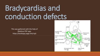
Bradycardias and
conduction defects
This was gathered with the help of
Medicos PDF app:
https://bookapp.page.link/rajit
Sinus bradycardia
Definition
Sinus rhythm
Causes
Myocardial: inferior MI, myocarditis.
CV drugs: β-blockers, α2-agonists (clonidine), rate-limiting CCBs, digoxin, amiodarone.
CNS drugs: opioids, BZD.
Sick sinus syndrome
Metabolic: ↑K+, hypothyroidism, hypothermia.
Physiological states: high levels of fitness, pain.
Anorexia nervosa.
Cushing reflex: response to ↑ICP.
Sick sinus syndrome
Aka sinus node dysfunction.
Usually due to idiopathic SA node fibrosis. May also be secondary to cardiomyopathy, infiltrative
disease (amyloidosis, sarcoidosis, haemochromatosis), drugs (digoxin, β-blockers), or metabolic
problems (↑K+, ↓Ca2+, ↓thyroid).
Causes sinus bradycardia, sinus arrest, or exit block. May also cause episodes of tachycardia
(tachy-brady syndrome, brady-tachy syndrome).
Presentation
Often asymptomatic. Otherwise:
Syncope
Fatigue
Palpitations
Sinoatrial exit block and sinus arrest
Failure of impulse formation (arrest) or conduction from (exit block) the SA node.
Absent P waves on ECG.
Escape rhythm may take over: junctional — narrow QRS 45–60bpm — or ventricular — wide
QRS 30–45 bpm.
Its causes overlap with those of sinus bradycardia, including sick sinus, ↑K+, inferior MI, and CV
drugs.
Atrioventricular (AV) block
Aka heart block.
Causes
Idiopathic
RCA infarct (inferior MI), as this supplies the AV node.
Myocarditis
Drugs: β-blockers, calcium channel blockers, adenosine, digoxin, cholinesterase inhibitors.
1st degree AV block
Prolonged PR interval (>0.2 seconds, 5 small squares).
No treatment required.
2nd degree AV block
Intermittent conduction of the P wave to the ventricles.
The conduction ratio is the number of P waves to QRS complexes e.g. 4:3.
A conduction ratio of 2:1 is untypable as it is hard to determine if there is progressive PR
prolongation.
High grade 2nd degree block is when 2 consecutive P waves fail to conduct to the ventricles.
The P waves are regular, distinguishing it from ectopic atrial contractions.
2nd degree AV block type 1
Aka Mobitz type 1, or Wenckebach.
Progressively prolonged PR interval until a P wave fails to transmit to the ventricles.
No treatment required.
Can sometimes be hard to distinguish from Mobitz type 2 when increases in PR are small. Look
for the biggest increase, which is between the 1st and 2nd PR after the missed QRS.
2nd degree AV block type 2
Aka Mobitz type 2.
Constant PR interval but intermittent failure to transmit to the ventricles.
High risk of progression to 3rd degree block so often requires pacemaker treatment.
3rd degree AV block
Aka complete heart block.
No transmission of P waves into ventricles, with a ventricular escape rhythm taking over.
QRS is usually wide, but occasionally the bundle of His provides the pacemaker and thus the QRS
is narrow.
HR 20–40.
This is one cause of AV dissociation. Others include accelerated idioventricular rhythm (ectopic
focus in ventricles with HR 50–110) and VT.
Requires pacemaker.
Bundle branch blocks
General features
Blockage in the bundle branches, which lie between the bundle of His and the Purkinje fibres.
Depolarisation instead spreads via the (slower) myocardium, causing broad QRS complexes.
The altered depolarisation sequence also leads to altered repolarisation, and hence ST-T
changes.
Left bundle branch block (LBBB)
Causes:
Anterior MI (LAD). May be the initial ECG sign.
HTN
Myocarditis
Cardiomyopathy
Aortic valve disease.
ECG:
Deep, wide S in V1 and RSR’ (M-shaped) in V6: SLaM (LBBB).
V1: delayed LV depolarisation results in a deep, wide S wave.
V6: right to left septal depolarisation, instead of the usual left to right, leads to initial R wave in
V6 followed by a dip during RV depolarisation, then 2nd R wave as depolarisation reaches the LV.
Same pattern seen in lead I. Often the middle notch of the M is very small, such that it simply
looks like a broad R wave.
Discordant T waves in V1 and V6.
Criteria: {broad QRS} + {broad R in V6} + {broad S in V1 or 2}.
Right bundle branch block (RBBB)
Causes:
Increased RV pressure: primary pulmonary HTN, cor pulmonale, PE.
Acquired heart disease: anterior MI (LAD), myocarditis, cardiomyopathy.
Congenital
Iatrogenic e.g. cardiac catheterisation.
Can also be a normal ECG variant in healthy individuals.
ECG:
rSR in V1 (M-shaped) and QRS (W-shaped) in V6: MaRroW (RBBB).
Electrophysiology: the initial rS (V1) and QR (V6) reflect a normal left to right septal
depolarisation and LV depolarisation. The delayed RV depolarisation leads to a 2nd broad R
wave in V1 and a late ‘slurred’ S wave in V6.
Instead of rSR, sometimes V1 simply has one large R ± a small notch as it rises.
Criteria: {broad QRS} + {slurred S V6 and/or rSR V1} + {overall +ve QRS in V1}.
Left anterior and posterior fascicular
block
Blockage in one of the two branches of the left bundle branch.
LAFB is much commoner, and in isolation may simply be a benign feature of aging. Other causes
include anterior MI, IHD, aortic valve disease, HTN, or cardiomyopathy.
LPFB is associated with inferior MI or cardiomyopathy.
Aka left anterior and posterior hemiblocks.
ECG
QRS normal or slightly prolonged (80–120 ms).
LAFB:
Left axis deviation.
Small Q and tall R (qR pattern) in lateral leads (I and aVL) with prolonged R peak time (>45 ms) in
aVL.
Small R and deep S (rS pattern) in inferior leads.
LPFB is the opposite:
Right axis deviation.
Small R and deep S in lateral leads.
Small Q and tall R in inferior leads, with prolonged R peak time in aVF.
Bifasciular and trifascicular block
Bifascicular block:
RBBB plus {LAFB or LPFB}. Conduction is via the single remaining fascicle.
ECG: RBBB plus left or right axis deviation.
Causes: IHD (50%), HTN (25%), aortic stenosis, anterior MI, congenital, ↑K+.
Clinical significance uncertain, but carries a 1% annual risk of progression to complete heart
block.
Trifascicular block:
May not be a clinically useful term. It implies blockage in right bundle plus both fascicles, which
is essentially just 3rd degree AV block.
In practice, it may be used to describe an incomplete trifascicular block where there is still
partial/intermittent transmission in one of the fascicles, plus associated 1st/2nd degree AV
block. Resulting ECG shows RBBB, LAFB or LPFB, and prolonged PR.
Escape rhythms and ectopic beats
Definitions
Escape rhythm: a non-sinus pacemaker takes over from a non-functioning SA node. Beat
occurs after the next expected sinus beat. HR is <60, except in ‘accelerated’ escape rhythm,
which is 60–100.
Ectopic beat: a non-sinus beat occurs before the next expected sinus beat. Often irregular
ventricular ectopics, which are non-pathological; can also be regular e.g. ventricular bigeminy.
ECG findings in escape rhythms
Atrial escape rhythms: HR 40–60, P wave may be inverted.
Junctional escape rhythms: HR 40–60, P wave hidden in QRS complex
Ventricular escape rhythms: HR 20–40, broad QRS.
Cardiac axis deviation
Cardiac axis which is less than -30° (left axis deviation, LAD) or greater than +90°
(right axis deviation, RAD).
ECG
Look at the QRS complexes in the limb leads:
In LAD, they’re Leaving (QRS pointing away from each other): +ve QRS (dominant R) in I and aVL,
-ve QRS (dominant S) in II and aVF.
In RAD, they’re Romantic (QRS pointing towards each other): -ve QRS (dominant S) in I and aVL,
+ve QRS (dominant R) in III and aVF.
Causes
LAD:
Left anterior fascicular block.
LBBB
LVH
Inferior MI
RAD:
Left posterior fascicular block.
RVH
Lateral MI
Lung disease: PE, COPD.
↑K+
May be a normal variant.
WPW syndrome and ventricular ectopics can cause either.
Acute management of bradycardia
1. Atropine 500 mcg IV if there is cardiac ischaemia, syncope, SBP <90, or HF.
2. Further measures if there is inadequate response or risk of asystole: further atropine (up to
3 mg), transcutaenous pacing, adrenaline infusion, or isoprenaline infusion (β1 agonist). Risk
of asystole is defined as severe AV block (3rd degree or 2nd degree type 2), recent asystole,
or ventricular pauses (> 3 s).
3. Definitive management with transvenous and/or permanent pacemaker.
Pacemakers and ICDs
Implantable devices used to control cardiac rhythm, collectively known as cardiac conduction
devices.
Devices and indications
Implantable cardioverter-defibrillators (ICDs) are used to prevent sudden cardiac
death (SCD) in:
{LVSD with EF ❤5%} plus {wide QRS [120–149 ms] or high SCD risk}.
Sustained VT causing syncope or haemodynamic instability.
Congenital high risk conditions e.g. long QT, Brugada, HCM.
Secondary prevention: post VF or VT cardiac arrest.
Permanent pacemakers (PPMs) are used to maintain an adequate heart rate in:
AV block: 3rd degree or 2nd degree type 2.
Sinus node dysfunction with symptomatic bradycardia.
Carotid sinus syndrome.
Cardiac resynchronization therapy (CRT, aka biventricular pacemaker):
Indication: {LVSD NYHA class 2–4 with EF ❤5%} plus {very wide QRS [>150 ms] or LBBB}.
Can be pacer only (CRT-P) or include an ICD function (CRT-D).
Structure and mechanism
Pulse generator — comprising a battery, control circuits, and transmitter/receiver — is placed in
the infraclavicular area (subcutaneously or submuscularly). Requires reimplantation every 5–10
years due to battery lifespan.
Pacing leads (one or two) extend from the generator, transvenously, into the right atrium and/or
ventricle (plus left ventricle in CRT), with the tips implanted in the myocardium. These leads
both sense cardiac depolarization and deliver cardiac stimulation.
PPMs can provide either a fixed impulse rate (‘asynchronous’), or an impulse in response to
absent depolarization (‘synchronous’).
ICDs respond to ventricular tachycardias with a defibrillation shock. Many devices also have a
pacer function, both to treat co-morbid arrhythmias and to deliver antitachycardia pacing before
shocking.
Pacemaker codes and modes
Standard 5 letter code to describe PPMs, with often just the first 3 used:
1. Where it’s pacing: Atria, Ventricles, or Dual (A+V).
2. Where it’s sensing: Atria, Ventricles, or Dual (A+V).
3. Response to sensing depolarization: Triggers pacing, Inhibits pacing (i.e. doesn’t pace), Dual
(triggers and inhibits).
4. Rate modulation: ability to adjust HR in response to physiological need.
5. Anti-tachycardia function: Pacing, Shock, or Dual (P+S).
Common modes:
VVI: no pacing if ventricular depolarization detected, otherwise it paces. AAI is the same for the
atria.
DDD: senses both A and V, and takes over if either don’t work.
VDD: used in AV block, as it senses both A and V but only paces V.
VOO: asynchronous pacing, which should be used during surgery as diathermy may affect
device.
Interpreting pacemaker ECGs
Most pacemaker leads sit in RV causing LBBB pattern, though a minority are LV and RBBB.
If patient’s heart rate is above PPM threshold, pacing spikes will be appropriately absent.
See pacemaker and ICD complications for abnormal ECG findings in presence of PPM.
Device interrogation and manipulation
Investigate cardiac symptoms in PPM/ICD patients as usual, including with an ECG, but specific device
interrogation may also be needed:
Should be done by specialists using specialist devices. Even in asymptomatic patients it is done
regularly e.g. 3-monthly.
Pacemaker/ICD magnets allow basic device manipulation. When placed over a PPM, it will revert
to asynchronous mode (good if device is undersensing or overpacing), and when placed over an
ICD, it will prevent shocks (but not pacing).
Pacemaker and ICD complications
General
Acute (post-placement): pneumothorax, infection, bleeding (including pocket haematoma).
Device-related pain.
ICD malfunctions
Inappropriate ICD shocks: may be triggered by atrial arrhythmias (AF, SVT) or device
malfunction.
Failure to shock. If this occurs, treat ventricular dysrhythmias as usual e.g. external defibrillation,
anti-arrhythmic drugs.
PPM malfunctions
Bradycardia
Failure to output/pace: no impulse (e.g. due to device malfunction, battery failure) and hence
no pacing artefact on ECG.
Failure to capture i.e. no response from heart (e.g. due to poor lead contact, cardiac problem).
ECG shows pacing spikes not followed by atrial or ventricular activity.
Oversensing: noise (e.g. movement artefacts) misinterpreted as cardiac activity and hence PPM
fails to pace.
Tachycardia
Pacemaker-mediated tachycardia: PPM forms re-entrant loop. Less common with new devices.
Sensor-induced tachycardia: noise (e.g. movement artefacts) misinterpreted as physiologically
increased heart rate and PPM increases rate. Occurs in newer devices which allow
physiologically-varied heart rate in response to need.
Can also be due to all the usual causes of tachycardia e.g. physiological response, SVT.
Other dysrhythmias
Undersensing (e.g. due to poor lead contact), leading to asynchronous pacing. Suggested by
pacing spikes within or just after QRS.
Pacemaker syndrome: AV dyssyncrhony due to PPM failing to perfectly replicate normal cardiac
contraction. Causes reduced cardiac output, fatigue, dizziness, and palpitations.
Thank You
This information was taken with the help of Medicos PDF app. You can download the app from
Google Play Store. There are tons of slides, book and articles that a medical student should
need.
◦ https://bookapp.page.link/rajit

More Related Content

What's hot

Ecg quiz
Ecg quizEcg quiz
Ecg quiz
shafei lashin
 
Ecg changes in chamber enlargement
Ecg changes in chamber enlargementEcg changes in chamber enlargement
Ecg changes in chamber enlargement
Anirudhya J
 
Aortic stenosis - Echocardiography
Aortic stenosis - EchocardiographyAortic stenosis - Echocardiography
Aortic stenosis - Echocardiography
Ankur Gupta
 
Bradyarrhythmias
BradyarrhythmiasBradyarrhythmias
Bradyarrhythmias
Abraham Paul
 
16 arrhythmias2009
16 arrhythmias200916 arrhythmias2009
16 arrhythmias2009
internalmed
 
Ventricular tachycardia
Ventricular tachycardiaVentricular tachycardia
Ventricular tachycardia
Praveen Nagula
 
Heart blocks
Heart blocks Heart blocks
Heart blocks
MEEQAT HOSPITAL
 
Vt vs svt ab copy
Vt vs svt ab   copyVt vs svt ab   copy
Vt vs svt ab copy
Ginu George
 
Tachyarrhythmias
TachyarrhythmiasTachyarrhythmias
Tachyarrhythmias
SCGH ED CME
 
Cardiac arrhythmias
Cardiac arrhythmiasCardiac arrhythmias
Cardiac arrhythmias
Farhan Ali
 
ECG BASICS IN DETAIL
ECG BASICS IN DETAILECG BASICS IN DETAIL
Arrhythmia
ArrhythmiaArrhythmia
Arrhythmia
Amira Badr
 
ECG & Heart block [doctors online]
ECG & Heart block [doctors online]ECG & Heart block [doctors online]
ECG & Heart block [doctors online]
Chandan Banerjee
 
Ecg criteria of chamber enlargement
Ecg criteria of chamber enlargementEcg criteria of chamber enlargement
Ecg criteria of chamber enlargement
Adarsh
 
ST Segment Elevations in ECG
ST Segment Elevations in ECGST Segment Elevations in ECG
ST Segment Elevations in ECG
Chew Keng Sheng
 
Heart failure - Echocardiography
Heart failure - EchocardiographyHeart failure - Echocardiography
Heart failure - Echocardiography
Praveen Nagula
 
Conduction disturbances
Conduction disturbancesConduction disturbances
Conduction disturbances
shyam771
 
Ecg ecg abnormalities
Ecg ecg abnormalitiesEcg ecg abnormalities
Ecg ecg abnormalities
jhundaily
 
Arrythmia
ArrythmiaArrythmia
Arrythmia
Muhammad Saim
 
Interpretation of common ecg abnormalities
Interpretation of common ecg  abnormalitiesInterpretation of common ecg  abnormalities
Interpretation of common ecg abnormalities
Dr. Lazaro Nicanor Rodriguez Gonzalez
 

What's hot (20)

Ecg quiz
Ecg quizEcg quiz
Ecg quiz
 
Ecg changes in chamber enlargement
Ecg changes in chamber enlargementEcg changes in chamber enlargement
Ecg changes in chamber enlargement
 
Aortic stenosis - Echocardiography
Aortic stenosis - EchocardiographyAortic stenosis - Echocardiography
Aortic stenosis - Echocardiography
 
Bradyarrhythmias
BradyarrhythmiasBradyarrhythmias
Bradyarrhythmias
 
16 arrhythmias2009
16 arrhythmias200916 arrhythmias2009
16 arrhythmias2009
 
Ventricular tachycardia
Ventricular tachycardiaVentricular tachycardia
Ventricular tachycardia
 
Heart blocks
Heart blocks Heart blocks
Heart blocks
 
Vt vs svt ab copy
Vt vs svt ab   copyVt vs svt ab   copy
Vt vs svt ab copy
 
Tachyarrhythmias
TachyarrhythmiasTachyarrhythmias
Tachyarrhythmias
 
Cardiac arrhythmias
Cardiac arrhythmiasCardiac arrhythmias
Cardiac arrhythmias
 
ECG BASICS IN DETAIL
ECG BASICS IN DETAILECG BASICS IN DETAIL
ECG BASICS IN DETAIL
 
Arrhythmia
ArrhythmiaArrhythmia
Arrhythmia
 
ECG & Heart block [doctors online]
ECG & Heart block [doctors online]ECG & Heart block [doctors online]
ECG & Heart block [doctors online]
 
Ecg criteria of chamber enlargement
Ecg criteria of chamber enlargementEcg criteria of chamber enlargement
Ecg criteria of chamber enlargement
 
ST Segment Elevations in ECG
ST Segment Elevations in ECGST Segment Elevations in ECG
ST Segment Elevations in ECG
 
Heart failure - Echocardiography
Heart failure - EchocardiographyHeart failure - Echocardiography
Heart failure - Echocardiography
 
Conduction disturbances
Conduction disturbancesConduction disturbances
Conduction disturbances
 
Ecg ecg abnormalities
Ecg ecg abnormalitiesEcg ecg abnormalities
Ecg ecg abnormalities
 
Arrythmia
ArrythmiaArrythmia
Arrythmia
 
Interpretation of common ecg abnormalities
Interpretation of common ecg  abnormalitiesInterpretation of common ecg  abnormalities
Interpretation of common ecg abnormalities
 

Similar to Bradycardias and conduction defects

Approach to bradyarrythmias1
Approach to bradyarrythmias1Approach to bradyarrythmias1
Approach to bradyarrythmias1
Bhargav Kiran
 
Beginners Guide for ECG Interpretation
Beginners Guide for ECG InterpretationBeginners Guide for ECG Interpretation
Beginners Guide for ECG Interpretation
meducationdotnet
 
A Guide TO ECG Interpretation
A Guide TO ECG InterpretationA Guide TO ECG Interpretation
A Guide TO ECG Interpretation
meducationdotnet
 
Complete EKG Interpretation Course
Complete EKG Interpretation Course Complete EKG Interpretation Course
Complete EKG Interpretation Course
Kerolus Shehata
 
ECG interpretation
ECG interpretationECG interpretation
ECG interpretation
AndrewCrofton
 
ECG Interpretation
ECG InterpretationECG Interpretation
ECG Interpretation
drmainuddin
 
Bradycardias & heart block Jane Nader.pdf
Bradycardias & heart block Jane Nader.pdfBradycardias & heart block Jane Nader.pdf
Bradycardias & heart block Jane Nader.pdf
Jane390174
 
Ecg step by step not by me
Ecg step by step not by meEcg step by step not by me
Ecg step by step not by me
Ahmed Ghany
 
Ecg 2
Ecg 2Ecg 2
Ecgrevised2 090808155046 Phpapp01
Ecgrevised2 090808155046 Phpapp01Ecgrevised2 090808155046 Phpapp01
Ecgrevised2 090808155046 Phpapp01
mahipal33
 
Cardiac Arrythmias
Cardiac ArrythmiasCardiac Arrythmias
Cardiac Arrythmias
meducationdotnet
 
Arrhythmia diagnosis and management
Arrhythmia diagnosis and managementArrhythmia diagnosis and management
Arrhythmia diagnosis and management
Pallab Nath
 
Arrhythmias July 09
Arrhythmias July 09Arrhythmias July 09
Arrhythmias July 09
Michael LaCombe
 
ECG Basics
ECG BasicsECG Basics
Arrhythmia.pdf
Arrhythmia.pdfArrhythmia.pdf
Arrhythmia.pdf
DrAliAlsaady1
 
How to read ECG systematically with practice strips
How to read ECG systematically with practice strips How to read ECG systematically with practice strips
How to read ECG systematically with practice strips
Khaled AlKhodari
 
ELECTROCARDIOGRAM
ELECTROCARDIOGRAMELECTROCARDIOGRAM
ELECTROCARDIOGRAM
DJ CrissCross
 
Arrhythmia Recognition & Management
Arrhythmia Recognition & ManagementArrhythmia Recognition & Management
Arrhythmia Recognition & Management
yuyuricci
 
Arrhythmias
ArrhythmiasArrhythmias
Arrhythmias
Rashad Siddiqi
 
ECG Notes
ECG Notes ECG Notes
ECG Notes
Hind waleed
 

Similar to Bradycardias and conduction defects (20)

Approach to bradyarrythmias1
Approach to bradyarrythmias1Approach to bradyarrythmias1
Approach to bradyarrythmias1
 
Beginners Guide for ECG Interpretation
Beginners Guide for ECG InterpretationBeginners Guide for ECG Interpretation
Beginners Guide for ECG Interpretation
 
A Guide TO ECG Interpretation
A Guide TO ECG InterpretationA Guide TO ECG Interpretation
A Guide TO ECG Interpretation
 
Complete EKG Interpretation Course
Complete EKG Interpretation Course Complete EKG Interpretation Course
Complete EKG Interpretation Course
 
ECG interpretation
ECG interpretationECG interpretation
ECG interpretation
 
ECG Interpretation
ECG InterpretationECG Interpretation
ECG Interpretation
 
Bradycardias & heart block Jane Nader.pdf
Bradycardias & heart block Jane Nader.pdfBradycardias & heart block Jane Nader.pdf
Bradycardias & heart block Jane Nader.pdf
 
Ecg step by step not by me
Ecg step by step not by meEcg step by step not by me
Ecg step by step not by me
 
Ecg 2
Ecg 2Ecg 2
Ecg 2
 
Ecgrevised2 090808155046 Phpapp01
Ecgrevised2 090808155046 Phpapp01Ecgrevised2 090808155046 Phpapp01
Ecgrevised2 090808155046 Phpapp01
 
Cardiac Arrythmias
Cardiac ArrythmiasCardiac Arrythmias
Cardiac Arrythmias
 
Arrhythmia diagnosis and management
Arrhythmia diagnosis and managementArrhythmia diagnosis and management
Arrhythmia diagnosis and management
 
Arrhythmias July 09
Arrhythmias July 09Arrhythmias July 09
Arrhythmias July 09
 
ECG Basics
ECG BasicsECG Basics
ECG Basics
 
Arrhythmia.pdf
Arrhythmia.pdfArrhythmia.pdf
Arrhythmia.pdf
 
How to read ECG systematically with practice strips
How to read ECG systematically with practice strips How to read ECG systematically with practice strips
How to read ECG systematically with practice strips
 
ELECTROCARDIOGRAM
ELECTROCARDIOGRAMELECTROCARDIOGRAM
ELECTROCARDIOGRAM
 
Arrhythmia Recognition & Management
Arrhythmia Recognition & ManagementArrhythmia Recognition & Management
Arrhythmia Recognition & Management
 
Arrhythmias
ArrhythmiasArrhythmias
Arrhythmias
 
ECG Notes
ECG Notes ECG Notes
ECG Notes
 

More from AayushPokharel10

Anorexia nervosa
Anorexia nervosaAnorexia nervosa
Anorexia nervosa
AayushPokharel10
 
Schizophrenia
SchizophreniaSchizophrenia
Schizophrenia
AayushPokharel10
 
Myeloma
MyelomaMyeloma
Hodgkin's lymphoma
Hodgkin's lymphomaHodgkin's lymphoma
Hodgkin's lymphoma
AayushPokharel10
 
Speech problems
Speech problemsSpeech problems
Speech problems
AayushPokharel10
 
Visual problems, nystagmus, and vertigo
Visual problems, nystagmus, and vertigoVisual problems, nystagmus, and vertigo
Visual problems, nystagmus, and vertigo
AayushPokharel10
 
Cranial nerve problems
Cranial nerve problemsCranial nerve problems
Cranial nerve problems
AayushPokharel10
 
Neurological examination
Neurological examinationNeurological examination
Neurological examination
AayushPokharel10
 
Hodgkin's lymphoma
Hodgkin's lymphomaHodgkin's lymphoma
Hodgkin's lymphoma
AayushPokharel10
 
Labour complications
Labour complicationsLabour complications
Labour complications
AayushPokharel10
 
Gestational diabetes (gdm)
Gestational diabetes (gdm)Gestational diabetes (gdm)
Gestational diabetes (gdm)
AayushPokharel10
 
Ectopic pregnancy
Ectopic pregnancyEctopic pregnancy
Ectopic pregnancy
AayushPokharel10
 
Pelvic inflammatory disease (pid)
Pelvic inflammatory disease (pid)Pelvic inflammatory disease (pid)
Pelvic inflammatory disease (pid)
AayushPokharel10
 
Dysphagia
DysphagiaDysphagia
Dysphagia
AayushPokharel10
 
Cardiomyopathy
CardiomyopathyCardiomyopathy
Cardiomyopathy
AayushPokharel10
 
Burns
BurnsBurns
Leg ulcers
Leg ulcersLeg ulcers
Leg ulcers
AayushPokharel10
 
Rash
RashRash
Cardiac medications
Cardiac medicationsCardiac medications
Cardiac medications
AayushPokharel10
 
Cardiovascular examination
Cardiovascular examinationCardiovascular examination
Cardiovascular examination
AayushPokharel10
 

More from AayushPokharel10 (20)

Anorexia nervosa
Anorexia nervosaAnorexia nervosa
Anorexia nervosa
 
Schizophrenia
SchizophreniaSchizophrenia
Schizophrenia
 
Myeloma
MyelomaMyeloma
Myeloma
 
Hodgkin's lymphoma
Hodgkin's lymphomaHodgkin's lymphoma
Hodgkin's lymphoma
 
Speech problems
Speech problemsSpeech problems
Speech problems
 
Visual problems, nystagmus, and vertigo
Visual problems, nystagmus, and vertigoVisual problems, nystagmus, and vertigo
Visual problems, nystagmus, and vertigo
 
Cranial nerve problems
Cranial nerve problemsCranial nerve problems
Cranial nerve problems
 
Neurological examination
Neurological examinationNeurological examination
Neurological examination
 
Hodgkin's lymphoma
Hodgkin's lymphomaHodgkin's lymphoma
Hodgkin's lymphoma
 
Labour complications
Labour complicationsLabour complications
Labour complications
 
Gestational diabetes (gdm)
Gestational diabetes (gdm)Gestational diabetes (gdm)
Gestational diabetes (gdm)
 
Ectopic pregnancy
Ectopic pregnancyEctopic pregnancy
Ectopic pregnancy
 
Pelvic inflammatory disease (pid)
Pelvic inflammatory disease (pid)Pelvic inflammatory disease (pid)
Pelvic inflammatory disease (pid)
 
Dysphagia
DysphagiaDysphagia
Dysphagia
 
Cardiomyopathy
CardiomyopathyCardiomyopathy
Cardiomyopathy
 
Burns
BurnsBurns
Burns
 
Leg ulcers
Leg ulcersLeg ulcers
Leg ulcers
 
Rash
RashRash
Rash
 
Cardiac medications
Cardiac medicationsCardiac medications
Cardiac medications
 
Cardiovascular examination
Cardiovascular examinationCardiovascular examination
Cardiovascular examination
 

Recently uploaded

Top Effective Soaps for Fungal Skin Infections in India
Top Effective Soaps for Fungal Skin Infections in IndiaTop Effective Soaps for Fungal Skin Infections in India
Top Effective Soaps for Fungal Skin Infections in India
SwisschemDerma
 
Aortic Association CBL Pilot April 19 – 20 Bern
Aortic Association CBL Pilot April 19 – 20 BernAortic Association CBL Pilot April 19 – 20 Bern
Aortic Association CBL Pilot April 19 – 20 Bern
suvadeepdas911
 
REGULATION FOR COMBINATION PRODUCTS AND MEDICAL DEVICES.pptx
REGULATION FOR COMBINATION PRODUCTS AND MEDICAL DEVICES.pptxREGULATION FOR COMBINATION PRODUCTS AND MEDICAL DEVICES.pptx
REGULATION FOR COMBINATION PRODUCTS AND MEDICAL DEVICES.pptx
LaniyaNasrink
 
Muscles of Mastication by Dr. Rabia Inam Gandapore.pptx
Muscles of Mastication by Dr. Rabia Inam Gandapore.pptxMuscles of Mastication by Dr. Rabia Inam Gandapore.pptx
Muscles of Mastication by Dr. Rabia Inam Gandapore.pptx
Dr. Rabia Inam Gandapore
 
Best Ayurvedic medicine for Gas and Indigestion
Best Ayurvedic medicine for Gas and IndigestionBest Ayurvedic medicine for Gas and Indigestion
Best Ayurvedic medicine for Gas and Indigestion
Swastik Ayurveda
 
THERAPEUTIC ANTISENSE MOLECULES .pptx
THERAPEUTIC ANTISENSE MOLECULES    .pptxTHERAPEUTIC ANTISENSE MOLECULES    .pptx
THERAPEUTIC ANTISENSE MOLECULES .pptx
70KRISHPATEL
 
Does Over-Masturbation Contribute to Chronic Prostatitis.pptx
Does Over-Masturbation Contribute to Chronic Prostatitis.pptxDoes Over-Masturbation Contribute to Chronic Prostatitis.pptx
Does Over-Masturbation Contribute to Chronic Prostatitis.pptx
walterHu5
 
TEST BANK For Basic and Clinical Pharmacology, 14th Edition by Bertram G. Kat...
TEST BANK For Basic and Clinical Pharmacology, 14th Edition by Bertram G. Kat...TEST BANK For Basic and Clinical Pharmacology, 14th Edition by Bertram G. Kat...
TEST BANK For Basic and Clinical Pharmacology, 14th Edition by Bertram G. Kat...
rightmanforbloodline
 
Local Advanced Lung Cancer: Artificial Intelligence, Synergetics, Complex Sys...
Local Advanced Lung Cancer: Artificial Intelligence, Synergetics, Complex Sys...Local Advanced Lung Cancer: Artificial Intelligence, Synergetics, Complex Sys...
Local Advanced Lung Cancer: Artificial Intelligence, Synergetics, Complex Sys...
Oleg Kshivets
 
Netter's Atlas of Human Anatomy 7.ed.pdf
Netter's Atlas of Human Anatomy 7.ed.pdfNetter's Atlas of Human Anatomy 7.ed.pdf
Netter's Atlas of Human Anatomy 7.ed.pdf
BrissaOrtiz3
 
Ketone bodies and metabolism-biochemistry
Ketone bodies and metabolism-biochemistryKetone bodies and metabolism-biochemistry
Ketone bodies and metabolism-biochemistry
Dhayanithi C
 
Hemodialysis: Chapter 4, Dialysate Circuit - Dr.Gawad
Hemodialysis: Chapter 4, Dialysate Circuit - Dr.GawadHemodialysis: Chapter 4, Dialysate Circuit - Dr.Gawad
Hemodialysis: Chapter 4, Dialysate Circuit - Dr.Gawad
NephroTube - Dr.Gawad
 
Vestibulocochlear Nerve by Dr. Rabia Inam Gandapore.pptx
Vestibulocochlear Nerve by Dr. Rabia Inam Gandapore.pptxVestibulocochlear Nerve by Dr. Rabia Inam Gandapore.pptx
Vestibulocochlear Nerve by Dr. Rabia Inam Gandapore.pptx
Dr. Rabia Inam Gandapore
 
CHEMOTHERAPY_RDP_CHAPTER 3_ANTIFUNGAL AGENT.pdf
CHEMOTHERAPY_RDP_CHAPTER 3_ANTIFUNGAL AGENT.pdfCHEMOTHERAPY_RDP_CHAPTER 3_ANTIFUNGAL AGENT.pdf
CHEMOTHERAPY_RDP_CHAPTER 3_ANTIFUNGAL AGENT.pdf
rishi2789
 
Efficacy of Avartana Sneha in Ayurveda
Efficacy of Avartana Sneha in AyurvedaEfficacy of Avartana Sneha in Ayurveda
Efficacy of Avartana Sneha in Ayurveda
Dr. Jyothirmai Paindla
 
Role of Mukta Pishti in the Management of Hyperthyroidism
Role of Mukta Pishti in the Management of HyperthyroidismRole of Mukta Pishti in the Management of Hyperthyroidism
Role of Mukta Pishti in the Management of Hyperthyroidism
Dr. Jyothirmai Paindla
 
share - Lions, tigers, AI and health misinformation, oh my!.pptx
share - Lions, tigers, AI and health misinformation, oh my!.pptxshare - Lions, tigers, AI and health misinformation, oh my!.pptx
share - Lions, tigers, AI and health misinformation, oh my!.pptx
Tina Purnat
 
Top-Vitamin-Supplement-Brands-in-India List
Top-Vitamin-Supplement-Brands-in-India ListTop-Vitamin-Supplement-Brands-in-India List
Top-Vitamin-Supplement-Brands-in-India List
SwisschemDerma
 
K CỔ TỬ CUNG.pdf tự ghi chép, chữ hơi xấu
K CỔ TỬ CUNG.pdf tự ghi chép, chữ hơi xấuK CỔ TỬ CUNG.pdf tự ghi chép, chữ hơi xấu
K CỔ TỬ CUNG.pdf tự ghi chép, chữ hơi xấu
HongBiThi1
 
CHEMOTHERAPY_RDP_CHAPTER 6_Anti Malarial Drugs.pdf
CHEMOTHERAPY_RDP_CHAPTER 6_Anti Malarial Drugs.pdfCHEMOTHERAPY_RDP_CHAPTER 6_Anti Malarial Drugs.pdf
CHEMOTHERAPY_RDP_CHAPTER 6_Anti Malarial Drugs.pdf
rishi2789
 

Recently uploaded (20)

Top Effective Soaps for Fungal Skin Infections in India
Top Effective Soaps for Fungal Skin Infections in IndiaTop Effective Soaps for Fungal Skin Infections in India
Top Effective Soaps for Fungal Skin Infections in India
 
Aortic Association CBL Pilot April 19 – 20 Bern
Aortic Association CBL Pilot April 19 – 20 BernAortic Association CBL Pilot April 19 – 20 Bern
Aortic Association CBL Pilot April 19 – 20 Bern
 
REGULATION FOR COMBINATION PRODUCTS AND MEDICAL DEVICES.pptx
REGULATION FOR COMBINATION PRODUCTS AND MEDICAL DEVICES.pptxREGULATION FOR COMBINATION PRODUCTS AND MEDICAL DEVICES.pptx
REGULATION FOR COMBINATION PRODUCTS AND MEDICAL DEVICES.pptx
 
Muscles of Mastication by Dr. Rabia Inam Gandapore.pptx
Muscles of Mastication by Dr. Rabia Inam Gandapore.pptxMuscles of Mastication by Dr. Rabia Inam Gandapore.pptx
Muscles of Mastication by Dr. Rabia Inam Gandapore.pptx
 
Best Ayurvedic medicine for Gas and Indigestion
Best Ayurvedic medicine for Gas and IndigestionBest Ayurvedic medicine for Gas and Indigestion
Best Ayurvedic medicine for Gas and Indigestion
 
THERAPEUTIC ANTISENSE MOLECULES .pptx
THERAPEUTIC ANTISENSE MOLECULES    .pptxTHERAPEUTIC ANTISENSE MOLECULES    .pptx
THERAPEUTIC ANTISENSE MOLECULES .pptx
 
Does Over-Masturbation Contribute to Chronic Prostatitis.pptx
Does Over-Masturbation Contribute to Chronic Prostatitis.pptxDoes Over-Masturbation Contribute to Chronic Prostatitis.pptx
Does Over-Masturbation Contribute to Chronic Prostatitis.pptx
 
TEST BANK For Basic and Clinical Pharmacology, 14th Edition by Bertram G. Kat...
TEST BANK For Basic and Clinical Pharmacology, 14th Edition by Bertram G. Kat...TEST BANK For Basic and Clinical Pharmacology, 14th Edition by Bertram G. Kat...
TEST BANK For Basic and Clinical Pharmacology, 14th Edition by Bertram G. Kat...
 
Local Advanced Lung Cancer: Artificial Intelligence, Synergetics, Complex Sys...
Local Advanced Lung Cancer: Artificial Intelligence, Synergetics, Complex Sys...Local Advanced Lung Cancer: Artificial Intelligence, Synergetics, Complex Sys...
Local Advanced Lung Cancer: Artificial Intelligence, Synergetics, Complex Sys...
 
Netter's Atlas of Human Anatomy 7.ed.pdf
Netter's Atlas of Human Anatomy 7.ed.pdfNetter's Atlas of Human Anatomy 7.ed.pdf
Netter's Atlas of Human Anatomy 7.ed.pdf
 
Ketone bodies and metabolism-biochemistry
Ketone bodies and metabolism-biochemistryKetone bodies and metabolism-biochemistry
Ketone bodies and metabolism-biochemistry
 
Hemodialysis: Chapter 4, Dialysate Circuit - Dr.Gawad
Hemodialysis: Chapter 4, Dialysate Circuit - Dr.GawadHemodialysis: Chapter 4, Dialysate Circuit - Dr.Gawad
Hemodialysis: Chapter 4, Dialysate Circuit - Dr.Gawad
 
Vestibulocochlear Nerve by Dr. Rabia Inam Gandapore.pptx
Vestibulocochlear Nerve by Dr. Rabia Inam Gandapore.pptxVestibulocochlear Nerve by Dr. Rabia Inam Gandapore.pptx
Vestibulocochlear Nerve by Dr. Rabia Inam Gandapore.pptx
 
CHEMOTHERAPY_RDP_CHAPTER 3_ANTIFUNGAL AGENT.pdf
CHEMOTHERAPY_RDP_CHAPTER 3_ANTIFUNGAL AGENT.pdfCHEMOTHERAPY_RDP_CHAPTER 3_ANTIFUNGAL AGENT.pdf
CHEMOTHERAPY_RDP_CHAPTER 3_ANTIFUNGAL AGENT.pdf
 
Efficacy of Avartana Sneha in Ayurveda
Efficacy of Avartana Sneha in AyurvedaEfficacy of Avartana Sneha in Ayurveda
Efficacy of Avartana Sneha in Ayurveda
 
Role of Mukta Pishti in the Management of Hyperthyroidism
Role of Mukta Pishti in the Management of HyperthyroidismRole of Mukta Pishti in the Management of Hyperthyroidism
Role of Mukta Pishti in the Management of Hyperthyroidism
 
share - Lions, tigers, AI and health misinformation, oh my!.pptx
share - Lions, tigers, AI and health misinformation, oh my!.pptxshare - Lions, tigers, AI and health misinformation, oh my!.pptx
share - Lions, tigers, AI and health misinformation, oh my!.pptx
 
Top-Vitamin-Supplement-Brands-in-India List
Top-Vitamin-Supplement-Brands-in-India ListTop-Vitamin-Supplement-Brands-in-India List
Top-Vitamin-Supplement-Brands-in-India List
 
K CỔ TỬ CUNG.pdf tự ghi chép, chữ hơi xấu
K CỔ TỬ CUNG.pdf tự ghi chép, chữ hơi xấuK CỔ TỬ CUNG.pdf tự ghi chép, chữ hơi xấu
K CỔ TỬ CUNG.pdf tự ghi chép, chữ hơi xấu
 
CHEMOTHERAPY_RDP_CHAPTER 6_Anti Malarial Drugs.pdf
CHEMOTHERAPY_RDP_CHAPTER 6_Anti Malarial Drugs.pdfCHEMOTHERAPY_RDP_CHAPTER 6_Anti Malarial Drugs.pdf
CHEMOTHERAPY_RDP_CHAPTER 6_Anti Malarial Drugs.pdf
 

Bradycardias and conduction defects

  • 1. Bradycardias and conduction defects This was gathered with the help of Medicos PDF app: https://bookapp.page.link/rajit
  • 2. Sinus bradycardia Definition Sinus rhythm Causes Myocardial: inferior MI, myocarditis. CV drugs: β-blockers, α2-agonists (clonidine), rate-limiting CCBs, digoxin, amiodarone. CNS drugs: opioids, BZD. Sick sinus syndrome Metabolic: ↑K+, hypothyroidism, hypothermia. Physiological states: high levels of fitness, pain. Anorexia nervosa. Cushing reflex: response to ↑ICP.
  • 3. Sick sinus syndrome Aka sinus node dysfunction. Usually due to idiopathic SA node fibrosis. May also be secondary to cardiomyopathy, infiltrative disease (amyloidosis, sarcoidosis, haemochromatosis), drugs (digoxin, β-blockers), or metabolic problems (↑K+, ↓Ca2+, ↓thyroid). Causes sinus bradycardia, sinus arrest, or exit block. May also cause episodes of tachycardia (tachy-brady syndrome, brady-tachy syndrome).
  • 5. Sinoatrial exit block and sinus arrest Failure of impulse formation (arrest) or conduction from (exit block) the SA node. Absent P waves on ECG. Escape rhythm may take over: junctional — narrow QRS 45–60bpm — or ventricular — wide QRS 30–45 bpm. Its causes overlap with those of sinus bradycardia, including sick sinus, ↑K+, inferior MI, and CV drugs.
  • 6. Atrioventricular (AV) block Aka heart block. Causes Idiopathic RCA infarct (inferior MI), as this supplies the AV node. Myocarditis Drugs: β-blockers, calcium channel blockers, adenosine, digoxin, cholinesterase inhibitors.
  • 7. 1st degree AV block Prolonged PR interval (>0.2 seconds, 5 small squares). No treatment required.
  • 8. 2nd degree AV block Intermittent conduction of the P wave to the ventricles. The conduction ratio is the number of P waves to QRS complexes e.g. 4:3. A conduction ratio of 2:1 is untypable as it is hard to determine if there is progressive PR prolongation. High grade 2nd degree block is when 2 consecutive P waves fail to conduct to the ventricles. The P waves are regular, distinguishing it from ectopic atrial contractions.
  • 9. 2nd degree AV block type 1 Aka Mobitz type 1, or Wenckebach. Progressively prolonged PR interval until a P wave fails to transmit to the ventricles. No treatment required. Can sometimes be hard to distinguish from Mobitz type 2 when increases in PR are small. Look for the biggest increase, which is between the 1st and 2nd PR after the missed QRS.
  • 10. 2nd degree AV block type 2 Aka Mobitz type 2. Constant PR interval but intermittent failure to transmit to the ventricles. High risk of progression to 3rd degree block so often requires pacemaker treatment.
  • 11. 3rd degree AV block Aka complete heart block. No transmission of P waves into ventricles, with a ventricular escape rhythm taking over. QRS is usually wide, but occasionally the bundle of His provides the pacemaker and thus the QRS is narrow. HR 20–40. This is one cause of AV dissociation. Others include accelerated idioventricular rhythm (ectopic focus in ventricles with HR 50–110) and VT. Requires pacemaker.
  • 12. Bundle branch blocks General features Blockage in the bundle branches, which lie between the bundle of His and the Purkinje fibres. Depolarisation instead spreads via the (slower) myocardium, causing broad QRS complexes. The altered depolarisation sequence also leads to altered repolarisation, and hence ST-T changes.
  • 13. Left bundle branch block (LBBB) Causes: Anterior MI (LAD). May be the initial ECG sign. HTN Myocarditis Cardiomyopathy Aortic valve disease.
  • 14. ECG: Deep, wide S in V1 and RSR’ (M-shaped) in V6: SLaM (LBBB). V1: delayed LV depolarisation results in a deep, wide S wave. V6: right to left septal depolarisation, instead of the usual left to right, leads to initial R wave in V6 followed by a dip during RV depolarisation, then 2nd R wave as depolarisation reaches the LV. Same pattern seen in lead I. Often the middle notch of the M is very small, such that it simply looks like a broad R wave. Discordant T waves in V1 and V6. Criteria: {broad QRS} + {broad R in V6} + {broad S in V1 or 2}.
  • 15. Right bundle branch block (RBBB) Causes: Increased RV pressure: primary pulmonary HTN, cor pulmonale, PE. Acquired heart disease: anterior MI (LAD), myocarditis, cardiomyopathy. Congenital Iatrogenic e.g. cardiac catheterisation. Can also be a normal ECG variant in healthy individuals.
  • 16. ECG: rSR in V1 (M-shaped) and QRS (W-shaped) in V6: MaRroW (RBBB). Electrophysiology: the initial rS (V1) and QR (V6) reflect a normal left to right septal depolarisation and LV depolarisation. The delayed RV depolarisation leads to a 2nd broad R wave in V1 and a late ‘slurred’ S wave in V6. Instead of rSR, sometimes V1 simply has one large R ± a small notch as it rises. Criteria: {broad QRS} + {slurred S V6 and/or rSR V1} + {overall +ve QRS in V1}.
  • 17. Left anterior and posterior fascicular block Blockage in one of the two branches of the left bundle branch. LAFB is much commoner, and in isolation may simply be a benign feature of aging. Other causes include anterior MI, IHD, aortic valve disease, HTN, or cardiomyopathy. LPFB is associated with inferior MI or cardiomyopathy. Aka left anterior and posterior hemiblocks.
  • 18. ECG QRS normal or slightly prolonged (80–120 ms). LAFB: Left axis deviation. Small Q and tall R (qR pattern) in lateral leads (I and aVL) with prolonged R peak time (>45 ms) in aVL. Small R and deep S (rS pattern) in inferior leads. LPFB is the opposite: Right axis deviation. Small R and deep S in lateral leads. Small Q and tall R in inferior leads, with prolonged R peak time in aVF.
  • 19. Bifasciular and trifascicular block Bifascicular block: RBBB plus {LAFB or LPFB}. Conduction is via the single remaining fascicle. ECG: RBBB plus left or right axis deviation. Causes: IHD (50%), HTN (25%), aortic stenosis, anterior MI, congenital, ↑K+. Clinical significance uncertain, but carries a 1% annual risk of progression to complete heart block.
  • 20. Trifascicular block: May not be a clinically useful term. It implies blockage in right bundle plus both fascicles, which is essentially just 3rd degree AV block. In practice, it may be used to describe an incomplete trifascicular block where there is still partial/intermittent transmission in one of the fascicles, plus associated 1st/2nd degree AV block. Resulting ECG shows RBBB, LAFB or LPFB, and prolonged PR.
  • 21. Escape rhythms and ectopic beats Definitions Escape rhythm: a non-sinus pacemaker takes over from a non-functioning SA node. Beat occurs after the next expected sinus beat. HR is <60, except in ‘accelerated’ escape rhythm, which is 60–100. Ectopic beat: a non-sinus beat occurs before the next expected sinus beat. Often irregular ventricular ectopics, which are non-pathological; can also be regular e.g. ventricular bigeminy.
  • 22. ECG findings in escape rhythms Atrial escape rhythms: HR 40–60, P wave may be inverted. Junctional escape rhythms: HR 40–60, P wave hidden in QRS complex Ventricular escape rhythms: HR 20–40, broad QRS.
  • 23. Cardiac axis deviation Cardiac axis which is less than -30° (left axis deviation, LAD) or greater than +90° (right axis deviation, RAD). ECG Look at the QRS complexes in the limb leads: In LAD, they’re Leaving (QRS pointing away from each other): +ve QRS (dominant R) in I and aVL, -ve QRS (dominant S) in II and aVF. In RAD, they’re Romantic (QRS pointing towards each other): -ve QRS (dominant S) in I and aVL, +ve QRS (dominant R) in III and aVF.
  • 24. Causes LAD: Left anterior fascicular block. LBBB LVH Inferior MI RAD: Left posterior fascicular block. RVH Lateral MI Lung disease: PE, COPD. ↑K+ May be a normal variant. WPW syndrome and ventricular ectopics can cause either.
  • 25. Acute management of bradycardia 1. Atropine 500 mcg IV if there is cardiac ischaemia, syncope, SBP <90, or HF. 2. Further measures if there is inadequate response or risk of asystole: further atropine (up to 3 mg), transcutaenous pacing, adrenaline infusion, or isoprenaline infusion (β1 agonist). Risk of asystole is defined as severe AV block (3rd degree or 2nd degree type 2), recent asystole, or ventricular pauses (> 3 s). 3. Definitive management with transvenous and/or permanent pacemaker.
  • 26. Pacemakers and ICDs Implantable devices used to control cardiac rhythm, collectively known as cardiac conduction devices.
  • 27. Devices and indications Implantable cardioverter-defibrillators (ICDs) are used to prevent sudden cardiac death (SCD) in: {LVSD with EF ❤5%} plus {wide QRS [120–149 ms] or high SCD risk}. Sustained VT causing syncope or haemodynamic instability. Congenital high risk conditions e.g. long QT, Brugada, HCM. Secondary prevention: post VF or VT cardiac arrest. Permanent pacemakers (PPMs) are used to maintain an adequate heart rate in: AV block: 3rd degree or 2nd degree type 2. Sinus node dysfunction with symptomatic bradycardia. Carotid sinus syndrome. Cardiac resynchronization therapy (CRT, aka biventricular pacemaker): Indication: {LVSD NYHA class 2–4 with EF ❤5%} plus {very wide QRS [>150 ms] or LBBB}. Can be pacer only (CRT-P) or include an ICD function (CRT-D).
  • 28. Structure and mechanism Pulse generator — comprising a battery, control circuits, and transmitter/receiver — is placed in the infraclavicular area (subcutaneously or submuscularly). Requires reimplantation every 5–10 years due to battery lifespan. Pacing leads (one or two) extend from the generator, transvenously, into the right atrium and/or ventricle (plus left ventricle in CRT), with the tips implanted in the myocardium. These leads both sense cardiac depolarization and deliver cardiac stimulation. PPMs can provide either a fixed impulse rate (‘asynchronous’), or an impulse in response to absent depolarization (‘synchronous’). ICDs respond to ventricular tachycardias with a defibrillation shock. Many devices also have a pacer function, both to treat co-morbid arrhythmias and to deliver antitachycardia pacing before shocking.
  • 29. Pacemaker codes and modes Standard 5 letter code to describe PPMs, with often just the first 3 used: 1. Where it’s pacing: Atria, Ventricles, or Dual (A+V). 2. Where it’s sensing: Atria, Ventricles, or Dual (A+V). 3. Response to sensing depolarization: Triggers pacing, Inhibits pacing (i.e. doesn’t pace), Dual (triggers and inhibits). 4. Rate modulation: ability to adjust HR in response to physiological need. 5. Anti-tachycardia function: Pacing, Shock, or Dual (P+S).
  • 30. Common modes: VVI: no pacing if ventricular depolarization detected, otherwise it paces. AAI is the same for the atria. DDD: senses both A and V, and takes over if either don’t work. VDD: used in AV block, as it senses both A and V but only paces V. VOO: asynchronous pacing, which should be used during surgery as diathermy may affect device.
  • 31. Interpreting pacemaker ECGs Most pacemaker leads sit in RV causing LBBB pattern, though a minority are LV and RBBB. If patient’s heart rate is above PPM threshold, pacing spikes will be appropriately absent. See pacemaker and ICD complications for abnormal ECG findings in presence of PPM.
  • 32. Device interrogation and manipulation Investigate cardiac symptoms in PPM/ICD patients as usual, including with an ECG, but specific device interrogation may also be needed: Should be done by specialists using specialist devices. Even in asymptomatic patients it is done regularly e.g. 3-monthly. Pacemaker/ICD magnets allow basic device manipulation. When placed over a PPM, it will revert to asynchronous mode (good if device is undersensing or overpacing), and when placed over an ICD, it will prevent shocks (but not pacing).
  • 33. Pacemaker and ICD complications General Acute (post-placement): pneumothorax, infection, bleeding (including pocket haematoma). Device-related pain. ICD malfunctions Inappropriate ICD shocks: may be triggered by atrial arrhythmias (AF, SVT) or device malfunction. Failure to shock. If this occurs, treat ventricular dysrhythmias as usual e.g. external defibrillation, anti-arrhythmic drugs.
  • 34. PPM malfunctions Bradycardia Failure to output/pace: no impulse (e.g. due to device malfunction, battery failure) and hence no pacing artefact on ECG. Failure to capture i.e. no response from heart (e.g. due to poor lead contact, cardiac problem). ECG shows pacing spikes not followed by atrial or ventricular activity. Oversensing: noise (e.g. movement artefacts) misinterpreted as cardiac activity and hence PPM fails to pace.
  • 35. Tachycardia Pacemaker-mediated tachycardia: PPM forms re-entrant loop. Less common with new devices. Sensor-induced tachycardia: noise (e.g. movement artefacts) misinterpreted as physiologically increased heart rate and PPM increases rate. Occurs in newer devices which allow physiologically-varied heart rate in response to need. Can also be due to all the usual causes of tachycardia e.g. physiological response, SVT.
  • 36. Other dysrhythmias Undersensing (e.g. due to poor lead contact), leading to asynchronous pacing. Suggested by pacing spikes within or just after QRS. Pacemaker syndrome: AV dyssyncrhony due to PPM failing to perfectly replicate normal cardiac contraction. Causes reduced cardiac output, fatigue, dizziness, and palpitations.
  • 37. Thank You This information was taken with the help of Medicos PDF app. You can download the app from Google Play Store. There are tons of slides, book and articles that a medical student should need. ◦ https://bookapp.page.link/rajit