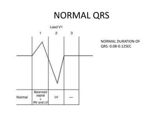
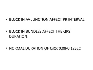
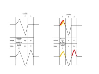
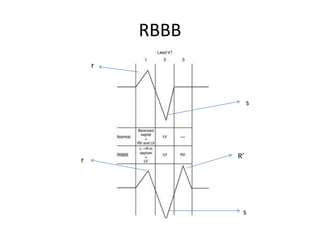
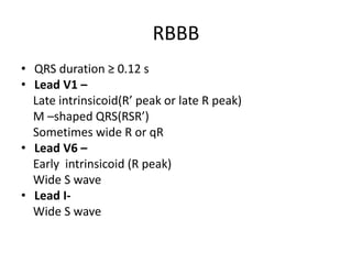
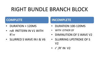
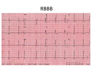
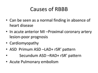
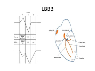
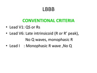
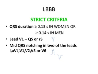
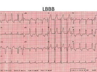
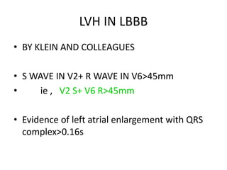
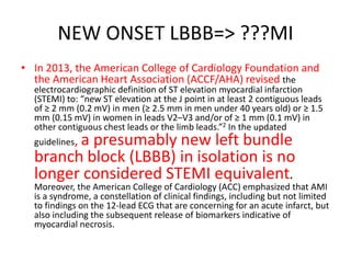
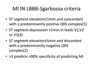
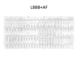
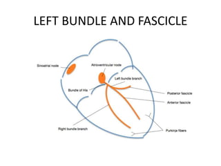
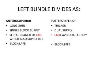
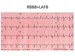
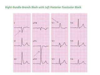
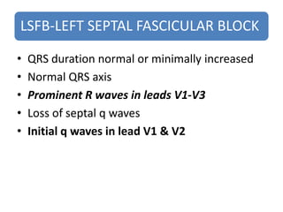
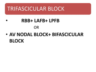
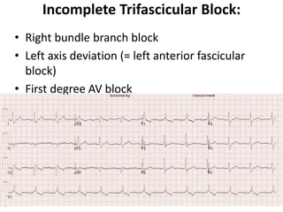
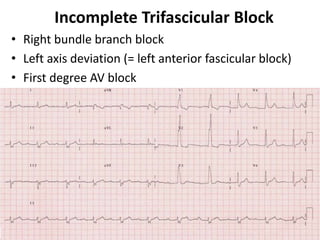
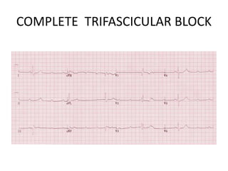
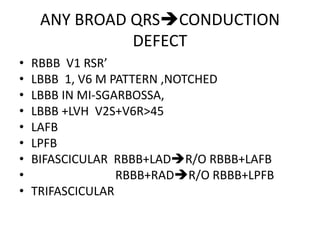
ECG Lecture: Sinus arrest, sinoatrial exit block, AV block and escape rhythmsMichael-Joseph Agbayani
Simple ECG lecture about sinus arrest, sinoatrial exit block, AV block and escape rhythms. Slideshow was made with an audience of medical professionals in mind.
ECG Lecture: Sinus arrest, sinoatrial exit block, AV block and escape rhythmsMichael-Joseph Agbayani
Simple ECG lecture about sinus arrest, sinoatrial exit block, AV block and escape rhythms. Slideshow was made with an audience of medical professionals in mind.
Biatrial enlargement is diagnosed when criteria for both right and left atrial enlargement are present on the same ECG.
The diagnosis of biatrial enlargement requires criteria for LAE and RAE to be met in either lead II, lead V1 or a combination of leads.
A lecture on the echocardiographic evaluation of hypertrophic cardiomyopathy. Starts with an overview of the topic then a systematic approach to diagnosis and then a differential diagnosis followed by take-home messages and conclusion.
ventricular premature complexes and idioventricular rhythm identification is important in the ICU ..they may run into arryhthmias..look over my seminar...
any queries...
AV nodal reentrant tachycardia (AVNRT), or atrioventricular nodal reentrant tachycardia, is a type of tachycardia (fast rhythm) of the heart. It is a type of supraventricular tachycardia (SVT), meaning that it originates from a location within the heart above the bundle of His. AV nodal reentrant tachycardia is the most common regular supraventricular tachycardia. It is more common in women than men (approximately 75% of cases occur in females). The main symptom is palpitations. Treatment may be with specific physical maneuvers, medication, or, rarely, synchronized cardioversion. Frequent attacks may require radiofrequency ablation, in which the abnormally conducting tissue in the heart is destroyed.
AVNRT occurs when a reentry circuit forms within or just next to the atrioventricular node. The circuit usually involves two anatomical pathways: the fast pathway and the slow pathway, which are both in the right atrium. The slow pathway (which is usually targeted for ablation) is located inferior and slightly posterior to the AV node, often following the anterior margin of the coronary sinus. The fast pathway is usually located just superior and posterior to the AV node. These pathways are formed from tissue that behaves very much like the AV node, and some authors regard them as part of the AV node.
The fast and slow pathways should not be confused with the accessory pathways that give rise to Wolff-Parkinson-White syndrome (WPW syndrome) or atrioventricular reciprocating tachycardia (AVRT). In AVNRT, the fast and slow pathways are located within the right atrium close to or within the AV node and exhibit electrophysiologic properties similar to AV nodal tissue. Accessory pathways that give rise to WPW syndrome and AVRT are located in the atrioventricular valvular rings. They provide a direct connection between the atria and ventricles, and have electrophysiologic properties similar to ventricular myocardium.
An electrocardiogram (ECG or EKG) records the electrical signal from your heart to check for different heart conditions. Electrodes are placed on your chest to record your heart's electrical signals, which cause your heart to beat. The signals are shown as waves on an attached computer monitor or printer
MANAGEMENT OF ATRIOVENTRICULAR CONDUCTION BLOCK.pdfJim Jacob Roy
Cardiac conduction defects can occur due to various causes.
Atrioventricular conduction blocks ( AV blocks ) are classified into 3 types.
This document describes the acute management of AV block.
Couples presenting to the infertility clinic- Do they really have infertility...Sujoy Dasgupta
Dr Sujoy Dasgupta presented the study on "Couples presenting to the infertility clinic- Do they really have infertility? – The unexplored stories of non-consummation" in the 13th Congress of the Asia Pacific Initiative on Reproduction (ASPIRE 2024) at Manila on 24 May, 2024.
TEST BANK for Operations Management, 14th Edition by William J. Stevenson, Ve...kevinkariuki227
TEST BANK for Operations Management, 14th Edition by William J. Stevenson, Verified Chapters 1 - 19, Complete Newest Version.pdf
Ozempic: Preoperative Management of Patients on GLP-1 Receptor Agonists Saeid Safari
Preoperative Management of Patients on GLP-1 Receptor Agonists like Ozempic and Semiglutide
ASA GUIDELINE
NYSORA Guideline
2 Case Reports of Gastric Ultrasound
Pulmonary Thromboembolism - etilogy, types, medical- Surgical and nursing man...VarunMahajani
Disruption of blood supply to lung alveoli due to blockage of one or more pulmonary blood vessels is called as Pulmonary thromboembolism. In this presentation we will discuss its causes, types and its management in depth.
Tom Selleck Health: A Comprehensive Look at the Iconic Actor’s Wellness Journeygreendigital
Tom Selleck, an enduring figure in Hollywood. has captivated audiences for decades with his rugged charm, iconic moustache. and memorable roles in television and film. From his breakout role as Thomas Magnum in Magnum P.I. to his current portrayal of Frank Reagan in Blue Bloods. Selleck's career has spanned over 50 years. But beyond his professional achievements. fans have often been curious about Tom Selleck Health. especially as he has aged in the public eye.
Follow us on: Pinterest
Introduction
Many have been interested in Tom Selleck health. not only because of his enduring presence on screen but also because of the challenges. and lifestyle choices he has faced and made over the years. This article delves into the various aspects of Tom Selleck health. exploring his fitness regimen, diet, mental health. and the challenges he has encountered as he ages. We'll look at how he maintains his well-being. the health issues he has faced, and his approach to ageing .
Early Life and Career
Childhood and Athletic Beginnings
Tom Selleck was born on January 29, 1945, in Detroit, Michigan, and grew up in Sherman Oaks, California. From an early age, he was involved in sports, particularly basketball. which played a significant role in his physical development. His athletic pursuits continued into college. where he attended the University of Southern California (USC) on a basketball scholarship. This early involvement in sports laid a strong foundation for his physical health and disciplined lifestyle.
Transition to Acting
Selleck's transition from an athlete to an actor came with its physical demands. His first significant role in "Magnum P.I." required him to perform various stunts and maintain a fit appearance. This role, which he played from 1980 to 1988. necessitated a rigorous fitness routine to meet the show's demands. setting the stage for his long-term commitment to health and wellness.
Fitness Regimen
Workout Routine
Tom Selleck health and fitness regimen has evolved. adapting to his changing roles and age. During his "Magnum, P.I." days. Selleck's workouts were intense and focused on building and maintaining muscle mass. His routine included weightlifting, cardiovascular exercises. and specific training for the stunts he performed on the show.
Selleck adjusted his fitness routine as he aged to suit his body's needs. Today, his workouts focus on maintaining flexibility, strength, and cardiovascular health. He incorporates low-impact exercises such as swimming, walking, and light weightlifting. This balanced approach helps him stay fit without putting undue strain on his joints and muscles.
Importance of Flexibility and Mobility
In recent years, Selleck has emphasized the importance of flexibility and mobility in his fitness regimen. Understanding the natural decline in muscle mass and joint flexibility with age. he includes stretching and yoga in his routine. These practices help prevent injuries, improve posture, and maintain mobilit
Report Back from SGO 2024: What’s the Latest in Cervical Cancer?bkling
Are you curious about what’s new in cervical cancer research or unsure what the findings mean? Join Dr. Emily Ko, a gynecologic oncologist at Penn Medicine, to learn about the latest updates from the Society of Gynecologic Oncology (SGO) 2024 Annual Meeting on Women’s Cancer. Dr. Ko will discuss what the research presented at the conference means for you and answer your questions about the new developments.
These simplified slides by Dr. Sidra Arshad present an overview of the non-respiratory functions of the respiratory tract.
Learning objectives:
1. Enlist the non-respiratory functions of the respiratory tract
2. Briefly explain how these functions are carried out
3. Discuss the significance of dead space
4. Differentiate between minute ventilation and alveolar ventilation
5. Describe the cough and sneeze reflexes
Study Resources:
1. Chapter 39, Guyton and Hall Textbook of Medical Physiology, 14th edition
2. Chapter 34, Ganong’s Review of Medical Physiology, 26th edition
3. Chapter 17, Human Physiology by Lauralee Sherwood, 9th edition
4. Non-respiratory functions of the lungs https://academic.oup.com/bjaed/article/13/3/98/278874
The prostate is an exocrine gland of the male mammalian reproductive system
It is a walnut-sized gland that forms part of the male reproductive system and is located in front of the rectum and just below the urinary bladder
Function is to store and secrete a clear, slightly alkaline fluid that constitutes 10-30% of the volume of the seminal fluid that along with the spermatozoa, constitutes semen
A healthy human prostate measures (4cm-vertical, by 3cm-horizontal, 2cm ant-post ).
It surrounds the urethra just below the urinary bladder. It has anterior, median, posterior and two lateral lobes
It’s work is regulated by androgens which are responsible for male sex characteristics
Generalised disease of the prostate due to hormonal derangement which leads to non malignant enlargement of the gland (increase in the number of epithelial cells and stromal tissue)to cause compression of the urethra leading to symptoms (LUTS
Prix Galien International 2024 Forum ProgramLevi Shapiro
June 20, 2024, Prix Galien International and Jerusalem Ethics Forum in ROME. Detailed agenda including panels:
- ADVANCES IN CARDIOLOGY: A NEW PARADIGM IS COMING
- WOMEN’S HEALTH: FERTILITY PRESERVATION
- WHAT’S NEW IN THE TREATMENT OF INFECTIOUS,
ONCOLOGICAL AND INFLAMMATORY SKIN DISEASES?
- ARTIFICIAL INTELLIGENCE AND ETHICS
- GENE THERAPY
- BEYOND BORDERS: GLOBAL INITIATIVES FOR DEMOCRATIZING LIFE SCIENCE TECHNOLOGIES AND PROMOTING ACCESS TO HEALTHCARE
- ETHICAL CHALLENGES IN LIFE SCIENCES
- Prix Galien International Awards Ceremony
10. RBBB
• QRS duration ≥ 0.12 s
• Lead V1 –
Late intrinsicoid(R’ peak or late R peak)
M –shaped QRS(RSR’)
Sometimes wide R or qR
• Lead V6 –
Early intrinsicoid (R peak)
Wide S wave
• Lead I-
Wide S wave
11. RIGHT BUNDLE BRANCH BLOCK
COMPLETE
• DURATION > 120MS
• rsR PATTERN IN V1 WITH
R’>r
• SLURRED S WAVE IN I & V6
INCOMPLETE
• DURATION 100-120MS
• WITH EITHER OF
• DIMINUTION OF S WAVE V2
• SLURRING UPSTROKE OF S
V2
• r’ /R’ IN V2
13. Causes of RBBB
• Can be seen as a normal finding in absence of
heart disease
• In acute anterior MI –Proximal coronary artery
lesion-poor prognosis
• Cardiomyopathy
• ASD Primum ASD –LAD+ rSR’ pattern
• Secundum ASD –RAD+ rSR’ pattern
• Acute Pulmonary embolism
15. LBBB
CONVENTIONAL CRITERIA
• Lead V1: QS or Rs
• Lead V6: Late intrinsicoid (R or R’ peak),
No Q waves, monophasic R
• Lead I : Monophasic R wave ,No Q
16. STRICT CRITERIA
• QRS duration ≥ 0.13 s IN WOMEN OR
≥ 0.14 s IN MEN
• Lead V1 – QS or rS
• Mid QRS notching in two of the leads
I,aVL,V1,V2,V5 or V6
LBBB
17. ST T CHANGES IN LBBB
IMPORTANT NOTE
• ST SEGMENT AND T WAVE ARE IN OPPOSITE
DIRECTIONS TO TERMINAL QRS DEFLECTION
19. INCOMPLETE LBBB
• EARLIEST CHANGE:
DISAPPEARANCE OF INITIAL
q WAVE OF QR COMPLEX IN LEFT LEADS
TALL R
• SMALL INITIAL R WAVE IN RIGHT LEADS
DISAPPEAR QS COMPLEX
21. CAUSES OF LBBB
• IHD
• CARDIOMYOPATHY
• HYPERTENSIVE HEART DISEASE
• ACUTE MI-can be the presenting ecg change
• Aortic stenosis
• INTRINSIC DISEASES OF CONDUCTION SYSTEM-
Primary degenerative disease (fibrosis) of the
conducting system (Lenegre disease)
• Digoxin toxicity
22. LVH IN LBBB
• BY KLEIN AND COLLEAGUES
• S WAVE IN V2+ R WAVE IN V6>45mm
• ie , V2 S+ V6 R>45mm
• Evidence of left atrial enlargement with QRS
complex>0.16s
23. NEW ONSET LBBB=> ???MI
• In 2013, the American College of Cardiology Foundation and
the American Heart Association (ACCF/AHA) revised the
electrocardiographic definition of ST elevation myocardial infarction
(STEMI) to: “new ST elevation at the J point in at least 2 contiguous leads
of ≥ 2 mm (0.2 mV) in men (≥ 2.5 mm in men under 40 years old) or ≥ 1.5
mm (0.15 mV) in women in leads V2–V3 and/or of ≥ 1 mm (0.1 mV) in
other contiguous chest leads or the limb leads.”2 In the updated
guidelines, a presumably new left bundle
branch block (LBBB) in isolation is no
longer considered STEMI equivalent.
Moreover, the American College of Cardiology (ACC) emphasized that AMI
is a syndrome, a constellation of clinical findings, including but not limited
to findings on the 12-lead ECG that are concerning for an acute infarct, but
also including the subsequent release of biomarkers indicative of
myocardial necrosis.
25. MI IN LBBB-Sgarbossa criteria
• ST segment elevation≥1mm and concordant
with a predominantly positive QRS complex(5)
• ST segment depression ≥1mm in leads V1,V2
or V3(3)
• ST segment elevation≥5mm and discordant
with a predominantly negative QRS
complex(2)
• >3 predicts >90% specificity of predicting MI
26. Modified Sgarbossa Criteria
• A. ≥ 1 lead with ≥1 mm of concordant ST
elevation
• B. ≥ 1 lead of V1-V3 with ≥ 1 mm of
concordant ST depression
• C. ≥ 1 lead anywhere with ≥ 1 mm STE and
proportionally excessive discordant STE, as
defined by ≥ 25% of the depth of the
preceding S-wave.
• Any of above three STEMI equivalent
33. LEFT BUNDLE DIVIDES AS:
ANTEROSUPERIOR
• LONG ,THIN
• SINGLE BLOOD SUPPLY
• SEPTAL BRANCH OF LAD
WHICH ALSO SUPPLY RBB
• BLOCK:LAFB
POSTEROINFERIOR
• THICKER
• DUAL SUPPLY
• LAD+ AV NODAL ARTERY
• BLOCK:LPFB
34. LAFB
• Left axis deviation (usually ≥ -30°) !MCC!
• Small Q (qR complexes)in leads I and
aVL;small r(rS complexes)in II,III,aVF
• Minimal QRS prolongation (0.020s) from
baseline
• Late intrinsicoid (R wave peak) deflection in
aVL (>0.045s)
• Increased QRS voltage in limb leads
35. T wave changes -LAFB
• In any intraventricular conduction defect T
wave vector goes in opposite direction of main
QRS vector
• LAFB: T low or inverted in I ,aVL
T upright in II,III,aVF
37. LAFB+LVH
• ECG S/O LAFB WITH
• S WAVE IN LEAD III > 1.5mm(0.15mV)
• Can also due to advanced LAFB
38. LPFB
• Right axis deviation(usually ≥ +120°)
• Small R in leads I and aVL(rS complexes);small
Q in II,III,aVF(qR complexes)
• Usually normal QRS duration
• Late intrinsicoid (R wave peak) deflection in
aVF (>0.045s)
• Increased QRS voltage in limb leads
• No evidence of RVH
39. T wave changes LPFB
• T inverted in II,III,aVF
• Upright T waves in lead I
43. LPFB+RBBB
Very rarely occurs
Consider only if no clinical e/o RVH
• V1 shows changes typical of RBBB
• I,aVL shows rS s/o LPFB
• Duration > 0.12s
• QRS Axis: should be > +90°
44.
45. SEPTAL FASCICLE??
• BELIEVED TO BE RESPONSIBLE FOR INITIAL
LEFT TO RIGHT SEPTAL ACTIVATION
• INITIAL 35-40ms OF QRS COMPLEX
46. LSFB-LEFT SEPTAL FASCICULAR BLOCK
• QRS duration normal or minimally increased
• Normal QRS axis
• Prominent R waves in leads V1-V3
• Loss of septal q waves
• Initial q waves in lead V1 & V2
48. INCOMPLETE TRIFASCICULAR
• BIFASCICULAR BLOCK WITH EVIDENCE OF
DELAYED CONDUCTION IN AV NODE
• FIXED BLOCK OF ONE FASCICLE WITH
INTERMITTENT FAILURE OF OTHER TWO
50. Incomplete Trifascicular Block:
• Right bundle branch block
• Left axis deviation (= left anterior fascicular
block)
• First degree AV block
51. Incomplete Trifascicular Block
• Right bundle branch block
• Left axis deviation (= left anterior fascicular block)
• First degree AV block
•
52. COMPLETE TRIFASCICULAR
• 3RD DEGREE AV BLOCK WITH FEATURES OF
BIFASCICLULAR BLOCK
• ESCAPE RHYTHM ARISES FROM LEFT
ANTERIOR OR POSTERIOR FASCICLE
PRODUCING QRS COMPLEXES WITH
APPEARANCES OF RBBB PLUS LAFB OR LPFB
RESPECTIVELY