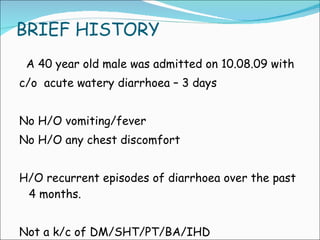
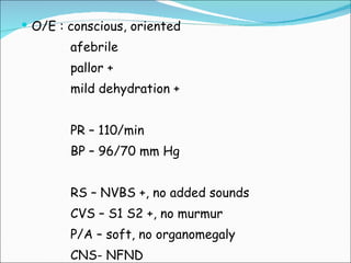
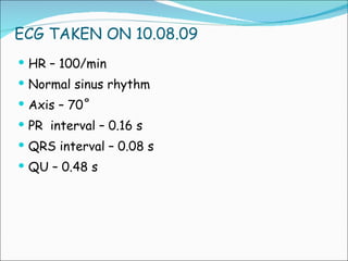
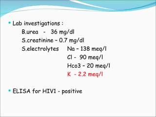
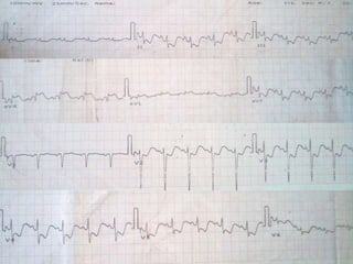
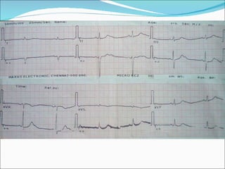
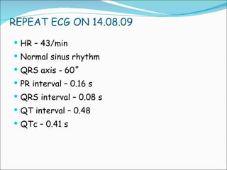
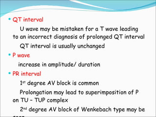
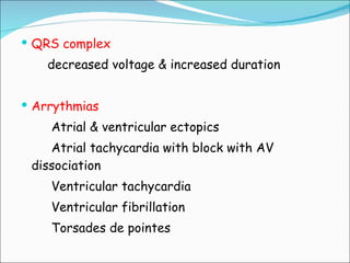
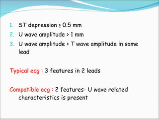
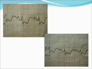
DR. PRIYA KUBENDIRAN M-1 UNIT PROF DR.RUCKMANI’S UNIT
BRIEF HISTORY A 40 year old male was admitted on 10.08.09 with  c/o  acute watery diarrhoea – 3 days No H/O vomiting/fever No H/O any chest discomfort  H/O recurrent episodes of diarrhoea over the past 4 months. Not a k/c of DM/SHT/PT/BA/IHD
O/E : conscious, oriented afebrile pallor + mild dehydration + PR – 110/min BP – 96/70 mm Hg RS – NVBS +, no added sounds CVS – S1 S2 +, no murmur P/A – soft, no organomegaly CNS- NFND
 
ECG TAKEN ON 10.08.09 HR – 100/min Normal sinus rhythm Axis – 70˚ PR  interval – 0.16 s QRS interval – 0.08 s QU – 0.48 s
Lab investigations : B.urea  -  36 mg/dl S.creatinine – 0.7 mg/dl S.electrolytes  Na – 138 meq/l Cl -  90 meq/l Hco3 – 20 meq/l K  - 2.2 meq/l ELISA for HIV1 - positive
 
Cardiac evaluation  Echocardiogram: No RWMA Normal Echo study
After adequate hydration, i.v antibiotics and corrective measures for hypokalemia oral KCl 15 ml tds i.v  -  20 mEq/day 3 days later repeat s.potassium –  4.2 mEq/L A repeat ecg was taken
 
REPEAT ECG ON 14.08.09 HR – 43/min Normal sinus rhythm QRS axis - 60˚ PR interval – 0.16 s QRS interval – 0.08 s QT interval – 0.48 QTc – 0.41 s
ECG CHANGES IN HYPOKALEMIA The ecg changes are due to DELAYED  VENTRICULAR  REPOLARIZATION Diagnosis is therefore based on ST segment, T wave & U wave abnormalities
T wave -progressive dimunition in amplitude, may eventually disappear - a remnant of T maybe visible as a slight irregularity (minimal upward bump) on ST segment U wave progressive increase in amplitude & maintains its rounded appearance may get superimposed on T  -TU complex ST segment ST depression is seen in all leads which may be horizontal or concave upwards
Prominent U waves combined with ST segment depression & flattened T waves –  ROLLER COASTER EFFECT
QT interval U wave may be mistaken for a T wave leading to an incorrect diagnosis of prolonged QT interval QT interval is usually unchanged P wave increase in amplitude/ duration PR interval 1 st  degree AV block is common Prolongation may lead to superimposition of P on TU – TUP complex 2 nd  degree AV block of Wenkebach type may be seen
QRS complex decreased voltage & increased duration Arrythmias Atrial & ventricular ectopics Atrial tachycardia with block with AV dissociation Ventricular tachycardia Ventricular fibrillation Torsades de pointes
ST depression ≥ 0.5 mm U wave amplitude > 1 mm U wave amplitude > T wave amplitude in same lead Typical ecg :  3 features in 2 leads Compatible ecg :  2 features- U wave related characteristics is present


ECG: Hypokalemia

  • 1.
    DR. PRIYA KUBENDIRANM-1 UNIT PROF DR.RUCKMANI’S UNIT
  • 2.
    BRIEF HISTORY A40 year old male was admitted on 10.08.09 with c/o acute watery diarrhoea – 3 days No H/O vomiting/fever No H/O any chest discomfort H/O recurrent episodes of diarrhoea over the past 4 months. Not a k/c of DM/SHT/PT/BA/IHD
  • 3.
    O/E : conscious,oriented afebrile pallor + mild dehydration + PR – 110/min BP – 96/70 mm Hg RS – NVBS +, no added sounds CVS – S1 S2 +, no murmur P/A – soft, no organomegaly CNS- NFND
  • 4.
  • 5.
    ECG TAKEN ON10.08.09 HR – 100/min Normal sinus rhythm Axis – 70˚ PR interval – 0.16 s QRS interval – 0.08 s QU – 0.48 s
  • 6.
    Lab investigations :B.urea - 36 mg/dl S.creatinine – 0.7 mg/dl S.electrolytes Na – 138 meq/l Cl - 90 meq/l Hco3 – 20 meq/l K - 2.2 meq/l ELISA for HIV1 - positive
  • 7.
  • 8.
    Cardiac evaluation Echocardiogram: No RWMA Normal Echo study
  • 9.
    After adequate hydration,i.v antibiotics and corrective measures for hypokalemia oral KCl 15 ml tds i.v - 20 mEq/day 3 days later repeat s.potassium – 4.2 mEq/L A repeat ecg was taken
  • 10.
  • 11.
    REPEAT ECG ON14.08.09 HR – 43/min Normal sinus rhythm QRS axis - 60˚ PR interval – 0.16 s QRS interval – 0.08 s QT interval – 0.48 QTc – 0.41 s
  • 12.
    ECG CHANGES INHYPOKALEMIA The ecg changes are due to DELAYED VENTRICULAR REPOLARIZATION Diagnosis is therefore based on ST segment, T wave & U wave abnormalities
  • 13.
    T wave -progressivedimunition in amplitude, may eventually disappear - a remnant of T maybe visible as a slight irregularity (minimal upward bump) on ST segment U wave progressive increase in amplitude & maintains its rounded appearance may get superimposed on T -TU complex ST segment ST depression is seen in all leads which may be horizontal or concave upwards
  • 14.
    Prominent U wavescombined with ST segment depression & flattened T waves – ROLLER COASTER EFFECT
  • 15.
    QT interval Uwave may be mistaken for a T wave leading to an incorrect diagnosis of prolonged QT interval QT interval is usually unchanged P wave increase in amplitude/ duration PR interval 1 st degree AV block is common Prolongation may lead to superimposition of P on TU – TUP complex 2 nd degree AV block of Wenkebach type may be seen
  • 16.
    QRS complex decreasedvoltage & increased duration Arrythmias Atrial & ventricular ectopics Atrial tachycardia with block with AV dissociation Ventricular tachycardia Ventricular fibrillation Torsades de pointes
  • 17.
    ST depression ≥0.5 mm U wave amplitude > 1 mm U wave amplitude > T wave amplitude in same lead Typical ecg : 3 features in 2 leads Compatible ecg : 2 features- U wave related characteristics is present
  • 18.
  • 19.