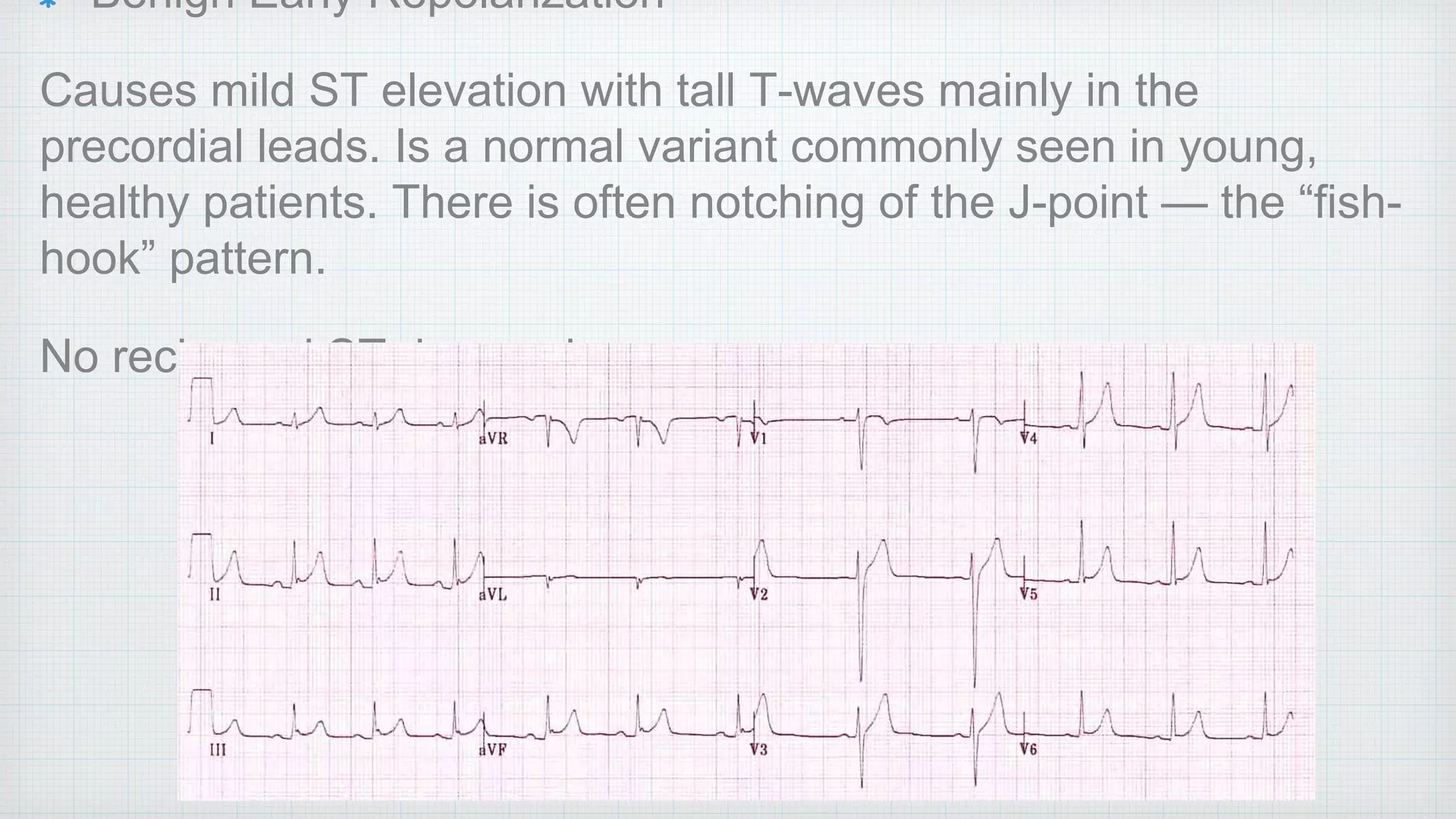
SlideShare a Scribd company logo
ECG Abnormalities In 
Pulmonary Medicine 
Dr. Manjit S. Tendolkar 
Dept of Chest Medicine, 
Seth G. S. Medical College and K.E.M. Hospital, Mumbai.
Normal ECG
P-wave = <0.12 sec & <2.5 mm height (limb) <1.5 mm (precordial) 
P-R interval = 0.12-0.2 sec 
QRS = 0.12 sec 
QTc = 0.42 sec (QT/sq root RR)
P Wave 
P wave is always positive in lead II and 
always negative in lead aVR during sinus 
rhythm
P-Pulmonale 
The presence of tall, peaked P waves 
(>2.5 mm) in lead II is a sign of right atrial 
enlargement, usually due to pulmonary 
hypertension (e.g. cor pulmonale from 
chronic respiratory disease).
Q wave 
Considered Pathological if 
• > 40 ms (1 mm) wide 
• > 2 mm deep 
• > 25% of depth of QRS complex 
• Seen in leads V1-3 
Signifies Old MI
R wave 
Abnormalities: 
1. Dominant R wave in V1 
2. Dominant R wave in aVR (TCA, 
Dextrocardia) 
3. Poor R wave progression
Dominant R wave in V1 
Normal in Children and Young Adults 
Right Ventricular Hypertrophy 
- Pulmonary Embolus 
- RBBB 
Posterior Wall MI 
Right Ventricular Hypertrophy
Poor R wave Progression 
Defined as R wave < 3mm height in V3 
Causes: 
• LVH 
• Prior Antero Septal MI 
• Inaccurate Lead Placement 
• Normal Variant
T wave 
Upright in all leads except aVR and V1 
Amplitude: < 5 mm in limb leads ; < 10 mm in 
precordial leads
Peaked T wave (narrow & symmetrically 
peaked) 
Hyperkalemia
Hyperacute T wave (broad & 
asymmetrically peaked) 
Early stages of ST elevated 
MI, often preceding 
occurrence of ST elevation 
and Q waves.
T wave Inversion 
Normal in children 
Pulmonary embolism 
Ventricular hypertrophy 
(Strain Pattern) 
Raised ICP 
T wave inversion in lead III 
is normal variant. 
Pathological T wave 
inversion is usually 
symmetrical and deep (> 3 
mm)
Biphasic T wave 
Ischaemia ( up-down ) 
Hypokalemia ( down-up 
)
Camel Hump T waves 
Prominent U wave fused to 
end of T wave ( severe 
Hypokalemia) 
Hidden P wave embedded in 
T wave ( Sinus Tachycardia, 
Heart Blocks)
U wave 
Normally, inversely proportional to heart rate. 
Grows bigger as heart rate slows down. (visible at 
HR < 65) 
Normal, < 25 % T wave voltage or < 1-2 mm 
amplitude
Prominent U wave 
Severe Hypokalemia 
Digoxin
Abnormalities of 
Segments & Intervals
QRS 
Narrow Complex <100 ms ( 
Supraventricular) 
Broad Complex >120 ms ( 
Ventricular or aberrant supra 
ventricular conduction - 
Hyperkalemia,BBB) 
Sinus rhythm with frequent ventricular ectopics
RBBB 
• In RBBB, activation of the right ventricle is delayed as depolarisation has to spread across the 
septum from the left ventricle. 
• The left ventricle is activated normally, meaning that the early part of the QRS complex is 
unchanged. 
• The delayed right ventricular activation produces a secondary R wave (R’) in the right 
precordial leads (V1-3) and a wide, slurred S wave in the lateral leads.
Diagnostic Criteria Of RBBB 
• Broad QRS > 120 ms 
• RSR’ pattern in V1-3 (‘M-shaped’ QRS complex) 
• Wide, slurred S wave in the lateral leads (I, aVL, V5-6)
Causes of RBBB 
Cor Pulmonale / Right Ventricular Hypertrophy 
Pulmonary Emboli 
IHD
Incomplete RBBB 
QRS < 120 ms 
Normal Variant, Common in Children.
LBBB 
Causes: 
Hyperkalemia 
Digoxin Toxicity
Hypokalaemia 
Decreased extracellular potassium causes myocardial 
hyperexcitability with the potential to develop re-entrant 
arrhythmias 
• Hypokalaemia is defined as a potassium level < 3.5 mEq/L 
• Moderate hypokalemia is a serum level of < 3.0 mEq/L 
• Severe hypokalemia is defined as a level < 2.5 mEq/L
Changes appear when K+ falls below about 2.7 mmol/l 
• Increased amplitude and width of the P wave 
• Prolongation of the PR interval 
• T wave flattening and inversion 
• ST depression 
• Prominent U waves (best seen in the precordial leads) 
• Apparent long QT interval due to fusion of the T and U 
waves (= long QU interval)
With worsening hypokalaemia: 
• Frequent supraventricular and ventricular 
ectopics 
• Supraventricular tachyarrhythmias: AF, atrial 
flutter, atrial tachycardia 
• Potential to develop life-threatening ventricular 
arrhythmias, e.g. VT, VF and Torsades de Pointes
Hyperkalemia 
• Increased extracellular potassium reduces myocardial excitability, with depression of both pacemaking and 
conducting tissues. 
• Progressively worsening hyperkalaemia leads to suppression of impulse generation by the SA node and 
reduced conduction by the AV node and His-Purkinje system, resulting in bradycardia and conduction blocks and 
ultimately cardiac arrest. 
• Hyperkalaemia is defined as a potassium level > 5.5 mEq/L 
• Moderate hyperkalaemia is a serum potassium > 6.0 mEq/L 
• Severe hyperkalaemia is a serum potassium > 7.0 mE/L
Serum potassium > 5.5 mEq/L is associated 
with repolarization abnormalities: 
• Peaked T waves (usually the earliest sign of 
hyperkalaemia)
Serum potassium > 6.5 mEq/L is associated 
with progressive paralysis of the atria: 
• P wave widens and flattens 
• PR segment lengthens 
• P waves eventually disappear
Serum potassium > 7.0 mEq/L is associated with conduction 
abnormalities and bradycardia: 
• Prolonged QRS interval with bizarre QRS morphology 
• High-grade AV block with slow junctional and ventricular escape 
rhythms 
• Any kind of conduction block (bundle branch blocks, fascicular 
blocks) 
• Sinus bradycardia or slow AF 
• Development of a sine wave appearance (a pre-terminal rhythm)
Serum potassium level of > 9.0 mEq/Lcauses cardiac 
arrest due to: 
• Asystole 
• Ventricular fibrillation 
• PEA with bizarre, wide complex rhythm
QTc Interval 
Prolonged QTc (>440 ms) 
• Hypokalaemia 
• Hypomagnesaemia 
• Hypocalcaemia 
• Hypothermia 
• Myocardial ischemia
ST Segment Elevation 
Acute STEMI may produce ST elevation with either concave, convex or 
obliquely straight morphology 
Reciprocal ST depression in opposite leads.
Benign Early Repolarization 
Causes mild ST elevation with tall T-waves mainly in the 
precordial leads. Is a normal variant commonly seen in young, 
healthy patients. There is often notching of the J-point — the “fish-hook” 
pattern. 
No reciprocal ST depression.
Less common causes of ST elevation: 
• Pulmonary embolism and acute cor pulmonale 
(usually in lead III) 
• J-waves (hypothermia, hypercalcaemia) 
• Hyperkalaemia
ST Segment Depression • ST depression can be either upsloping, downsloping, or 
horizontal. 
• Horizontal or downsloping ST depression ≥ 0.5 mm at the J-point 
in ≥ 2 contiguous leads indicates myocardial ischaemia 
(according to the 2007 Task Force Criteria). 
• Upsloping ST depression is non-specific for myocardial 
ischaemia.
Hypokalaemia causes widespread downsloping ST 
depression with T-wave flattening/inversion, prominent 
U waves and a prolonged QU interval.
Treatment with digoxin causes downsloping ST 
depression with a “sagging” morphology, reminiscent 
of Salvador Dali’s moustache.
Thank You

More Related Content

What's hot

Anesthesia for children with Congenital Heart Disease
Anesthesia for children with Congenital Heart DiseaseAnesthesia for children with Congenital Heart Disease
Anesthesia for children with Congenital Heart Disease
cairo1957
 
Stent Thrombosis
Stent ThrombosisStent Thrombosis
Stent Thrombosis
Dr.Sayeedur Rumi
 
INTERESTING ECGS
INTERESTING ECGSINTERESTING ECGS
INTERESTING ECGS
Praveen Nagula
 
Prosthetic valve thrombosis
Prosthetic valve thrombosisProsthetic valve thrombosis
Prosthetic valve thrombosis
kazi alam nowaz
 
Transesophageal echocardiography(TEE)
Transesophageal echocardiography(TEE)Transesophageal echocardiography(TEE)
Transesophageal echocardiography(TEE)
LPS Institute of Cardiology Kanpur UP India
 
Ecg test 2
Ecg test 2Ecg test 2
Ecg test 2
hospital
 
Diastolic Dysfunction 2016
Diastolic Dysfunction 2016Diastolic Dysfunction 2016
Diastolic Dysfunction 2016
drabhishekbabbu
 
Vascular closure device.pptx
Vascular closure device.pptxVascular closure device.pptx
Vascular closure device.pptx
RohitWalse2
 
Echo in prosthetic valve evaluation
Echo in prosthetic valve evaluationEcho in prosthetic valve evaluation
Echo in prosthetic valve evaluation
Sruthi Meenaxshi
 
QUIZ
QUIZ QUIZ
Tte and tee assessment for asd closure 2
Tte and tee assessment for asd closure 2Tte and tee assessment for asd closure 2
Tte and tee assessment for asd closure 2
Rahul Chalwade
 
Critical Care Summit Egypt 2015 Common Arrhythmias in the ICU
Critical Care Summit Egypt 2015 Common Arrhythmias in the ICUCritical Care Summit Egypt 2015 Common Arrhythmias in the ICU
Critical Care Summit Egypt 2015 Common Arrhythmias in the ICU
Dr.Mahmoud Abbas
 
Localization of WPW( accessory Pathway) by surface ECG
Localization of WPW( accessory Pathway) by surface ECGLocalization of WPW( accessory Pathway) by surface ECG
Localization of WPW( accessory Pathway) by surface ECG
Raghu Kishore Galla
 
Coronary air embolism
Coronary air embolismCoronary air embolism
Coronary air embolism
Dr Siva subramaniyan
 
ECHO ASSESSMENT OF ASD FOR DEVICE CLOSURE
ECHO ASSESSMENT OF ASD FOR DEVICE CLOSURE ECHO ASSESSMENT OF ASD FOR DEVICE CLOSURE
ECHO ASSESSMENT OF ASD FOR DEVICE CLOSURE
SR,CARDIOLOGY,JIPMER,PUDUCHERRY
 
Coronary Intramural Hematoma
Coronary Intramural HematomaCoronary Intramural Hematoma
Coronary Intramural Hematoma
Satyam Rajvanshi
 
Stemi equivalents
Stemi equivalentsStemi equivalents
Stemi equivalents
Mohamed Hamoda
 
Transesophageal echocardiography
Transesophageal echocardiographyTransesophageal echocardiography
Transesophageal echocardiography
Amit Gulati
 
Invasive Hemodynamics: Assessment and interpretation
Invasive Hemodynamics: Assessment and interpretationInvasive Hemodynamics: Assessment and interpretation
Invasive Hemodynamics: Assessment and interpretation
Kerolus Shehata
 

What's hot (20)

Anesthesia for children with Congenital Heart Disease
Anesthesia for children with Congenital Heart DiseaseAnesthesia for children with Congenital Heart Disease
Anesthesia for children with Congenital Heart Disease
 
Stent Thrombosis
Stent ThrombosisStent Thrombosis
Stent Thrombosis
 
INTERESTING ECGS
INTERESTING ECGSINTERESTING ECGS
INTERESTING ECGS
 
Prosthetic valve thrombosis
Prosthetic valve thrombosisProsthetic valve thrombosis
Prosthetic valve thrombosis
 
Transesophageal echocardiography(TEE)
Transesophageal echocardiography(TEE)Transesophageal echocardiography(TEE)
Transesophageal echocardiography(TEE)
 
Ecg test 2
Ecg test 2Ecg test 2
Ecg test 2
 
Diastolic Dysfunction 2016
Diastolic Dysfunction 2016Diastolic Dysfunction 2016
Diastolic Dysfunction 2016
 
PTMC/PBMC
PTMC/PBMCPTMC/PBMC
PTMC/PBMC
 
Vascular closure device.pptx
Vascular closure device.pptxVascular closure device.pptx
Vascular closure device.pptx
 
Echo in prosthetic valve evaluation
Echo in prosthetic valve evaluationEcho in prosthetic valve evaluation
Echo in prosthetic valve evaluation
 
QUIZ
QUIZ QUIZ
QUIZ
 
Tte and tee assessment for asd closure 2
Tte and tee assessment for asd closure 2Tte and tee assessment for asd closure 2
Tte and tee assessment for asd closure 2
 
Critical Care Summit Egypt 2015 Common Arrhythmias in the ICU
Critical Care Summit Egypt 2015 Common Arrhythmias in the ICUCritical Care Summit Egypt 2015 Common Arrhythmias in the ICU
Critical Care Summit Egypt 2015 Common Arrhythmias in the ICU
 
Localization of WPW( accessory Pathway) by surface ECG
Localization of WPW( accessory Pathway) by surface ECGLocalization of WPW( accessory Pathway) by surface ECG
Localization of WPW( accessory Pathway) by surface ECG
 
Coronary air embolism
Coronary air embolismCoronary air embolism
Coronary air embolism
 
ECHO ASSESSMENT OF ASD FOR DEVICE CLOSURE
ECHO ASSESSMENT OF ASD FOR DEVICE CLOSURE ECHO ASSESSMENT OF ASD FOR DEVICE CLOSURE
ECHO ASSESSMENT OF ASD FOR DEVICE CLOSURE
 
Coronary Intramural Hematoma
Coronary Intramural HematomaCoronary Intramural Hematoma
Coronary Intramural Hematoma
 
Stemi equivalents
Stemi equivalentsStemi equivalents
Stemi equivalents
 
Transesophageal echocardiography
Transesophageal echocardiographyTransesophageal echocardiography
Transesophageal echocardiography
 
Invasive Hemodynamics: Assessment and interpretation
Invasive Hemodynamics: Assessment and interpretationInvasive Hemodynamics: Assessment and interpretation
Invasive Hemodynamics: Assessment and interpretation
 

Viewers also liked

ECG
ECGECG
ST Segment Elevations in ECG
ST Segment Elevations in ECGST Segment Elevations in ECG
ST Segment Elevations in ECGChew Keng Sheng
 
ECG interpretation: the basics
ECG interpretation: the basicsECG interpretation: the basics
ECG interpretation: the basics
Jamie Ranse
 
ECG
ECGECG
Cardiac arrhythmias
Cardiac arrhythmiasCardiac arrhythmias
Cardiac arrhythmias
Elhadi Hajow
 
Ecg in acs
Ecg in acsEcg in acs
16 arrhythmias2009
16 arrhythmias200916 arrhythmias2009
16 arrhythmias2009internalmed
 
Myocardial infarction
Myocardial infarctionMyocardial infarction
Myocardial infarction
Spriore
 

Viewers also liked (9)

ECG
ECGECG
ECG
 
ST Segment Elevations in ECG
ST Segment Elevations in ECGST Segment Elevations in ECG
ST Segment Elevations in ECG
 
ECG interpretation: the basics
ECG interpretation: the basicsECG interpretation: the basics
ECG interpretation: the basics
 
ECG
ECGECG
ECG
 
Cardiac arrhythmias
Cardiac arrhythmiasCardiac arrhythmias
Cardiac arrhythmias
 
Ecg in acs
Ecg in acsEcg in acs
Ecg in acs
 
Basics of ECG
Basics of ECGBasics of ECG
Basics of ECG
 
16 arrhythmias2009
16 arrhythmias200916 arrhythmias2009
16 arrhythmias2009
 
Myocardial infarction
Myocardial infarctionMyocardial infarction
Myocardial infarction
 

Similar to ECG Abnormalities in Pulmonary Medicine

Complete EKG Interpretation Course
Complete EKG Interpretation Course Complete EKG Interpretation Course
Complete EKG Interpretation Course
Kerolus Shehata
 
Interpretation of common ecg abnormalities
Interpretation of common ecg  abnormalitiesInterpretation of common ecg  abnormalities
Interpretation of common ecg abnormalities
Dr. Lazaro Nicanor Rodriguez Gonzalez
 
ECG Interpretation
ECG InterpretationECG Interpretation
ECG Interpretation
Alireza Bahmani
 
ECG session 3 د مازن الشباني.pdf
ECG session 3 د مازن الشباني.pdfECG session 3 د مازن الشباني.pdf
ECG session 3 د مازن الشباني.pdf
cww5y7w8j6
 
ECG, rapid review of essential topics
ECG, rapid review of essential topicsECG, rapid review of essential topics
ECG, rapid review of essential topics
Mahdi Najafi
 
ECG Interpretation
ECG InterpretationECG Interpretation
ECG Interpretation
drmainuddin
 
Basic ECG &rhythm interpretation
Basic ECG &rhythm interpretationBasic ECG &rhythm interpretation
Basic ECG &rhythm interpretation
Dr Abd Elaal Elbahnasy
 
Tutorial in ecg
Tutorial in ecgTutorial in ecg
Tutorial in ecg
Syahmi Mizan
 
Ecg fundamentals
Ecg fundamentalsEcg fundamentals
Ecg fundamentals
Dr Emad efat
 
ECG CME NURUL.pptx
ECG CME NURUL.pptxECG CME NURUL.pptx
ECG CME NURUL.pptx
shinichikudo300206
 
ECG interpretation in emergency settings
ECG interpretation in emergency settingsECG interpretation in emergency settings
ECG interpretation in emergency settings
AimanSaleh5
 
ELECTROCARDIOGRAM
ELECTROCARDIOGRAMELECTROCARDIOGRAM
ELECTROCARDIOGRAM
DJ CrissCross
 
Its all about ECG..from A to Z.its so easy to understand
Its all about ECG..from A to Z.its so easy to understandIts all about ECG..from A to Z.its so easy to understand
Its all about ECG..from A to Z.its so easy to understand
haravenkatdoddi
 
Ecg interpretation
Ecg interpretationEcg interpretation
Ecg interpretationSudhir Dev
 
Beginners Guide for ECG Interpretation
Beginners Guide for ECG InterpretationBeginners Guide for ECG Interpretation
Beginners Guide for ECG Interpretationmeducationdotnet
 
A Guide TO ECG Interpretation
A Guide TO ECG InterpretationA Guide TO ECG Interpretation
A Guide TO ECG Interpretationmeducationdotnet
 
Cardiac Arrhythmias
Cardiac ArrhythmiasCardiac Arrhythmias
Cardiac Arrhythmias
Kathiri Venkat
 

Similar to ECG Abnormalities in Pulmonary Medicine (20)

Complete EKG Interpretation Course
Complete EKG Interpretation Course Complete EKG Interpretation Course
Complete EKG Interpretation Course
 
Interpretation of common ecg abnormalities
Interpretation of common ecg  abnormalitiesInterpretation of common ecg  abnormalities
Interpretation of common ecg abnormalities
 
ECG Interpretation
ECG InterpretationECG Interpretation
ECG Interpretation
 
ECG session 3 د مازن الشباني.pdf
ECG session 3 د مازن الشباني.pdfECG session 3 د مازن الشباني.pdf
ECG session 3 د مازن الشباني.pdf
 
ECG, rapid review of essential topics
ECG, rapid review of essential topicsECG, rapid review of essential topics
ECG, rapid review of essential topics
 
ECG Interpretation
ECG InterpretationECG Interpretation
ECG Interpretation
 
ECG.ppt
ECG.pptECG.ppt
ECG.ppt
 
Basic ECG &rhythm interpretation
Basic ECG &rhythm interpretationBasic ECG &rhythm interpretation
Basic ECG &rhythm interpretation
 
Tutorial in ecg
Tutorial in ecgTutorial in ecg
Tutorial in ecg
 
Ecgs
EcgsEcgs
Ecgs
 
Ecg fundamentals
Ecg fundamentalsEcg fundamentals
Ecg fundamentals
 
ECG CME NURUL.pptx
ECG CME NURUL.pptxECG CME NURUL.pptx
ECG CME NURUL.pptx
 
Ecg basics
Ecg basicsEcg basics
Ecg basics
 
ECG interpretation in emergency settings
ECG interpretation in emergency settingsECG interpretation in emergency settings
ECG interpretation in emergency settings
 
ELECTROCARDIOGRAM
ELECTROCARDIOGRAMELECTROCARDIOGRAM
ELECTROCARDIOGRAM
 
Its all about ECG..from A to Z.its so easy to understand
Its all about ECG..from A to Z.its so easy to understandIts all about ECG..from A to Z.its so easy to understand
Its all about ECG..from A to Z.its so easy to understand
 
Ecg interpretation
Ecg interpretationEcg interpretation
Ecg interpretation
 
Beginners Guide for ECG Interpretation
Beginners Guide for ECG InterpretationBeginners Guide for ECG Interpretation
Beginners Guide for ECG Interpretation
 
A Guide TO ECG Interpretation
A Guide TO ECG InterpretationA Guide TO ECG Interpretation
A Guide TO ECG Interpretation
 
Cardiac Arrhythmias
Cardiac ArrhythmiasCardiac Arrhythmias
Cardiac Arrhythmias
 

Recently uploaded

Evaluation of antidepressant activity of clitoris ternatea in animals
Evaluation of antidepressant activity of clitoris ternatea in animalsEvaluation of antidepressant activity of clitoris ternatea in animals
Evaluation of antidepressant activity of clitoris ternatea in animals
Shweta
 
Triangles of Neck and Clinical Correlation by Dr. RIG.pptx
Triangles of Neck and Clinical Correlation by Dr. RIG.pptxTriangles of Neck and Clinical Correlation by Dr. RIG.pptx
Triangles of Neck and Clinical Correlation by Dr. RIG.pptx
Dr. Rabia Inam Gandapore
 
Novas diretrizes da OMS para os cuidados perinatais de mais qualidade
Novas diretrizes da OMS para os cuidados perinatais de mais qualidadeNovas diretrizes da OMS para os cuidados perinatais de mais qualidade
Novas diretrizes da OMS para os cuidados perinatais de mais qualidade
Prof. Marcus Renato de Carvalho
 
Knee anatomy and clinical tests 2024.pdf
Knee anatomy and clinical tests 2024.pdfKnee anatomy and clinical tests 2024.pdf
Knee anatomy and clinical tests 2024.pdf
vimalpl1234
 
KDIGO 2024 guidelines for diabetologists
KDIGO 2024 guidelines for diabetologistsKDIGO 2024 guidelines for diabetologists
KDIGO 2024 guidelines for diabetologists
د.محمود نجيب
 
Physiology of Chemical Sensation of smell.pdf
Physiology of Chemical Sensation of smell.pdfPhysiology of Chemical Sensation of smell.pdf
Physiology of Chemical Sensation of smell.pdf
MedicoseAcademics
 
For Better Surat #ℂall #Girl Service ❤85270-49040❤ Surat #ℂall #Girls
For Better Surat #ℂall #Girl Service ❤85270-49040❤ Surat #ℂall #GirlsFor Better Surat #ℂall #Girl Service ❤85270-49040❤ Surat #ℂall #Girls
For Better Surat #ℂall #Girl Service ❤85270-49040❤ Surat #ℂall #Girls
Savita Shen $i11
 
Sex determination from mandible pelvis and skull
Sex determination from mandible pelvis and skullSex determination from mandible pelvis and skull
Sex determination from mandible pelvis and skull
ShashankRoodkee
 
Dehradun #ℂall #gIRLS Oyo Hotel 9719300533 #ℂall #gIRL in Dehradun
Dehradun #ℂall #gIRLS Oyo Hotel 9719300533 #ℂall #gIRL in DehradunDehradun #ℂall #gIRLS Oyo Hotel 9719300533 #ℂall #gIRL in Dehradun
Dehradun #ℂall #gIRLS Oyo Hotel 9719300533 #ℂall #gIRL in Dehradun
chandankumarsmartiso
 
HOT NEW PRODUCT! BIG SALES FAST SHIPPING NOW FROM CHINA!! EU KU DB BK substit...
HOT NEW PRODUCT! BIG SALES FAST SHIPPING NOW FROM CHINA!! EU KU DB BK substit...HOT NEW PRODUCT! BIG SALES FAST SHIPPING NOW FROM CHINA!! EU KU DB BK substit...
HOT NEW PRODUCT! BIG SALES FAST SHIPPING NOW FROM CHINA!! EU KU DB BK substit...
GL Anaacs
 
New Drug Discovery and Development .....
New Drug Discovery and Development .....New Drug Discovery and Development .....
New Drug Discovery and Development .....
NEHA GUPTA
 
Are There Any Natural Remedies To Treat Syphilis.pdf
Are There Any Natural Remedies To Treat Syphilis.pdfAre There Any Natural Remedies To Treat Syphilis.pdf
Are There Any Natural Remedies To Treat Syphilis.pdf
Little Cross Family Clinic
 
NVBDCP.pptx Nation vector borne disease control program
NVBDCP.pptx Nation vector borne disease control programNVBDCP.pptx Nation vector borne disease control program
NVBDCP.pptx Nation vector borne disease control program
Sapna Thakur
 
Superficial & Deep Fascia of the NECK.pptx
Superficial & Deep Fascia of the NECK.pptxSuperficial & Deep Fascia of the NECK.pptx
Superficial & Deep Fascia of the NECK.pptx
Dr. Rabia Inam Gandapore
 
Alcohol_Dr. Jeenal Mistry MD Pharmacology.pdf
Alcohol_Dr. Jeenal Mistry MD Pharmacology.pdfAlcohol_Dr. Jeenal Mistry MD Pharmacology.pdf
Alcohol_Dr. Jeenal Mistry MD Pharmacology.pdf
Dr Jeenal Mistry
 
Report Back from SGO 2024: What’s the Latest in Cervical Cancer?
Report Back from SGO 2024: What’s the Latest in Cervical Cancer?Report Back from SGO 2024: What’s the Latest in Cervical Cancer?
Report Back from SGO 2024: What’s the Latest in Cervical Cancer?
bkling
 
Lung Cancer: Artificial Intelligence, Synergetics, Complex System Analysis, S...
Lung Cancer: Artificial Intelligence, Synergetics, Complex System Analysis, S...Lung Cancer: Artificial Intelligence, Synergetics, Complex System Analysis, S...
Lung Cancer: Artificial Intelligence, Synergetics, Complex System Analysis, S...
Oleg Kshivets
 
ARTIFICIAL INTELLIGENCE IN HEALTHCARE.pdf
ARTIFICIAL INTELLIGENCE IN  HEALTHCARE.pdfARTIFICIAL INTELLIGENCE IN  HEALTHCARE.pdf
ARTIFICIAL INTELLIGENCE IN HEALTHCARE.pdf
Anujkumaranit
 
Non-respiratory Functions of the Lungs.pdf
Non-respiratory Functions of the Lungs.pdfNon-respiratory Functions of the Lungs.pdf
Non-respiratory Functions of the Lungs.pdf
MedicoseAcademics
 
Hemodialysis: Chapter 3, Dialysis Water Unit - Dr.Gawad
Hemodialysis: Chapter 3, Dialysis Water Unit - Dr.GawadHemodialysis: Chapter 3, Dialysis Water Unit - Dr.Gawad
Hemodialysis: Chapter 3, Dialysis Water Unit - Dr.Gawad
NephroTube - Dr.Gawad
 

Recently uploaded (20)

Evaluation of antidepressant activity of clitoris ternatea in animals
Evaluation of antidepressant activity of clitoris ternatea in animalsEvaluation of antidepressant activity of clitoris ternatea in animals
Evaluation of antidepressant activity of clitoris ternatea in animals
 
Triangles of Neck and Clinical Correlation by Dr. RIG.pptx
Triangles of Neck and Clinical Correlation by Dr. RIG.pptxTriangles of Neck and Clinical Correlation by Dr. RIG.pptx
Triangles of Neck and Clinical Correlation by Dr. RIG.pptx
 
Novas diretrizes da OMS para os cuidados perinatais de mais qualidade
Novas diretrizes da OMS para os cuidados perinatais de mais qualidadeNovas diretrizes da OMS para os cuidados perinatais de mais qualidade
Novas diretrizes da OMS para os cuidados perinatais de mais qualidade
 
Knee anatomy and clinical tests 2024.pdf
Knee anatomy and clinical tests 2024.pdfKnee anatomy and clinical tests 2024.pdf
Knee anatomy and clinical tests 2024.pdf
 
KDIGO 2024 guidelines for diabetologists
KDIGO 2024 guidelines for diabetologistsKDIGO 2024 guidelines for diabetologists
KDIGO 2024 guidelines for diabetologists
 
Physiology of Chemical Sensation of smell.pdf
Physiology of Chemical Sensation of smell.pdfPhysiology of Chemical Sensation of smell.pdf
Physiology of Chemical Sensation of smell.pdf
 
For Better Surat #ℂall #Girl Service ❤85270-49040❤ Surat #ℂall #Girls
For Better Surat #ℂall #Girl Service ❤85270-49040❤ Surat #ℂall #GirlsFor Better Surat #ℂall #Girl Service ❤85270-49040❤ Surat #ℂall #Girls
For Better Surat #ℂall #Girl Service ❤85270-49040❤ Surat #ℂall #Girls
 
Sex determination from mandible pelvis and skull
Sex determination from mandible pelvis and skullSex determination from mandible pelvis and skull
Sex determination from mandible pelvis and skull
 
Dehradun #ℂall #gIRLS Oyo Hotel 9719300533 #ℂall #gIRL in Dehradun
Dehradun #ℂall #gIRLS Oyo Hotel 9719300533 #ℂall #gIRL in DehradunDehradun #ℂall #gIRLS Oyo Hotel 9719300533 #ℂall #gIRL in Dehradun
Dehradun #ℂall #gIRLS Oyo Hotel 9719300533 #ℂall #gIRL in Dehradun
 
HOT NEW PRODUCT! BIG SALES FAST SHIPPING NOW FROM CHINA!! EU KU DB BK substit...
HOT NEW PRODUCT! BIG SALES FAST SHIPPING NOW FROM CHINA!! EU KU DB BK substit...HOT NEW PRODUCT! BIG SALES FAST SHIPPING NOW FROM CHINA!! EU KU DB BK substit...
HOT NEW PRODUCT! BIG SALES FAST SHIPPING NOW FROM CHINA!! EU KU DB BK substit...
 
New Drug Discovery and Development .....
New Drug Discovery and Development .....New Drug Discovery and Development .....
New Drug Discovery and Development .....
 
Are There Any Natural Remedies To Treat Syphilis.pdf
Are There Any Natural Remedies To Treat Syphilis.pdfAre There Any Natural Remedies To Treat Syphilis.pdf
Are There Any Natural Remedies To Treat Syphilis.pdf
 
NVBDCP.pptx Nation vector borne disease control program
NVBDCP.pptx Nation vector borne disease control programNVBDCP.pptx Nation vector borne disease control program
NVBDCP.pptx Nation vector borne disease control program
 
Superficial & Deep Fascia of the NECK.pptx
Superficial & Deep Fascia of the NECK.pptxSuperficial & Deep Fascia of the NECK.pptx
Superficial & Deep Fascia of the NECK.pptx
 
Alcohol_Dr. Jeenal Mistry MD Pharmacology.pdf
Alcohol_Dr. Jeenal Mistry MD Pharmacology.pdfAlcohol_Dr. Jeenal Mistry MD Pharmacology.pdf
Alcohol_Dr. Jeenal Mistry MD Pharmacology.pdf
 
Report Back from SGO 2024: What’s the Latest in Cervical Cancer?
Report Back from SGO 2024: What’s the Latest in Cervical Cancer?Report Back from SGO 2024: What’s the Latest in Cervical Cancer?
Report Back from SGO 2024: What’s the Latest in Cervical Cancer?
 
Lung Cancer: Artificial Intelligence, Synergetics, Complex System Analysis, S...
Lung Cancer: Artificial Intelligence, Synergetics, Complex System Analysis, S...Lung Cancer: Artificial Intelligence, Synergetics, Complex System Analysis, S...
Lung Cancer: Artificial Intelligence, Synergetics, Complex System Analysis, S...
 
ARTIFICIAL INTELLIGENCE IN HEALTHCARE.pdf
ARTIFICIAL INTELLIGENCE IN  HEALTHCARE.pdfARTIFICIAL INTELLIGENCE IN  HEALTHCARE.pdf
ARTIFICIAL INTELLIGENCE IN HEALTHCARE.pdf
 
Non-respiratory Functions of the Lungs.pdf
Non-respiratory Functions of the Lungs.pdfNon-respiratory Functions of the Lungs.pdf
Non-respiratory Functions of the Lungs.pdf
 
Hemodialysis: Chapter 3, Dialysis Water Unit - Dr.Gawad
Hemodialysis: Chapter 3, Dialysis Water Unit - Dr.GawadHemodialysis: Chapter 3, Dialysis Water Unit - Dr.Gawad
Hemodialysis: Chapter 3, Dialysis Water Unit - Dr.Gawad
 

ECG Abnormalities in Pulmonary Medicine

  • 1. ECG Abnormalities In Pulmonary Medicine Dr. Manjit S. Tendolkar Dept of Chest Medicine, Seth G. S. Medical College and K.E.M. Hospital, Mumbai.
  • 3. P-wave = <0.12 sec & <2.5 mm height (limb) <1.5 mm (precordial) P-R interval = 0.12-0.2 sec QRS = 0.12 sec QTc = 0.42 sec (QT/sq root RR)
  • 4. P Wave P wave is always positive in lead II and always negative in lead aVR during sinus rhythm
  • 5. P-Pulmonale The presence of tall, peaked P waves (>2.5 mm) in lead II is a sign of right atrial enlargement, usually due to pulmonary hypertension (e.g. cor pulmonale from chronic respiratory disease).
  • 6. Q wave Considered Pathological if • > 40 ms (1 mm) wide • > 2 mm deep • > 25% of depth of QRS complex • Seen in leads V1-3 Signifies Old MI
  • 7. R wave Abnormalities: 1. Dominant R wave in V1 2. Dominant R wave in aVR (TCA, Dextrocardia) 3. Poor R wave progression
  • 8. Dominant R wave in V1 Normal in Children and Young Adults Right Ventricular Hypertrophy - Pulmonary Embolus - RBBB Posterior Wall MI Right Ventricular Hypertrophy
  • 9. Poor R wave Progression Defined as R wave < 3mm height in V3 Causes: • LVH • Prior Antero Septal MI • Inaccurate Lead Placement • Normal Variant
  • 10. T wave Upright in all leads except aVR and V1 Amplitude: < 5 mm in limb leads ; < 10 mm in precordial leads
  • 11. Peaked T wave (narrow & symmetrically peaked) Hyperkalemia
  • 12. Hyperacute T wave (broad & asymmetrically peaked) Early stages of ST elevated MI, often preceding occurrence of ST elevation and Q waves.
  • 13. T wave Inversion Normal in children Pulmonary embolism Ventricular hypertrophy (Strain Pattern) Raised ICP T wave inversion in lead III is normal variant. Pathological T wave inversion is usually symmetrical and deep (> 3 mm)
  • 14. Biphasic T wave Ischaemia ( up-down ) Hypokalemia ( down-up )
  • 15. Camel Hump T waves Prominent U wave fused to end of T wave ( severe Hypokalemia) Hidden P wave embedded in T wave ( Sinus Tachycardia, Heart Blocks)
  • 16. U wave Normally, inversely proportional to heart rate. Grows bigger as heart rate slows down. (visible at HR < 65) Normal, < 25 % T wave voltage or < 1-2 mm amplitude
  • 17. Prominent U wave Severe Hypokalemia Digoxin
  • 19. QRS Narrow Complex <100 ms ( Supraventricular) Broad Complex >120 ms ( Ventricular or aberrant supra ventricular conduction - Hyperkalemia,BBB) Sinus rhythm with frequent ventricular ectopics
  • 20. RBBB • In RBBB, activation of the right ventricle is delayed as depolarisation has to spread across the septum from the left ventricle. • The left ventricle is activated normally, meaning that the early part of the QRS complex is unchanged. • The delayed right ventricular activation produces a secondary R wave (R’) in the right precordial leads (V1-3) and a wide, slurred S wave in the lateral leads.
  • 21. Diagnostic Criteria Of RBBB • Broad QRS > 120 ms • RSR’ pattern in V1-3 (‘M-shaped’ QRS complex) • Wide, slurred S wave in the lateral leads (I, aVL, V5-6)
  • 22. Causes of RBBB Cor Pulmonale / Right Ventricular Hypertrophy Pulmonary Emboli IHD
  • 23. Incomplete RBBB QRS < 120 ms Normal Variant, Common in Children.
  • 24. LBBB Causes: Hyperkalemia Digoxin Toxicity
  • 25. Hypokalaemia Decreased extracellular potassium causes myocardial hyperexcitability with the potential to develop re-entrant arrhythmias • Hypokalaemia is defined as a potassium level < 3.5 mEq/L • Moderate hypokalemia is a serum level of < 3.0 mEq/L • Severe hypokalemia is defined as a level < 2.5 mEq/L
  • 26. Changes appear when K+ falls below about 2.7 mmol/l • Increased amplitude and width of the P wave • Prolongation of the PR interval • T wave flattening and inversion • ST depression • Prominent U waves (best seen in the precordial leads) • Apparent long QT interval due to fusion of the T and U waves (= long QU interval)
  • 27. With worsening hypokalaemia: • Frequent supraventricular and ventricular ectopics • Supraventricular tachyarrhythmias: AF, atrial flutter, atrial tachycardia • Potential to develop life-threatening ventricular arrhythmias, e.g. VT, VF and Torsades de Pointes
  • 28. Hyperkalemia • Increased extracellular potassium reduces myocardial excitability, with depression of both pacemaking and conducting tissues. • Progressively worsening hyperkalaemia leads to suppression of impulse generation by the SA node and reduced conduction by the AV node and His-Purkinje system, resulting in bradycardia and conduction blocks and ultimately cardiac arrest. • Hyperkalaemia is defined as a potassium level > 5.5 mEq/L • Moderate hyperkalaemia is a serum potassium > 6.0 mEq/L • Severe hyperkalaemia is a serum potassium > 7.0 mE/L
  • 29. Serum potassium > 5.5 mEq/L is associated with repolarization abnormalities: • Peaked T waves (usually the earliest sign of hyperkalaemia)
  • 30. Serum potassium > 6.5 mEq/L is associated with progressive paralysis of the atria: • P wave widens and flattens • PR segment lengthens • P waves eventually disappear
  • 31. Serum potassium > 7.0 mEq/L is associated with conduction abnormalities and bradycardia: • Prolonged QRS interval with bizarre QRS morphology • High-grade AV block with slow junctional and ventricular escape rhythms • Any kind of conduction block (bundle branch blocks, fascicular blocks) • Sinus bradycardia or slow AF • Development of a sine wave appearance (a pre-terminal rhythm)
  • 32. Serum potassium level of > 9.0 mEq/Lcauses cardiac arrest due to: • Asystole • Ventricular fibrillation • PEA with bizarre, wide complex rhythm
  • 33. QTc Interval Prolonged QTc (>440 ms) • Hypokalaemia • Hypomagnesaemia • Hypocalcaemia • Hypothermia • Myocardial ischemia
  • 34. ST Segment Elevation Acute STEMI may produce ST elevation with either concave, convex or obliquely straight morphology Reciprocal ST depression in opposite leads.
  • 35. Benign Early Repolarization Causes mild ST elevation with tall T-waves mainly in the precordial leads. Is a normal variant commonly seen in young, healthy patients. There is often notching of the J-point — the “fish-hook” pattern. No reciprocal ST depression.
  • 36. Less common causes of ST elevation: • Pulmonary embolism and acute cor pulmonale (usually in lead III) • J-waves (hypothermia, hypercalcaemia) • Hyperkalaemia
  • 37. ST Segment Depression • ST depression can be either upsloping, downsloping, or horizontal. • Horizontal or downsloping ST depression ≥ 0.5 mm at the J-point in ≥ 2 contiguous leads indicates myocardial ischaemia (according to the 2007 Task Force Criteria). • Upsloping ST depression is non-specific for myocardial ischaemia.
  • 38. Hypokalaemia causes widespread downsloping ST depression with T-wave flattening/inversion, prominent U waves and a prolonged QU interval.
  • 39. Treatment with digoxin causes downsloping ST depression with a “sagging” morphology, reminiscent of Salvador Dali’s moustache.