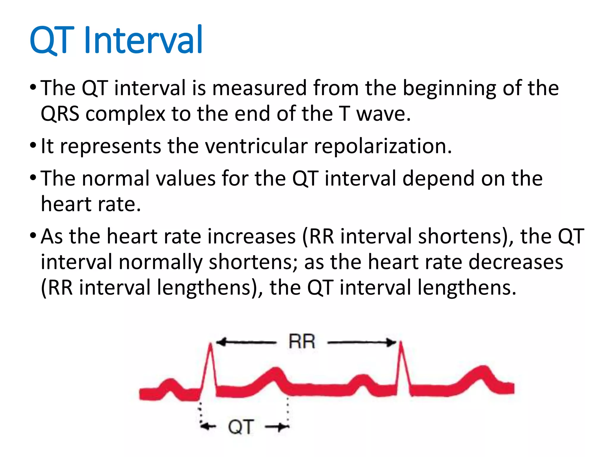
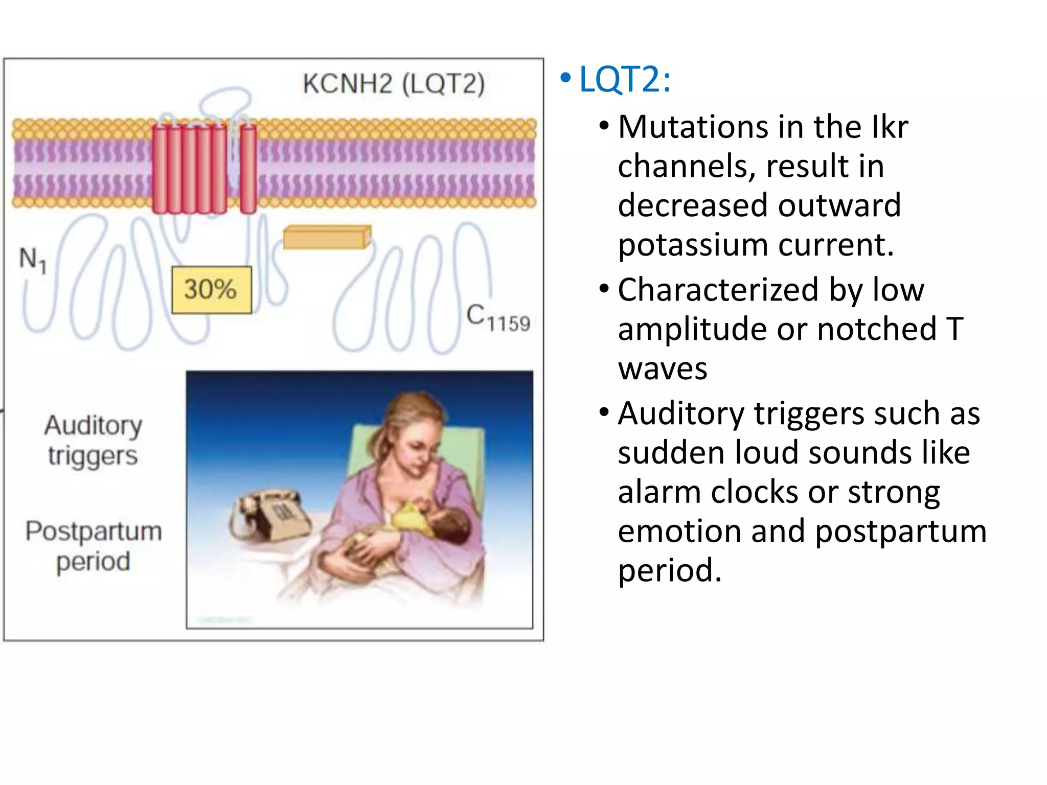
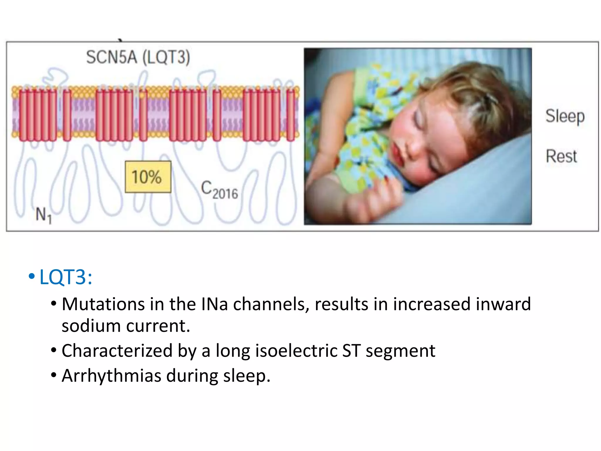
The Long QT Syndrome
(LQTS)
______________________________________________________
Dr. Sayeedur Rahman Khan Rumi
dr.rumibd@gmail.com
MD (Cardiology) Final Part Student
National Heart Foundation Hospital and Research Institute
QT Interval
•The QT interval is measured from the beginning of the
QRS complex to the end of the T wave.
•It represents the ventricular repolarization.
•The normal values for the QT interval depend on the
heart rate.
•As the heart rate increases (RR interval shortens), the QT
interval normally shortens; as the heart rate decreases
(RR interval lengthens), the QT interval lengthens.
QTc
•Because of this problem, another set of indexes of the
QT interval have been devised.
•These indexes are called rate-corrected QT or QTc
intervals.
•A widely used one (Bazett formula) is the square root
method, obtained by dividing the actual QT interval by
the square root of the RR interval.
•QTc = QT /√RR
The long QT syndrome (LQTS)
•The long QT syndrome (LQTS) is an Inherited
Arrhythmogenic Diseases in the structurally normal
heart characterized by abnormally prolonged QT
interval with peculiar morphologic abnormalities of the
T wave.
•LQTS manifests with syncope and/or cardiac arrest
typically occurring in children or teenagers.
•The estimated prevalence of LQTS is between 1:7000
and 1:3000
Types of LQTS
•Two major phenotypic variants have been described in
the early 1960s:
1. One autosomal dominant (Romano-Ward syndrome)
and
2. One autosomal recessive (Jervell and Lange-Nielsen
syndrome) also presenting with sensorineural
deafness.
•Additional rare variants including extracardiac
involvement are the Andersen syndrome and the
Timothy syndrome.
• 12 mutations have been identified, but the most common subtypes
of LQTS are LQT1, LQT2, and LQT3
•LQT1
• Mutations in the Iks channels, result in decreased outward
potassium current.
• Typically have broad-based T waves
• Exercise-induce arrhythmias, especially during swimming.
• It accounts for 45% to 55% of patients.
•LQT2:
• Mutations in the Ikr
channels, result in
decreased outward
potassium current.
• Characterized by low
amplitude or notched T
waves
• Auditory triggers such as
sudden loud sounds like
alarm clocks or strong
emotion and postpartum
period.
•LQT3:
• Mutations in the INa channels, results in increased inward
sodium current.
• Characterized by a long isoelectric ST segment
• Arrhythmias during sleep.
Clinical Presentation
•Syncope is often triggered by the onset of rapid
polymorphic ventricular tachycardia (VT) (torsades
de pointes) that can degenerate into ventricular
fibrillation and cause sudden death.
•The mean age of onset of symptoms is 12 years,
and earlier onset is usually associated with a more
severe form of the disease.
Diagnosis
• The diagnosis of LQTS is based on the evaluation of the
electrocardiogram (ECG) and on the measurement of the
QT interval.
• QTc values >440 ms (in males) and >460 ms (in females)
are considered abnormal.
• The evidence of QTc interval >500 ms is associated with a
five-fold increased risk of events.
• Additional risk factors are female sex (approximately two-
fold increased risk) and the occurrence of a first cardiac
event in early childhood.
• Genotype specific ECG patterns in long QT syndrome.
A. LQT1 -early onset broad based T wave.
B. LQT2 - low amplitude and bifid T wave.
C. LQT3 - long isoelectric ST segment with a late-appearing T wave
Risk stratification in long QT syndrome (LQTS)
Risk categories in LQTS according to QTc duration, genotype, and sex.
Percentages on the left indicate the risk of a first cardiac event
(syncope or cardiac arrest) in patients younger than 40 years of age in
the absence of any LQTS active treatment.
Treatment
• According to current guidelines, all LQTS patients with a history of
syncope and asymptomatic individuals with definite QT
prolongation should be treated with β-blockers (class I
recommendation)
• The most frequently used drugs are nadolol (1-2.5 mg/kg/d),
propranolol (2-4 mg/kg/d), and metoprolol (2-4 mg/kg/d)
• Lifestyle changes aimed at avoiding conditions that can precipitate
arrhythmias are also indicated.
• Specifically, LQTS patients should avoid competitive sports, QT-
prolonging drugs, and lowered potassium plasma levels (for
example, during diarrhea and vomiting)
• Patient with syncope, history of aborted sudden death, or torsade
de pointes despite β-blocker therapy should undergo ICD
implantation. (Secondary prevention)
• Certain high-risk subgroups such as patients with LQT3 in whom β-
blockers may be less effective, patients with QTc > 550 milliseconds,
or female LQT2 patients with QTc > 500 milliseconds may benefit
from ICD implantation. (Primary prevention)
• In all instances, β-blockers should be continued after ICD implant to
reduce the risk of cardiac arrest and shocks from the device.
• In patients with evidence of pause-dependent onset of torsade de
pointes and polymorphic VT, the use of pacemaker may also be
indicated.
• Left cardiac sympathetic denervation can be used as an adjunctive
therapy to reduce recurrence of arrhythmias.
Thank You

Long QT Syndrome

  • 1.
    The Long QTSyndrome (LQTS) ______________________________________________________ Dr. Sayeedur Rahman Khan Rumi dr.rumibd@gmail.com MD (Cardiology) Final Part Student National Heart Foundation Hospital and Research Institute
  • 2.
    QT Interval •The QTinterval is measured from the beginning of the QRS complex to the end of the T wave. •It represents the ventricular repolarization. •The normal values for the QT interval depend on the heart rate. •As the heart rate increases (RR interval shortens), the QT interval normally shortens; as the heart rate decreases (RR interval lengthens), the QT interval lengthens.
  • 3.
    QTc •Because of thisproblem, another set of indexes of the QT interval have been devised. •These indexes are called rate-corrected QT or QTc intervals. •A widely used one (Bazett formula) is the square root method, obtained by dividing the actual QT interval by the square root of the RR interval. •QTc = QT /√RR
  • 4.
    The long QTsyndrome (LQTS) •The long QT syndrome (LQTS) is an Inherited Arrhythmogenic Diseases in the structurally normal heart characterized by abnormally prolonged QT interval with peculiar morphologic abnormalities of the T wave. •LQTS manifests with syncope and/or cardiac arrest typically occurring in children or teenagers. •The estimated prevalence of LQTS is between 1:7000 and 1:3000
  • 5.
    Types of LQTS •Twomajor phenotypic variants have been described in the early 1960s: 1. One autosomal dominant (Romano-Ward syndrome) and 2. One autosomal recessive (Jervell and Lange-Nielsen syndrome) also presenting with sensorineural deafness. •Additional rare variants including extracardiac involvement are the Andersen syndrome and the Timothy syndrome.
  • 6.
    • 12 mutationshave been identified, but the most common subtypes of LQTS are LQT1, LQT2, and LQT3 •LQT1 • Mutations in the Iks channels, result in decreased outward potassium current. • Typically have broad-based T waves • Exercise-induce arrhythmias, especially during swimming. • It accounts for 45% to 55% of patients.
  • 7.
    •LQT2: • Mutations inthe Ikr channels, result in decreased outward potassium current. • Characterized by low amplitude or notched T waves • Auditory triggers such as sudden loud sounds like alarm clocks or strong emotion and postpartum period.
  • 8.
    •LQT3: • Mutations inthe INa channels, results in increased inward sodium current. • Characterized by a long isoelectric ST segment • Arrhythmias during sleep.
  • 9.
    Clinical Presentation •Syncope isoften triggered by the onset of rapid polymorphic ventricular tachycardia (VT) (torsades de pointes) that can degenerate into ventricular fibrillation and cause sudden death. •The mean age of onset of symptoms is 12 years, and earlier onset is usually associated with a more severe form of the disease.
  • 10.
    Diagnosis • The diagnosisof LQTS is based on the evaluation of the electrocardiogram (ECG) and on the measurement of the QT interval. • QTc values >440 ms (in males) and >460 ms (in females) are considered abnormal. • The evidence of QTc interval >500 ms is associated with a five-fold increased risk of events. • Additional risk factors are female sex (approximately two- fold increased risk) and the occurrence of a first cardiac event in early childhood.
  • 11.
    • Genotype specificECG patterns in long QT syndrome. A. LQT1 -early onset broad based T wave. B. LQT2 - low amplitude and bifid T wave. C. LQT3 - long isoelectric ST segment with a late-appearing T wave
  • 12.
    Risk stratification inlong QT syndrome (LQTS) Risk categories in LQTS according to QTc duration, genotype, and sex. Percentages on the left indicate the risk of a first cardiac event (syncope or cardiac arrest) in patients younger than 40 years of age in the absence of any LQTS active treatment.
  • 13.
    Treatment • According tocurrent guidelines, all LQTS patients with a history of syncope and asymptomatic individuals with definite QT prolongation should be treated with β-blockers (class I recommendation) • The most frequently used drugs are nadolol (1-2.5 mg/kg/d), propranolol (2-4 mg/kg/d), and metoprolol (2-4 mg/kg/d) • Lifestyle changes aimed at avoiding conditions that can precipitate arrhythmias are also indicated. • Specifically, LQTS patients should avoid competitive sports, QT- prolonging drugs, and lowered potassium plasma levels (for example, during diarrhea and vomiting)
  • 14.
    • Patient withsyncope, history of aborted sudden death, or torsade de pointes despite β-blocker therapy should undergo ICD implantation. (Secondary prevention) • Certain high-risk subgroups such as patients with LQT3 in whom β- blockers may be less effective, patients with QTc > 550 milliseconds, or female LQT2 patients with QTc > 500 milliseconds may benefit from ICD implantation. (Primary prevention) • In all instances, β-blockers should be continued after ICD implant to reduce the risk of cardiac arrest and shocks from the device. • In patients with evidence of pause-dependent onset of torsade de pointes and polymorphic VT, the use of pacemaker may also be indicated. • Left cardiac sympathetic denervation can be used as an adjunctive therapy to reduce recurrence of arrhythmias.
  • 15.