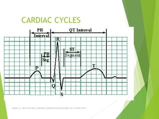
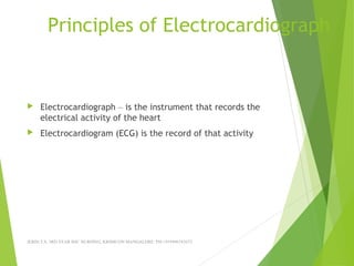
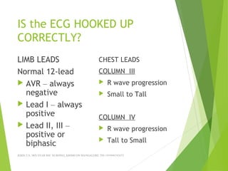
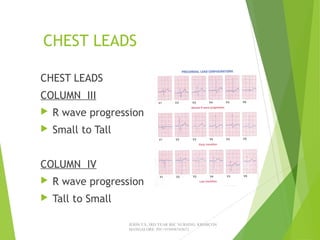
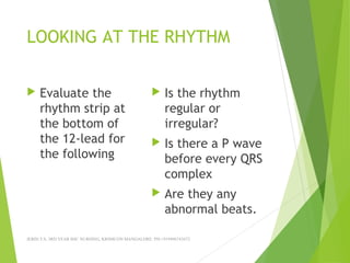
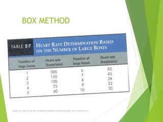
MAKING ECG’S EASY
APPLICATION FOR THE ECG
EVALUATING THE ECG
Upon completion one will be
able to:
 Describe what an ECG is.
 Describe the proper hook-up procedure for a 12-Lead
ECG
 Identify basic normal ECG waveform morphology.
 Distinguish between basic ECG arrhythmia and artifact.
JERIN.T.S, 3RD YEAR BSC NURSING, KRSMCON
MANGALORE. PH:+919496743672
QUICK REVIEW OF HEART
Purpose
 Pumps blood
Basic Anatomy
 4 chambers
 2 sides
 4 valves
JERIN.T.S, 3RD YEAR BSC NURSING, KRSMCON MANGALORE. PH:+919496743672
THE CONDUCTINGY SYSTEM
 SA Node
 Inter-nodal pathway
 AV Node
 Bundle of HIS
 Bundle Branches
 Purkinje Fibers
JERIN.T.S, 3RD YEAR BSC NURSING, KRSMCON
MANGALORE. PH:+919496743672
RELATIONSHIP
JERIN.T.S, 3RD YEAR BSC NURSING, KRSMCON
MANGALORE. PH:+919496743672
CARDIAC CYCLES
JERIN.T.S, 3RD YEAR BSC NURSING, KRSMCON MANGALORE. PH:+919496743672
12 LEAD ECG
Learning objectives:
Describe the correct
placement of all electrodes
Proper Skin preparation
Care of the ECG machine, wires and electrodes
JERIN.T.S, 3RD YEAR BSC NURSING, KRSMCON MANGALORE. PH:+919496743672
Principles of Electrocardiograph
 Electrocardiograph – is the instrument that records the
electrical activity of the heart
 Electrocardiogram (ECG) is the record of that activity
JERIN.T.S, 3RD YEAR BSC NURSING, KRSMCON MANGALORE. PH:+919496743672
HOOKING UP THE
12-LEAD ECG
 Proper skin prep
 Placement of the limb electrodes
 Placement of the chest electrodes
JERIN.T.S, 3RD YEAR BSC NURSING, KRSMCON MANGALORE. PH:+919496743672
SKIN PREPARATION
 REASON FOR SKIN PREPARATION
 5 STEP METHOD
 2 STEP METHOD
JERIN.T.S, 3RD YEAR BSC NURSING, KRSMCON MANGALORE. PH:+919496743672
ELECTRODE PLACEMENT
 LIMB LEADS
 CHEST LEAD
JERIN.T.S, 3RD YEAR BSC NURSING, KRSMCON MANGALORE. PH:+919496743672
LIMB LEADS
 Bipolar leads
I II III
 Augment leads
Avr Avl Avf
JERIN.T.S, 3RD YEAR BSC NURSING, KRSMCON
MANGALORE. PH:+919496743672
 
CHEST LEADS
 6 UNIPOLAR
LEADS
 V1
 V2
 V3
 V4
 V5
 V6
JERIN.T.S, 3RD YEAR BSC NURSING, KRSMCON MANGALORE. PH:+919496743672
LETS REVIEW
 What is an ECG
 What are the limb leads?
 What are the chest leads?
 Why do skin prep?
JERIN.T.S, 3RD YEAR BSC NURSING, KRSMCON MANGALORE. PH:+919496743672
12-LEAD ECG LAYOUT
JERIN.T.S, 3RD YEAR BSC NURSING, KRSMCON MANGALORE. PH:+919496743672
INFORMATION ON THE ECG
 Patients demographics
 Heart rate and measurements
 Speed ECG is recorded at
 Voltage ECG is recorded at
 What the filter is set on
JERIN.T.S, 3RD YEAR BSC NURSING, KRSMCON MANGALORE. PH:+919496743672
INFORMATION ON THE 12-
LEAD ECG
JERIN.T.S, 3RD YEAR BSC NURSING, KRSMCON MANGALORE. PH:+919496743672
WHAT YOU NEED TO LOOK
FOR
 Are the limb leads hooked up correctly?
 Are the chest leads hooked up correctly?
 Is the ECG free of artifact.
 Is this ECG a Critical Value
JERIN.T.S, 3RD YEAR BSC NURSING, KRSMCON MANGALORE. PH:+919496743672
IS the ECG HOOKED UP
CORRECTLY?
LIMB LEADS
Normal 12-lead
 AVR – always
negative
 Lead I – always
positive
 Lead II, III –
positive or
biphasic
CHEST LEADS
COLUMN III
 R wave progression
 Small to Tall
COLUMN IV
 R wave progression
 Tall to Small
JERIN.T.S, 3RD YEAR BSC NURSING, KRSMCON MANGALORE. PH:+919496743672
RULING OUT LIMB LEAD
REVERSAL
 Avr is always negative
 Lead I is always positive
 Lead II and III positive for the P wave and usually the
QRS complex
JERIN.T.S, 3RD YEAR BSC NURSING, KRSMCON MANGALORE. PH:+919496743672
CHEST LEADS
CHEST LEADS
COLUMN III
 R wave progression
 Small to Tall
COLUMN IV
 R wave progression
 Tall to Small
JERIN.T.S, 3RD YEAR BSC NURSING, KRSMCON
MANGALORE. PH:+919496743672
JERIN.T.S, 3RD YEAR BSC NURSING, KRSMCON MANGALORE. PH:+919496743672
LIMB LEADS
CHEST LEADS
JERIN.T.S, 3RD YEAR BSC NURSING, KRSMCON MANGALORE. PH:+919496743672
OTHER PROBLEMS WITH THE
ECG
 Artifact
 Electrical interference
 Somatic tremor
 Wandering baseline
JERIN.T.S, 3RD YEAR BSC NURSING, KRSMCON MANGALORE. PH:+919496743672
EINTHOVENS TRIANGLE
JERIN.T.S, 3RD YEAR BSC NURSING, KRSMCON MANGALORE. PH:+919496743672
ARTIFACT ON THE ECG
JERIN.T.S, 3RD YEAR BSC NURSING, KRSMCON MANGALORE. PH:+919496743672
FIND THE ARTEFACT
JERIN.T.S, 3RD YEAR BSC NURSING, KRSMCON MANGALORE. PH:+919496743672
WANDERIN BASELINE
JERIN.T.S, 3RD YEAR BSC NURSING, KRSMCON MANGALORE. PH:+919496743672
SOMATIC TREMOR
JERIN.T.S, 3RD YEAR BSC NURSING, KRSMCON MANGALORE. PH:+919496743672
ELECTRICAL INTERFERANCE
JERIN.T.S, 3RD YEAR BSC NURSING, KRSMCON MANGALORE. PH:+919496743672
BASIC CRITICAL VALUES
 Bradycardia – HR < 40bpm
 Tachycardia HR > 120bpm
 PVC’s - 4 or more in a row
 ST Elevation
JERIN.T.S, 3RD YEAR BSC NURSING, KRSMCON MANGALORE. PH:+919496743672
LOOKING AT THE RHYTHM
 Evaluate the
rhythm strip at
the bottom of
the 12-lead for
the following
 Is the rhythm
regular or
irregular?
 Is there a P wave
before every QRS
complex
 Are they any
abnormal beats.
JERIN.T.S, 3RD YEAR BSC NURSING, KRSMCON MANGALORE. PH:+919496743672
FINDING THE HEART RATE
 3 METHODS
 RATE RULER
 COUNTING
 BOX METHOD
JERIN.T.S, 3RD YEAR BSC NURSING, KRSMCON MANGALORE. PH:+919496743672
BOX METHOD
JERIN.T.S, 3RD YEAR BSC NURSING, KRSMCON MANGALORE. PH:+919496743672
ECTOPIC BEATS
JERIN.T.S, 3RD YEAR BSC NURSING, KRSMCON MANGALORE. PH:+919496743672
ECTOPIC BEATS
JERIN.T.S, 3RD YEAR BSC NURSING, KRSMCON MANGALORE. PH:+919496743672
LETS SUMMARIZE
How do we produce an excellent 12-lead ECG?
Proper skin prep
Correct electrode placement
Recognize and know how to correct problems
Recognize basic critical values
JERIN.T.S, 3RD YEAR BSC NURSING, KRSMCON MANGALORE. PH:+919496743672

Ecg made easy ppt

  • 1.
    MAKING ECG’S EASY APPLICATIONFOR THE ECG EVALUATING THE ECG
  • 2.
    Upon completion onewill be able to:  Describe what an ECG is.  Describe the proper hook-up procedure for a 12-Lead ECG  Identify basic normal ECG waveform morphology.  Distinguish between basic ECG arrhythmia and artifact. JERIN.T.S, 3RD YEAR BSC NURSING, KRSMCON MANGALORE. PH:+919496743672
  • 3.
    QUICK REVIEW OFHEART Purpose  Pumps blood Basic Anatomy  4 chambers  2 sides  4 valves JERIN.T.S, 3RD YEAR BSC NURSING, KRSMCON MANGALORE. PH:+919496743672
  • 4.
    THE CONDUCTINGY SYSTEM SA Node  Inter-nodal pathway  AV Node  Bundle of HIS  Bundle Branches  Purkinje Fibers JERIN.T.S, 3RD YEAR BSC NURSING, KRSMCON MANGALORE. PH:+919496743672
  • 5.
    RELATIONSHIP JERIN.T.S, 3RD YEARBSC NURSING, KRSMCON MANGALORE. PH:+919496743672
  • 6.
    CARDIAC CYCLES JERIN.T.S, 3RDYEAR BSC NURSING, KRSMCON MANGALORE. PH:+919496743672
  • 7.
    12 LEAD ECG Learningobjectives: Describe the correct placement of all electrodes Proper Skin preparation Care of the ECG machine, wires and electrodes JERIN.T.S, 3RD YEAR BSC NURSING, KRSMCON MANGALORE. PH:+919496743672
  • 8.
    Principles of Electrocardiograph Electrocardiograph – is the instrument that records the electrical activity of the heart  Electrocardiogram (ECG) is the record of that activity JERIN.T.S, 3RD YEAR BSC NURSING, KRSMCON MANGALORE. PH:+919496743672
  • 9.
    HOOKING UP THE 12-LEADECG  Proper skin prep  Placement of the limb electrodes  Placement of the chest electrodes JERIN.T.S, 3RD YEAR BSC NURSING, KRSMCON MANGALORE. PH:+919496743672
  • 10.
    SKIN PREPARATION  REASONFOR SKIN PREPARATION  5 STEP METHOD  2 STEP METHOD JERIN.T.S, 3RD YEAR BSC NURSING, KRSMCON MANGALORE. PH:+919496743672
  • 11.
    ELECTRODE PLACEMENT  LIMBLEADS  CHEST LEAD JERIN.T.S, 3RD YEAR BSC NURSING, KRSMCON MANGALORE. PH:+919496743672
  • 12.
    LIMB LEADS  Bipolarleads I II III  Augment leads Avr Avl Avf JERIN.T.S, 3RD YEAR BSC NURSING, KRSMCON MANGALORE. PH:+919496743672  
  • 13.
    CHEST LEADS  6UNIPOLAR LEADS  V1  V2  V3  V4  V5  V6 JERIN.T.S, 3RD YEAR BSC NURSING, KRSMCON MANGALORE. PH:+919496743672
  • 14.
    LETS REVIEW  Whatis an ECG  What are the limb leads?  What are the chest leads?  Why do skin prep? JERIN.T.S, 3RD YEAR BSC NURSING, KRSMCON MANGALORE. PH:+919496743672
  • 15.
    12-LEAD ECG LAYOUT JERIN.T.S,3RD YEAR BSC NURSING, KRSMCON MANGALORE. PH:+919496743672
  • 16.
    INFORMATION ON THEECG  Patients demographics  Heart rate and measurements  Speed ECG is recorded at  Voltage ECG is recorded at  What the filter is set on JERIN.T.S, 3RD YEAR BSC NURSING, KRSMCON MANGALORE. PH:+919496743672
  • 17.
    INFORMATION ON THE12- LEAD ECG JERIN.T.S, 3RD YEAR BSC NURSING, KRSMCON MANGALORE. PH:+919496743672
  • 18.
    WHAT YOU NEEDTO LOOK FOR  Are the limb leads hooked up correctly?  Are the chest leads hooked up correctly?  Is the ECG free of artifact.  Is this ECG a Critical Value JERIN.T.S, 3RD YEAR BSC NURSING, KRSMCON MANGALORE. PH:+919496743672
  • 19.
    IS the ECGHOOKED UP CORRECTLY? LIMB LEADS Normal 12-lead  AVR – always negative  Lead I – always positive  Lead II, III – positive or biphasic CHEST LEADS COLUMN III  R wave progression  Small to Tall COLUMN IV  R wave progression  Tall to Small JERIN.T.S, 3RD YEAR BSC NURSING, KRSMCON MANGALORE. PH:+919496743672
  • 20.
    RULING OUT LIMBLEAD REVERSAL  Avr is always negative  Lead I is always positive  Lead II and III positive for the P wave and usually the QRS complex JERIN.T.S, 3RD YEAR BSC NURSING, KRSMCON MANGALORE. PH:+919496743672
  • 21.
    CHEST LEADS CHEST LEADS COLUMNIII  R wave progression  Small to Tall COLUMN IV  R wave progression  Tall to Small JERIN.T.S, 3RD YEAR BSC NURSING, KRSMCON MANGALORE. PH:+919496743672
  • 22.
    JERIN.T.S, 3RD YEARBSC NURSING, KRSMCON MANGALORE. PH:+919496743672 LIMB LEADS
  • 23.
    CHEST LEADS JERIN.T.S, 3RDYEAR BSC NURSING, KRSMCON MANGALORE. PH:+919496743672
  • 24.
    JERIN.T.S, 3RD YEARBSC NURSING, KRSMCON MANGALORE. PH:+919496743672
  • 25.
    JERIN.T.S, 3RD YEARBSC NURSING, KRSMCON MANGALORE. PH:+919496743672
  • 26.
    JERIN.T.S, 3RD YEARBSC NURSING, KRSMCON MANGALORE. PH:+919496743672
  • 27.
    JERIN.T.S, 3RD YEARBSC NURSING, KRSMCON MANGALORE. PH:+919496743672
  • 28.
    OTHER PROBLEMS WITHTHE ECG  Artifact  Electrical interference  Somatic tremor  Wandering baseline JERIN.T.S, 3RD YEAR BSC NURSING, KRSMCON MANGALORE. PH:+919496743672
  • 29.
    EINTHOVENS TRIANGLE JERIN.T.S, 3RDYEAR BSC NURSING, KRSMCON MANGALORE. PH:+919496743672
  • 30.
    ARTIFACT ON THEECG JERIN.T.S, 3RD YEAR BSC NURSING, KRSMCON MANGALORE. PH:+919496743672
  • 31.
    FIND THE ARTEFACT JERIN.T.S,3RD YEAR BSC NURSING, KRSMCON MANGALORE. PH:+919496743672
  • 32.
    WANDERIN BASELINE JERIN.T.S, 3RDYEAR BSC NURSING, KRSMCON MANGALORE. PH:+919496743672
  • 33.
    SOMATIC TREMOR JERIN.T.S, 3RDYEAR BSC NURSING, KRSMCON MANGALORE. PH:+919496743672
  • 34.
    ELECTRICAL INTERFERANCE JERIN.T.S, 3RDYEAR BSC NURSING, KRSMCON MANGALORE. PH:+919496743672
  • 35.
    BASIC CRITICAL VALUES Bradycardia – HR < 40bpm  Tachycardia HR > 120bpm  PVC’s - 4 or more in a row  ST Elevation JERIN.T.S, 3RD YEAR BSC NURSING, KRSMCON MANGALORE. PH:+919496743672
  • 36.
    LOOKING AT THERHYTHM  Evaluate the rhythm strip at the bottom of the 12-lead for the following  Is the rhythm regular or irregular?  Is there a P wave before every QRS complex  Are they any abnormal beats. JERIN.T.S, 3RD YEAR BSC NURSING, KRSMCON MANGALORE. PH:+919496743672
  • 37.
    FINDING THE HEARTRATE  3 METHODS  RATE RULER  COUNTING  BOX METHOD JERIN.T.S, 3RD YEAR BSC NURSING, KRSMCON MANGALORE. PH:+919496743672
  • 38.
    BOX METHOD JERIN.T.S, 3RDYEAR BSC NURSING, KRSMCON MANGALORE. PH:+919496743672
  • 39.
    JERIN.T.S, 3RD YEARBSC NURSING, KRSMCON MANGALORE. PH:+919496743672
  • 40.
    JERIN.T.S, 3RD YEARBSC NURSING, KRSMCON MANGALORE. PH:+919496743672
  • 41.
    JERIN.T.S, 3RD YEARBSC NURSING, KRSMCON MANGALORE. PH:+919496743672
  • 42.
    JERIN.T.S, 3RD YEARBSC NURSING, KRSMCON MANGALORE. PH:+919496743672
  • 43.
    ECTOPIC BEATS JERIN.T.S, 3RDYEAR BSC NURSING, KRSMCON MANGALORE. PH:+919496743672
  • 44.
    ECTOPIC BEATS JERIN.T.S, 3RDYEAR BSC NURSING, KRSMCON MANGALORE. PH:+919496743672
  • 45.
    LETS SUMMARIZE How dowe produce an excellent 12-lead ECG? Proper skin prep Correct electrode placement Recognize and know how to correct problems Recognize basic critical values JERIN.T.S, 3RD YEAR BSC NURSING, KRSMCON MANGALORE. PH:+919496743672

Editor's Notes

  • #4 In an adult with a healthy heart, the heart rate is usually about 72 beats per minute. The excitatory and electrical conduction system of the heart is responsible for the contraction and relaxation of the heart muscle. The heart is divided into four chambers, but it functions as a two sided pump. Top is the right and left atria Bottom is the right and left ventricles The right side of the heart receives and pumps venous blood to the lungs. The left side of the heart receives (from the lungs) and pumps arterial blood to the body.
  • #5 The sinoatrial node (SA node) is the pacemaker where the electrical impulse is generated. This node is located along the posterior wall of the right atrium right beneath the opening of the superior vena cava. It is crescent shaped and about 3 mm wide and 1 cm long. The impulse travels from the SA node through the internodal pathways to the atrioventricular node (AV node). The AV node is responsible for conduction of the impulse from the atria to the ventricles. The impulse is delayed slightly at this point to allow complete emptying of the atria before the ventricles contract. The impulse continues through the AV bundle and down the left and right bundle branches of the Purkinje fibers. The Purkinje fibers conduct the impulse to all parts of the ventricles, causing contraction (Guyton, 1982).
  • #6 Lets look at how the conduction system related to what we record on the ECG. P wave: the sequential activation (depolarization) of the right and left atria QRS complex: right and left ventricular depolarization (normally the ventricles are activated simultaneously) ST-T wave ventricular repolarization U wave: origin for this wave is not clear - but probably represents &amp;quot;afterdepolarizations&amp;quot; in the ventricles  PR interval: time interval from onset of atrial depolarization (P wave) to onset of ventricular depolarization (QRS complex)QRS duration: duration of ventricular muscle depolarizationQT interval: duration of ventricular depolarization and repolarizationRR interval: duration of ventricular cardiac cycle (an indicator of ventricular rate)PP interval: duration of atrial cycle (an indicator of atrial rate)
  • #7 Recorded on graph paper. Time is measured across Voltage is measured up and down. Small squares 1mm high 0.04 secs wide 5 small squares = 1 large square 1 large square is 5mm high and 0.20 sec wide
  • #11 Both re-usable and disposable electrodes rely on the electrolyte in the electrodes to make an effective connection between the machines electrode and the patient’s electro-physiological signals.   These signals are not picked up just off the surface of the skin, where dead or dry skin, oils and hair all prevent the signals from being detected. The signals are under this surface, and thus we have to prepare the patient so that the electrolyte can reach the signals beneath.   This is achieved by: Removing oil, greasy and dirt from the skins sites with alcohol  
  • #12 ELECTRODE PLACEMENT The standard 12-Lead ECG is a collect of tracings of electrical activity occurring in the heart. Each lead provides a tracing, which is characteristic of a different view of the same electrical activity. Other words we take 12 different angles (pictures) of the same activity. In a standard 12-lead ECG there are 6 limb leads with a three-electrode connection made to the Right arm, Left arm and Left leg. The right leg is also hooked up to the machine but this is your ground lead and does not generate a signal on the ECG.
  • #14 The chest electrodes are labelled “V” and are numbered from 1 to 6. The placement of these electrodes needs to be exact to give the optimum information as possible. If the electrodes are placed incorrectly on the chest, the tracing will reveal duplication of some information, while other areas will not be represented properly. Incorrect placement of the electrodes can lead to serious errors of interpretation.  There are six chest leads: V1, V2,V3,V4, V5 and V6. V1 at the fourth intercostal space, at the right margin of the sternum V2at the fourth intercostal space, at the left margin of the sternum V3midway between the position of leads V2 and V4 (in a straight line) V4at the fifth intercostals space at the junction of the left midclavicular line V5 midway between the position of leads V4 and V6 (straight down from the axillary Line on the same horizontal position as V4 and V6 V6at the horizontal position of V4, at the left of the midaxillary line.
  • #23 Lets look at this ECG for the limb leads? Is lead I positive? Is AVR negative We have reversed our limb electrodes we have the right arm on the left arm and reverse.
  • #24 We need to have R wave progression In column III the R wave(first positive wave of the QRS) goes from short to tall V1 shortest R wave V2 R wave taller than in V1 and V3 has the tallest R wave. In column IV the R wave is always the shortest in V6.
  • #26 Is this ECG hooked up correctly for the limb leads? Is it for the chest leads?
  • #27 Lets evaluate this ECG to see if the Limb leads and Chest Leads are hooked up correctly?
  • #28 Is this ECG HOOKED UP CORRECTLY FOR LIMB AND CHEST?
  • #30 Lead I = right arm and left arm Lead II = right arm and left Leg Lead III = left arm and Left Leg.
  • #39 Look at the number of large boxes before two R waves and then using this method find the HR . I just remember that 2 ½ large boxes or less is critical value for tachycardia 7 or more large boxes is critical for bradycardia
  • #40 Look at the rhythm strip is it regular or irregular. How many large boxes are between two R waves = 5 = 60bpm
  • #41 Is the rhythm regular? What is the heart rate?