Downloaded 392 times
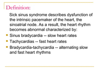


This document defines and describes sick sinus syndrome, which is a dysfunction of the sinoatrial node that can cause abnormal heart rhythms like bradycardia, tachycardia, and alternating slow and fast rhythms. It may be caused by certain drugs, aging, or underlying heart conditions. Symptoms can include fatigue, dizziness, and fainting. Diagnosis involves an electrocardiogram showing arrhythmias. Treatment options include medications or a pacemaker if symptoms are severe. The document also briefly discusses different types of heart block.


![Shadechapter08.ppt [read only]](https://cdn.slidesharecdn.com/ss_thumbnails/shadechapter08-150421102734-conversion-gate02-thumbnail.jpg?width=640&height=640&fit=bounds)