Dna replication.botany
•Download as PPTX, PDF•
8 likes•3,674 views

DNA replicates in a semi-conservative manner, as proven by Meselson and Stahl's experiment in 1958. Replication begins with initiation, where helicase unwinds the DNA double helix and primase lays down RNA primers. During elongation, DNA polymerase adds nucleotides to the 3' end of the primers on the leading and lagging strands. Okazaki fragments are formed and ligated on the lagging strand. Replication terminates when DNA polymerase reaches the telomeres at the end of the DNA strands.

Recommended
More Related Content
What's hot
What's hot (20)
Similar to Dna replication.botany
Similar to Dna replication.botany (20)
More from Jannat Iftikhar
More from Jannat Iftikhar (19)
Recently uploaded
Recently uploaded (20)
Dna replication.botany
- 2. University of the Punjab Lahore
- 3. DNA STRUCTURE In early 1900s scientist knew that chromosomes are made up of DNA and proteins containing genetic information. However, they didn’t know whether protein or DNA was actual genetic material.
- 4. DNA STRUCTURE In 1940s various researches showed that DNA was the genetic material. In early 1950s structure of DNA was determined.
- 5. STRUCTURE OF DNA James Watson & Francis Crick determined the structure of DNA in 1953.
- 6. STRUCTURE OF DNA DNA is polynucleotide; nucleotides are composed of a phosphate, a sugar and a nitrogen containing base.
- 7. STRUCTURE OF DNA Sugar in DNA is deoxyribose. Four nitrogen bases in DNA i. Adenine ii. Guanine iii. Thymine iv. cytosine
- 8. STRUCTURE OF DNA Watson and Crick showed that DNA is double helix in which A is paired with T G is paired with C This is called complementary base pairing because a purine is always paired with pyrimidine.
- 10. DOUBLE HELIX Each side of the double helix runs in opposite (anti-parallel) directions. The beauty of this structure is that it can unzip down the middle and each side can serve as a pattern or template for the other side.
- 12. REPLICATION Replica “copy”. DNA making copies of itself, we call it DNA replication .
- 13. WHY DNA REPLICATE ITSELF? To reproduce, a cell must copy and transmit its genetic information (DNA) to all of its progeny. To do so, DNA replicates. DNA carries information for making all of the cell’s protein.
- 14. REPLICATION IN DIFFERENT CELLS Different types of cells replicated their DNA at different rates. Hair cells, finger nails, bone marrow cells. constantly devide. Cells of brain, heart and muscles. cells go through several rounds of cell division and stop. Skin cells and liver cells. stop dividing, but can be induced to divide to repair injury.
- 15. WHERE REPLICATION OCCUR? In prokaryotes, DNA replication occurs in the cytoplasm. In eukaryotes, in the nucleus.
- 16. CLASSICAL MODELS FOR DNA REPLICATION Conservative Semi conservative Dispersive
- 17. CONSERVATIVE MODEL Conservative Model In this model the two parental DNA strands are back together after replication has occurred. That is, one daughter molecule contains both parental DNA strands, and the other daughter molecule contains DNA strands of all newly-synthesized material.
- 18. SEMI CONSERVATIVE MODEL Semi conservative Model In this model the two parental DNA strands separate and each of those strands then serves as a template for the synthesis of a new DNA strand. The result is two DNA double helices, both of which consist of one parental and one new strand.
- 19. DISPERSIVE MODEL Dispersive Model In this model the parental double helix is broken into double-stranded DNA segments that, as for the Conservative Model, act as templates for the synthesis of new double helix molecules. The segments then reassemble into complete DNA double helices, each with parental and progeny DNA segments interspersed.
- 20. CLASSICAL MODELS OF DNA REPLICATION
- 21. MESELSON AND STAHL EXPERIMENT Nobody knew for sure how DNA replication really worked until two scientists named Matthew Meselson and Franklin Stahl devised an ingenious experiment in 1958. Show that DNA follows semi conservative model to replicate itself.
- 22. MESELSON AND STAHL EXPERIMENT Hypothesis Experimental procedure Result
- 23. HYPOTHESIS Three hypotheses had been previously proposed for the method of replication of DNA. Semiconservative hypothesis, proposed by Watson and Crick. Conservative hypothesis proposed that the entire DNA molecule acted as a template. Dispersive hypothesis is exemplified by a model proposed by Max Delbruck.
- 25. RESULTS 1. Light DNA 2. Heavy DNA 3. Intermediate DNA 4. Light DNA & intermediate DNA
- 26. RESULTS Disproved conservative replication. Disproved dispersive replication. Proved that DNA replicates in semiconservative manner.
- 27. SEMI CONSERVATIVE REPLICATION Semiconservative replication describes the mechanism by which DNA is replicated in all known cells. This mechanism of replication was one of three models originally proposed for DNA replication.
- 28. REQUIREMENTS OF REPLICATION DNA template. Free 3’-OH group. Proteins of DNA replication
- 29. DNA TEMPLATE Template strand, that is to be copied. Each old strand act as a template.
- 30. FREE 3’-OH GROUP
- 31. PROTEINS OF REPLICATION 1. DNA Helicases 2. DNA single-stranded binding proteins 3. DNA Gyrase 4. DNA Polymerase 5. Primase 6. DNA Ligase
- 32. HELICASE DNA Helicases - These proteins bind to the double stranded DNA and stimulate the separation of the two strands.
- 33. DNA SINGLE-STRANDED BINDING PROTEINS DNA single-stranded binding proteins - These proteins bind to the DNA as a tetramer and stabilize the single-stranded structure that is generated by the action of the helicases. Replication is 100 times faster when these proteins are attached to the single-stranded DNA.
- 34. DNA GYRASE DNA Gyrase - This enzyme catalyzes the formation of negative supercoils that is thought to aid with the unwinding process.
- 35. DNA POLYMERASE DNA Polymerase - DNA Polymerase I (Pol I) was the first enzyme discovered with polymerase activity, and it is the best characterized enzyme. The DNA polymerases travel up the DNA molecule from an initiation site which is a region along the DNA that the enzyme complex can recognize. adds 5' C to 3' C in a phosphodiester linkage.
- 36. PRIMASE Primase - The requirement for a free 3' hydroxyl group is fulfilled by the RNA primers that are synthesized at the initiation sites by these enzymes.
- 37. DNA LIGASE DNA ligase- forms a covalent phosphodiester linkage between 3'-hydroxyl and 5'-phosphate groups.
- 38. DNA POLYMERASE FUNCTION Requires an RNA or DNA primer (RNA primer in eukaryotes). Reads DNA template in a 3'-->5- direction only Synthesizes new strand in 5'-->3' direction only - adds 5' phosphate to 3' hydroxyl group.
- 39. DIRECTION OF REPLICATION It replicates from 3’ to 5’ of the template strand. From 5’ to 3’ of the newly growing strand.
- 40. STEPS OF REPLICATION Initiation Elongation Termination
- 41. INITIATION 1. The first major step for the DNA Replication to take place is the breaking of hydrogen bonds between bases of the two antiparallel strands. 2. Helicase is the enzyme that splits the two strands
- 42. INITIATION 1. One of the most important steps of DNA Replication is the binding of RNA Primase in the initiation point of the 3'-5' parent chain. 2. RNA nucleotides are the primers (starters) for the binding of DNA nucleotides.
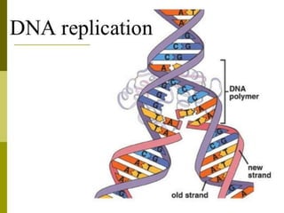
- 43. ELONGATION RNA primase lays down primers. Replication starts at primer and lays down nucleotides 5’ to 3’. Leading strand goes continuously, lagging strand goes discontinuously.
- 44. ELONGATION The elongation process is different for the 5'-3' and 3'-5' template. a)5'-3' Template: The 3'-5' proceeding daughter strand -that uses a 5'-3' template- is called leading strand because DNA Polymerase ä can "read" the template and continuously adds nucleotides (complementary to the nucleotides of the template, for example Adenine opposite to
- 45. LEADING STRAND Leading strand synthesis is continuous. From 3’ to 5’ of the template.
- 46. ELONGATION 5'-3'Template: The 5'-3' template cannot be "read" by DNA Polymerase ä. The replication of this template is complicated and the new strand is called lagging strand. In the lagging strand the RNA Primase adds more RNA Primers. DNA polymerase å reads the template and lengthens the bursts. The gap between two RNA primers is called "Okazaki Fragments".
- 47. LAGGING STRAND Lagging strand synthesis is discontinuous. Okazaki fragments. Ligase joins discontinuous fragments.
- 48. ELONGATION In the lagging strand the DNA Pol I - exonuclease- reads the fragments and removes the RNA Primers. The gaps are closed with the action of DNA Polymerase (adds complementary nucleotides to the gaps) and DNA Ligase (adds phosphate in the remaining gaps of the phosphate - sugar backbone).
- 49. TERMINATION The last step of DNA Replication is the Termination. RNA primer is removed. Replaced with DNA nucleotides. DNA ligase joins okazaki fragments with phosphodiester bonds. Helicase rewinds DNA together.
- 50. TERMINATION This process happens when the DNA Polymerase reaches to an end of the strands. These ends of linear (chromosomal) DNA consists of noncoding DNA that contains repeat sequences and are called telomeres. A part of the telomere is removed in every cycle of DNA Replication.
- 51. TERMINATION The DNA Replication is not completed before a mechanism of repair fixes possible errors caused during the replication. Enzymes like nucleases remove the wrong nucleotides and the DNA Polymerase fills the gaps.
- 52. TERMINATION Protein which binds to this sequence to physically stop DNA replication proceeding. This is named the DNA replication terminus site-binding protein or in other words, Ter- protein.
- 54. ,