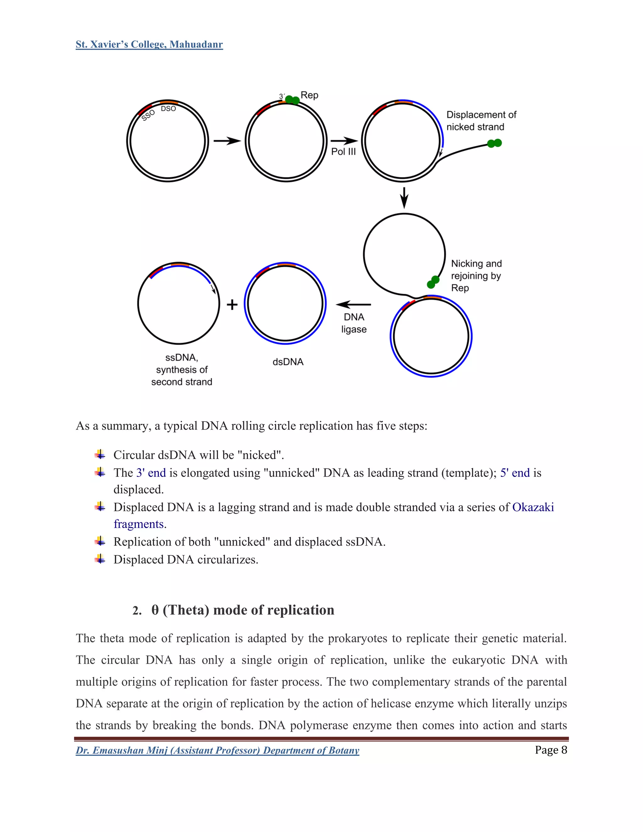
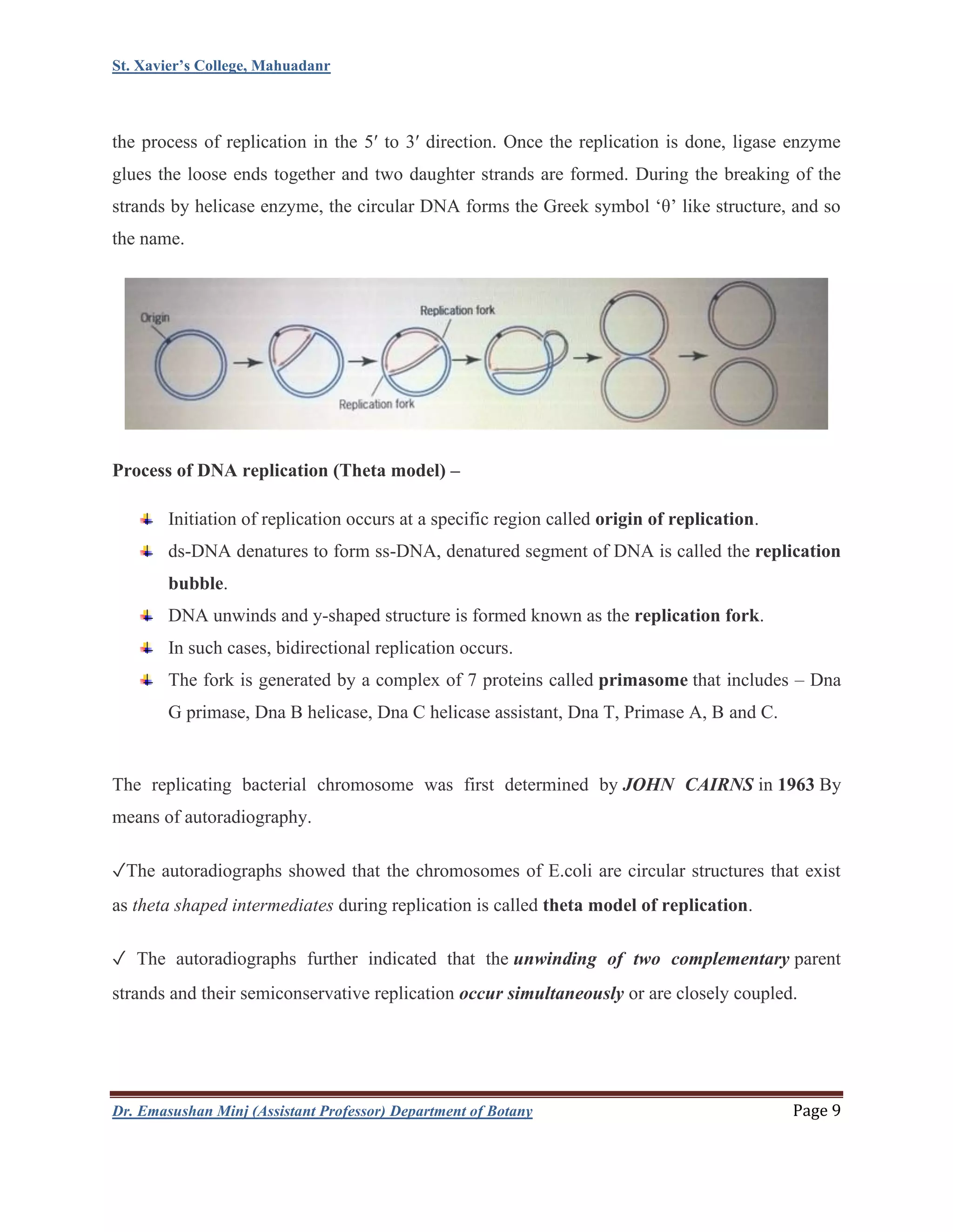
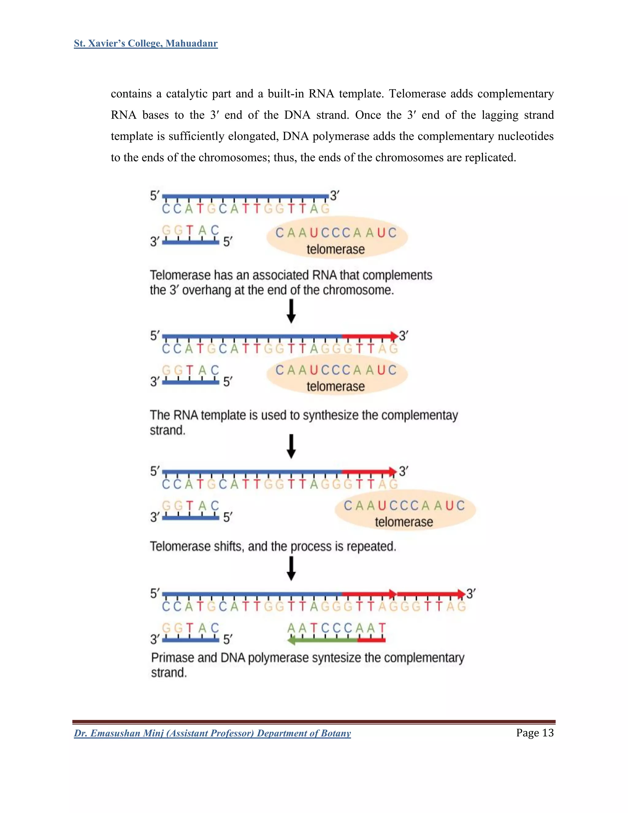
St. Xavier’s College, Mahuadanr
Dr. Emasushan Minj (Assistant Professor) Department of Botany Page 1
Unit 2
SEM-IV (C8)
DNA replication
⚫ DNA replication is the biological process of producing two identical replicas of DNA from
one original DNA molecule. DNA replication occurs in all living organisms acting as the
most essential part for biological inheritance.
⚫ In a cell, DNA replication begins at specific locations or origins of replication in
the genome.
⚫ Unwinding of DNA and synthesis of new strands takes place by an enzyme known
as helicase which results in the formation of replication forks, growing bi-directionally from
the origin.
⚫ Various proteins are associated with the replication fork to help in the initiation and
continuation of DNA synthesis.
⚫ Most prominently, DNA polymerase synthesizes the new strands by adding nucleotides that
complement each (template) strand.
⚫ DNA replication occurs during the S-stage of Interphase.
Replication process
DNA replication including all biological polymerization processes in three enzymatically
catalyzed and coordinated steps: initiation, elongation and termination.
Initiation
For a cell to divide, it must first replicate its DNA. Once DNA replication begins, it
proceeds to completion. Once replication is complete, it does not occur again in the same cell
cycle. This is made possible by the division of initiation of the pre-replication complex.
St. Xavier’s College, Mahuadanr
Dr. Emasushan Minj (Assistant Professor) Department of Botany Page 2
Sequence of events during initiation
Binding of DnaA protein to oriC in E.coli takes place and forms an initial complex.
Further DNA helicase is loaded which mediates unpairing of template stands and forms
open complex between DnaA and oriC.
Then primase binds to form primosome.
Synthesis of RNA primer takes place.
Initiation of DNA polymerization by DNA polymerase.
Elongation
DNA polymerases catalyze the step-by-step addition of deoxyribonucleotide units to a
DNA chain and it has 5′–3′ activity.
DNA replication systems require a free 3′ hydroxyl group before synthesis can be
initiated (note: the DNA template is read in 3′ to 5′ direction whereas a new strand is
synthesized in the 5′ to 3′ direction).
At each growing fork, on strand called the leading strand is synthesized continuously
from a single primer on the leading-strand template and grows in the 5’-3’ direction.
Growth of the leading strand proceeds in the same direction as the movement
of the growing fork.
Synthesis of the lagging strand is more complicated because DNA polymerases can add
nucleotides only to the 3’ end of a primer or growing DNA strand.
Movement of the growing fork unveils the template strand for lagging-strand synthesis in
the 5’-3’ direction.
After 1000 to 2000 nucleotides of the leading strand have been replicated, the first round
of discontinuous strand synthesis on the lagging strand can begin.
The short pieces of DNA called as Okazaki fragments are repeatedly synthesized on the
lagging-strand template.
St. Xavier’s College, Mahuadanr
Dr. Emasushan Minj (Assistant Professor) Department of Botany Page 3
Four distinct mechanisms for DNA synthesis are recognized:
1. All cellular life forms and many DNA viruses, phages and plasmids use a primase to
synthesize a short RNA primer with a free 3′ OH group which is subsequently elongated
by a DNA polymerase.
2. The retro elements (including retroviruses) employ a transfer RNA that primes DNA
replication by providing a free 3′ OH that is used for elongation by the reverse
transcriptase.
3. In the adenoviruses and the φ29 family of bacteriophages, the 3′ OH group is provided by
the side chain of an amino acid of the genome attached protein (the terminal protein) to
which nucleotides are added by the DNA polymerase to form a new strand.
4. In the single stranded DNA viruses—a group that includes the circoviruses,
the geminiviruses, the parvoviruses and others—and also the many phages
and plasmids that use the rolling circle replication (RCR) mechanism, the RCR
endonuclease creates a nick in the genome strand (single stranded viruses) or one of the
DNA strands (plasmids). The 5′ end of the nicked strand is transferred to
a tyrosine residue on the nuclease and the free 3′ OH group is then used by the DNA
polymerase to synthesize the new strand.
St. Xavier’s College, Mahuadanr
Dr. Emasushan Minj (Assistant Professor) Department of Botany Page 4
Semiconservative replication
⚫ It is crucial that the genetic material is reproduced accurately. When Watson and Crick
worked out the double-helix structure of DNA in 1953, they recognized that the
complementary nature of the two strands-A paired with T and G paired with C-might
play an important role in its replication. Because the two polynucleotide strands are
joined only Genetics by hydrogen bonds, they are able to separate without requiring
breakage of covalent bonds.
⚫ If the two strands of a parental double helix of DNA are separated, the base sequence of
each parental strand could serve as a template for the synthesis of a new complementary
strand, producing two identical progeny double helices. This process is called
semiconservative replication because the parental double helix is half conserved, each
parental single strand remaining intact.
St. Xavier’s College, Mahuadanr
Dr. Emasushan Minj (Assistant Professor) Department of Botany Page 5
⚫ The alternative methods are conservative and dispersive.
⚫ In conservative replication, the whole original double helix acts as a template for a new
one, one daughter molecule would consist of the original parental DNA, and the other
daughter would be totally new DNA. In dispersive replication, some parts of the original
double helix are conserved, and some parts are not. In this model, the parental double
helix is broken into double-stranded DNA segments and just like conservative mode of
replication acts as templates for the synthesis of new double-stranded DNA segments.
The segments then reassemble into complete DNA double helices, each with parental and
progeny DNA segments interspersed.
*The Meselson-Stahl experiment showed that DNA replicates by a semiconservative mechanism
In this experiment,
E. coli cells initially were grown in a medium containing ammonium salts prepared with
heavy nitrogen (15N) until the entire cellular DNA was labeled.
After the cells were transferred to a medium containing the normal light isotope (14N),
samples were removed periodically from the cultures and the DNA in each sample was
analyzed by equilibrium density-gradient centrifugation.
St. Xavier’s College, Mahuadanr
Dr. Emasushan Minj (Assistant Professor) Department of Botany Page 6
This technique can separate heavy-heavy (15N-15N), light-light (14N-14N), and heavy-light
(15N-14N) duplexes into distinct bands.
After one generation of growth, the entire extracted DNA had the density of 15N-14N
DNA.
After two generations, approximately half the DNA had the density of 15N-14N DNA; the
other half had the density of 15N-14N DNA.
With additional generations, a large fraction of the extracted DNA consisted of 14N-14N
duplexes; 15N-15N duplexes never appeared.
These results match the predicted pattern for the semi-conservative replication
mechanism.
Meselson and Stahl experimentally demonstrated the semi-conservative replication of
DNA in E. coli in 1958. If the parental DNA carries a heavy density label because the
organism has been grown in medium containing a suitable isotope (such as 15N), its
strands can be distinguished from those that are synthesized when the organism is
transferred to a medium containing normal light isotopes.
Various model of replication
1. Rolling circle replication
2. Theta mode of replication
3. Replication of linear ds-DNA
4. Replication of the 5’ end of linear chromosome (Telomere Replication)
1. Rolling circle replication
Rolling circle replication (RCA) is a process of unidirectional nucleic acid replication that can
rapidly synthesize multiple copies of circular molecules of DNA or RNA, such as plasmids,
the genomes of bacteriophages, and the circular RNA genome of viroids. Some eukaryotic
viruses also replicate their DNA or RNA via the rolling circle mechanism.
St. Xavier’s College, Mahuadanr
Dr. Emasushan Minj (Assistant Professor) Department of Botany Page 7
As a simplified version of natural rolling circle replication, an isothermal DNA amplification
technique, rolling circle amplification was developed. The RCA mechanism is widely used
in molecular biology & biomedical nanotechnology, especially in the field of biosensing.
Circular DNA Replication
• Rolling circle DNA replication is initiated by an initiator protein encoded by the plasmid
or bacteriophage DNA, which nicks one strand of the double-stranded, circular DNA
molecule at a site called the double-strand origin, or DSO.
• The initiator protein remains bound to the 5' phosphate end of the nicked strand, and the
free 3' hydroxyl end is released to serve as a primer for DNA synthesis by DNA
polymerase III.
• Using the unnicked strand as a template, replication proceeds around the circular DNA
molecule, displacing the nicked strand as single-stranded DNA.
• Displacement of the nicked strand is carried out by a host-encoded helicase called PcrA
(the abbreviation standing for plasmid copy reduced) in the presence of the plasmid
replication initiation protein.
• Continued DNA synthesis can produce multiple single-stranded linear copies of the
original DNA in a continuous head-to-tail series called a concatemer.
• These linear copies can be converted to double-stranded circular molecules through the
following process:
➢ First, the initiator protein makes another nick in the DNA to terminate
synthesis of the first (leading) strand.
➢ RNA polymerase and DNA polymerase III then replicate the single-
stranded origin (SSO) DNA to make another double-stranded circle.
➢ DNA polymerase I removes the primer, replacing it with DNA, and DNA
ligase joins the ends to make another molecule of double-stranded circular
DNA.
St. Xavier’s College, Mahuadanr
Dr. Emasushan Minj (Assistant Professor) Department of Botany Page 8
As a summary, a typical DNA rolling circle replication has five steps:
Circular dsDNA will be "nicked".
The 3' end is elongated using "unnicked" DNA as leading strand (template); 5' end is
displaced.
Displaced DNA is a lagging strand and is made double stranded via a series of Okazaki
fragments.
Replication of both "unnicked" and displaced ssDNA.
Displaced DNA circularizes.
2. θ (Theta) mode of replication
The theta mode of replication is adapted by the prokaryotes to replicate their genetic material.
The circular DNA has only a single origin of replication, unlike the eukaryotic DNA with
multiple origins of replication for faster process. The two complementary strands of the parental
DNA separate at the origin of replication by the action of helicase enzyme which literally unzips
the strands by breaking the bonds. DNA polymerase enzyme then comes into action and starts
St. Xavier’s College, Mahuadanr
Dr. Emasushan Minj (Assistant Professor) Department of Botany Page 9
the process of replication in the 5′ to 3′ direction. Once the replication is done, ligase enzyme
glues the loose ends together and two daughter strands are formed. During the breaking of the
strands by helicase enzyme, the circular DNA forms the Greek symbol ‘θ’ like structure, and so
the name.
Process of DNA replication (Theta model) –
Initiation of replication occurs at a specific region called origin of replication.
ds-DNA denatures to form ss-DNA, denatured segment of DNA is called the replication
bubble.
DNA unwinds and y-shaped structure is formed known as the replication fork.
In such cases, bidirectional replication occurs.
The fork is generated by a complex of 7 proteins called primasome that includes – Dna
G primase, Dna B helicase, Dna C helicase assistant, Dna T, Primase A, B and C.
The replicating bacterial chromosome was first determined by JOHN CAIRNS in 1963 By
means of autoradiography.
✓The autoradiographs showed that the chromosomes of E.coli are circular structures that exist
as theta shaped intermediates during replication is called theta model of replication.
✓ The autoradiographs further indicated that the unwinding of two complementary parent
strands and their semiconservative replication occur simultaneously or are closely coupled.
St. Xavier’s College, Mahuadanr
Dr. Emasushan Minj (Assistant Professor) Department of Botany Page 10
✓ Bacterial genomes are replicated bidirectionally from a single point, which means that two
replication forks should meet at a position diametrically opposite the origin of replication on the
genome map.
✓since the parental double helix must rotate 360° to unwind each helix is achieved by the action
of enzyme TOPOISOMERASE.
3. Replication of linear ds-DNA (bidirectional replication)
This is related to most nuclear dsDNA viruses, and many phages. Located in host cell nucleus
(eukaryots) or cytoplasm (prokaryots). This kind of replication is used by all cellular organisms
and some DNA viruses. It is the most classical way of replicating genomic nucleic acid.
St. Xavier’s College, Mahuadanr
Dr. Emasushan Minj (Assistant Professor) Department of Botany Page 11
DNA replication begins at specific locations in the genome, called “origins”.
A topoisomerase unwinds the DNA double-strand at the origin of replication.
ssDNA-binding proteins cover the single strand DNA created in the replication bundle.
A primase synthesizes short RNA primers that are then used by the DNA polymerase to
prime DNA synthesis.
The DNA polymerase and associated factors begins to elongate the leading strand at the
fork. For the lagging strand Okazaki fragments are elongated after sequential RNA
primer synthesis by the primase.
The lagging strand RNA primers are removed and Okazaki fragments ligated.
The replication forks go on until they reach the end of linear genome or until they meet at
the opposite side of a circular genome.
After synthesis, topoisomerase allows separation of the two strands resulting from the
replication.
St. Xavier’s College, Mahuadanr
Dr. Emasushan Minj (Assistant Professor) Department of Botany Page 12
4. Replication of the 5’ end of linear chromosome (Telomere
Replication)
Linear chromosomes have an end problem. After DNA replication, each newly synthesized DNA
strand is shorter at its 5′ end than at the parental DNA strand’s 5′ end. This produces a 3′
overhang at one end of each daughter DNA strand, such that the two daughter DNAs have their
3′ overhangs at opposite ends.
RNA primer synthesized during replication can be removed and replaced with DNA strands
except the RNA primer at the 5′ end of the newly synthesized strand. It can be removed but
cannot be replaced. RNase H and FEN1 are the enzymes remove RNA primers, but addition of
new DNA will take place by the DNA Polymerase only if the DNA Polymerase possess strand 5′
to it (“behind” it) to extend. Though, there is no more DNA in the 5′ direction DNA polymerase
cannot replace the RNA with DNA. Hence, both daughter DNA strands have an incomplete 5′
strand with 3′ overhang. Each daughter DNA would become shorter than the parental DNA, and
eventually entire DNA would be lost. To prevent this shortening, the ends of linear eukaryotic
chromosomes have special structures called telomeres.
Telomere Replication
The ends of the linear chromosomes are known as telomeres: repetitive sequences that
code for no particular gene and protect the important genes from being deleted as cells
divide and as DNA strands shorten during replication.
In humans, a six base pair sequence (i.e., TTAGGG) is repeated 100 to 1000 times. After
each round of DNA replication, some telomeric sequences are lost at the 5′ end of the
newly synthesized strand on each daughter DNA, but because these are noncoding
sequences, their loss does not adversely affect the cell. But these sequences are not
unlimited, therefore, after sufficient rounds of replication all the telomeric repeats are lost
and the risks of losing coding sequences of DNA after subsequent rounds.
The discovery of the enzyme telomerase helped in the understanding of how chromosome
ends are maintained. The telomerase enzyme attaches to the end of a chromosome and
St. Xavier’s College, Mahuadanr
Dr. Emasushan Minj (Assistant Professor) Department of Botany Page 13
contains a catalytic part and a built-in RNA template. Telomerase adds complementary
RNA bases to the 3′ end of the DNA strand. Once the 3′ end of the lagging strand
template is sufficiently elongated, DNA polymerase adds the complementary nucleotides
to the ends of the chromosomes; thus, the ends of the chromosomes are replicated.
St. Xavier’s College, Mahuadanr
Dr. Emasushan Minj (Assistant Professor) Department of Botany Page 14
Enzyme Function in DNA replication
DNA helicase
Also known as helix destabilizing enzyme. Helicase separates the two strands of DNA at
the Replication Fork behind the topoisomerase.
DNA polymerase
The enzyme responsible for catalyzing the addition of nucleotide substrates to DNA in
the 5′ to 3′ direction during DNA replication. Also performs proof-reading and error
correction. There exist many different types of DNA Polymerase, each of which
performs different functions in different types of cells.
DNA clamp
A protein which prevents elongating DNA polymerases from dissociating from the DNA
parent strand.
Single-strand DNA-
binding protein
Bind to ssDNA and prevent the DNA double helix from re-annealing after DNA
helicase unwinds it, thus maintaining the strand separation, and facilitating the synthesis
of the nascent strand.
Topoisomerase Relaxes the DNA from its super-coiled nature.
DNA gyrase Relieves strain of unwinding by DNA helicase; this is a specific type of topoisomerase
DNA ligase
Re-anneals the semi-conservative strands and joins Okazaki Fragments of the lagging
strand.
Primase
Provides a starting point of RNA (or DNA) for DNA polymerase to begin synthesis of
the new DNA strand.
Telomerase
Lengthens telomeric DNA by adding repetitive nucleotide sequences to the ends
of eukaryotic chromosomes. This allows germ cells and stem cells to avoid the
Hayflick limit on cell division.

DNA replication

  • 1.
    St. Xavier’s College,Mahuadanr Dr. Emasushan Minj (Assistant Professor) Department of Botany Page 1 Unit 2 SEM-IV (C8) DNA replication ⚫ DNA replication is the biological process of producing two identical replicas of DNA from one original DNA molecule. DNA replication occurs in all living organisms acting as the most essential part for biological inheritance. ⚫ In a cell, DNA replication begins at specific locations or origins of replication in the genome. ⚫ Unwinding of DNA and synthesis of new strands takes place by an enzyme known as helicase which results in the formation of replication forks, growing bi-directionally from the origin. ⚫ Various proteins are associated with the replication fork to help in the initiation and continuation of DNA synthesis. ⚫ Most prominently, DNA polymerase synthesizes the new strands by adding nucleotides that complement each (template) strand. ⚫ DNA replication occurs during the S-stage of Interphase. Replication process DNA replication including all biological polymerization processes in three enzymatically catalyzed and coordinated steps: initiation, elongation and termination. Initiation For a cell to divide, it must first replicate its DNA. Once DNA replication begins, it proceeds to completion. Once replication is complete, it does not occur again in the same cell cycle. This is made possible by the division of initiation of the pre-replication complex.
  • 2.
    St. Xavier’s College,Mahuadanr Dr. Emasushan Minj (Assistant Professor) Department of Botany Page 2 Sequence of events during initiation Binding of DnaA protein to oriC in E.coli takes place and forms an initial complex. Further DNA helicase is loaded which mediates unpairing of template stands and forms open complex between DnaA and oriC. Then primase binds to form primosome. Synthesis of RNA primer takes place. Initiation of DNA polymerization by DNA polymerase. Elongation DNA polymerases catalyze the step-by-step addition of deoxyribonucleotide units to a DNA chain and it has 5′–3′ activity. DNA replication systems require a free 3′ hydroxyl group before synthesis can be initiated (note: the DNA template is read in 3′ to 5′ direction whereas a new strand is synthesized in the 5′ to 3′ direction). At each growing fork, on strand called the leading strand is synthesized continuously from a single primer on the leading-strand template and grows in the 5’-3’ direction. Growth of the leading strand proceeds in the same direction as the movement of the growing fork. Synthesis of the lagging strand is more complicated because DNA polymerases can add nucleotides only to the 3’ end of a primer or growing DNA strand. Movement of the growing fork unveils the template strand for lagging-strand synthesis in the 5’-3’ direction. After 1000 to 2000 nucleotides of the leading strand have been replicated, the first round of discontinuous strand synthesis on the lagging strand can begin. The short pieces of DNA called as Okazaki fragments are repeatedly synthesized on the lagging-strand template.
  • 3.
    St. Xavier’s College,Mahuadanr Dr. Emasushan Minj (Assistant Professor) Department of Botany Page 3 Four distinct mechanisms for DNA synthesis are recognized: 1. All cellular life forms and many DNA viruses, phages and plasmids use a primase to synthesize a short RNA primer with a free 3′ OH group which is subsequently elongated by a DNA polymerase. 2. The retro elements (including retroviruses) employ a transfer RNA that primes DNA replication by providing a free 3′ OH that is used for elongation by the reverse transcriptase. 3. In the adenoviruses and the φ29 family of bacteriophages, the 3′ OH group is provided by the side chain of an amino acid of the genome attached protein (the terminal protein) to which nucleotides are added by the DNA polymerase to form a new strand. 4. In the single stranded DNA viruses—a group that includes the circoviruses, the geminiviruses, the parvoviruses and others—and also the many phages and plasmids that use the rolling circle replication (RCR) mechanism, the RCR endonuclease creates a nick in the genome strand (single stranded viruses) or one of the DNA strands (plasmids). The 5′ end of the nicked strand is transferred to a tyrosine residue on the nuclease and the free 3′ OH group is then used by the DNA polymerase to synthesize the new strand.
  • 4.
    St. Xavier’s College,Mahuadanr Dr. Emasushan Minj (Assistant Professor) Department of Botany Page 4 Semiconservative replication ⚫ It is crucial that the genetic material is reproduced accurately. When Watson and Crick worked out the double-helix structure of DNA in 1953, they recognized that the complementary nature of the two strands-A paired with T and G paired with C-might play an important role in its replication. Because the two polynucleotide strands are joined only Genetics by hydrogen bonds, they are able to separate without requiring breakage of covalent bonds. ⚫ If the two strands of a parental double helix of DNA are separated, the base sequence of each parental strand could serve as a template for the synthesis of a new complementary strand, producing two identical progeny double helices. This process is called semiconservative replication because the parental double helix is half conserved, each parental single strand remaining intact.
  • 5.
    St. Xavier’s College,Mahuadanr Dr. Emasushan Minj (Assistant Professor) Department of Botany Page 5 ⚫ The alternative methods are conservative and dispersive. ⚫ In conservative replication, the whole original double helix acts as a template for a new one, one daughter molecule would consist of the original parental DNA, and the other daughter would be totally new DNA. In dispersive replication, some parts of the original double helix are conserved, and some parts are not. In this model, the parental double helix is broken into double-stranded DNA segments and just like conservative mode of replication acts as templates for the synthesis of new double-stranded DNA segments. The segments then reassemble into complete DNA double helices, each with parental and progeny DNA segments interspersed. *The Meselson-Stahl experiment showed that DNA replicates by a semiconservative mechanism In this experiment, E. coli cells initially were grown in a medium containing ammonium salts prepared with heavy nitrogen (15N) until the entire cellular DNA was labeled. After the cells were transferred to a medium containing the normal light isotope (14N), samples were removed periodically from the cultures and the DNA in each sample was analyzed by equilibrium density-gradient centrifugation.
  • 6.
    St. Xavier’s College,Mahuadanr Dr. Emasushan Minj (Assistant Professor) Department of Botany Page 6 This technique can separate heavy-heavy (15N-15N), light-light (14N-14N), and heavy-light (15N-14N) duplexes into distinct bands. After one generation of growth, the entire extracted DNA had the density of 15N-14N DNA. After two generations, approximately half the DNA had the density of 15N-14N DNA; the other half had the density of 15N-14N DNA. With additional generations, a large fraction of the extracted DNA consisted of 14N-14N duplexes; 15N-15N duplexes never appeared. These results match the predicted pattern for the semi-conservative replication mechanism. Meselson and Stahl experimentally demonstrated the semi-conservative replication of DNA in E. coli in 1958. If the parental DNA carries a heavy density label because the organism has been grown in medium containing a suitable isotope (such as 15N), its strands can be distinguished from those that are synthesized when the organism is transferred to a medium containing normal light isotopes. Various model of replication 1. Rolling circle replication 2. Theta mode of replication 3. Replication of linear ds-DNA 4. Replication of the 5’ end of linear chromosome (Telomere Replication) 1. Rolling circle replication Rolling circle replication (RCA) is a process of unidirectional nucleic acid replication that can rapidly synthesize multiple copies of circular molecules of DNA or RNA, such as plasmids, the genomes of bacteriophages, and the circular RNA genome of viroids. Some eukaryotic viruses also replicate their DNA or RNA via the rolling circle mechanism.
  • 7.
    St. Xavier’s College,Mahuadanr Dr. Emasushan Minj (Assistant Professor) Department of Botany Page 7 As a simplified version of natural rolling circle replication, an isothermal DNA amplification technique, rolling circle amplification was developed. The RCA mechanism is widely used in molecular biology & biomedical nanotechnology, especially in the field of biosensing. Circular DNA Replication • Rolling circle DNA replication is initiated by an initiator protein encoded by the plasmid or bacteriophage DNA, which nicks one strand of the double-stranded, circular DNA molecule at a site called the double-strand origin, or DSO. • The initiator protein remains bound to the 5' phosphate end of the nicked strand, and the free 3' hydroxyl end is released to serve as a primer for DNA synthesis by DNA polymerase III. • Using the unnicked strand as a template, replication proceeds around the circular DNA molecule, displacing the nicked strand as single-stranded DNA. • Displacement of the nicked strand is carried out by a host-encoded helicase called PcrA (the abbreviation standing for plasmid copy reduced) in the presence of the plasmid replication initiation protein. • Continued DNA synthesis can produce multiple single-stranded linear copies of the original DNA in a continuous head-to-tail series called a concatemer. • These linear copies can be converted to double-stranded circular molecules through the following process: ➢ First, the initiator protein makes another nick in the DNA to terminate synthesis of the first (leading) strand. ➢ RNA polymerase and DNA polymerase III then replicate the single- stranded origin (SSO) DNA to make another double-stranded circle. ➢ DNA polymerase I removes the primer, replacing it with DNA, and DNA ligase joins the ends to make another molecule of double-stranded circular DNA.
  • 8.
    St. Xavier’s College,Mahuadanr Dr. Emasushan Minj (Assistant Professor) Department of Botany Page 8 As a summary, a typical DNA rolling circle replication has five steps: Circular dsDNA will be "nicked". The 3' end is elongated using "unnicked" DNA as leading strand (template); 5' end is displaced. Displaced DNA is a lagging strand and is made double stranded via a series of Okazaki fragments. Replication of both "unnicked" and displaced ssDNA. Displaced DNA circularizes. 2. θ (Theta) mode of replication The theta mode of replication is adapted by the prokaryotes to replicate their genetic material. The circular DNA has only a single origin of replication, unlike the eukaryotic DNA with multiple origins of replication for faster process. The two complementary strands of the parental DNA separate at the origin of replication by the action of helicase enzyme which literally unzips the strands by breaking the bonds. DNA polymerase enzyme then comes into action and starts
  • 9.
    St. Xavier’s College,Mahuadanr Dr. Emasushan Minj (Assistant Professor) Department of Botany Page 9 the process of replication in the 5′ to 3′ direction. Once the replication is done, ligase enzyme glues the loose ends together and two daughter strands are formed. During the breaking of the strands by helicase enzyme, the circular DNA forms the Greek symbol ‘θ’ like structure, and so the name. Process of DNA replication (Theta model) – Initiation of replication occurs at a specific region called origin of replication. ds-DNA denatures to form ss-DNA, denatured segment of DNA is called the replication bubble. DNA unwinds and y-shaped structure is formed known as the replication fork. In such cases, bidirectional replication occurs. The fork is generated by a complex of 7 proteins called primasome that includes – Dna G primase, Dna B helicase, Dna C helicase assistant, Dna T, Primase A, B and C. The replicating bacterial chromosome was first determined by JOHN CAIRNS in 1963 By means of autoradiography. ✓The autoradiographs showed that the chromosomes of E.coli are circular structures that exist as theta shaped intermediates during replication is called theta model of replication. ✓ The autoradiographs further indicated that the unwinding of two complementary parent strands and their semiconservative replication occur simultaneously or are closely coupled.
  • 10.
    St. Xavier’s College,Mahuadanr Dr. Emasushan Minj (Assistant Professor) Department of Botany Page 10 ✓ Bacterial genomes are replicated bidirectionally from a single point, which means that two replication forks should meet at a position diametrically opposite the origin of replication on the genome map. ✓since the parental double helix must rotate 360° to unwind each helix is achieved by the action of enzyme TOPOISOMERASE. 3. Replication of linear ds-DNA (bidirectional replication) This is related to most nuclear dsDNA viruses, and many phages. Located in host cell nucleus (eukaryots) or cytoplasm (prokaryots). This kind of replication is used by all cellular organisms and some DNA viruses. It is the most classical way of replicating genomic nucleic acid.
  • 11.
    St. Xavier’s College,Mahuadanr Dr. Emasushan Minj (Assistant Professor) Department of Botany Page 11 DNA replication begins at specific locations in the genome, called “origins”. A topoisomerase unwinds the DNA double-strand at the origin of replication. ssDNA-binding proteins cover the single strand DNA created in the replication bundle. A primase synthesizes short RNA primers that are then used by the DNA polymerase to prime DNA synthesis. The DNA polymerase and associated factors begins to elongate the leading strand at the fork. For the lagging strand Okazaki fragments are elongated after sequential RNA primer synthesis by the primase. The lagging strand RNA primers are removed and Okazaki fragments ligated. The replication forks go on until they reach the end of linear genome or until they meet at the opposite side of a circular genome. After synthesis, topoisomerase allows separation of the two strands resulting from the replication.
  • 12.
    St. Xavier’s College,Mahuadanr Dr. Emasushan Minj (Assistant Professor) Department of Botany Page 12 4. Replication of the 5’ end of linear chromosome (Telomere Replication) Linear chromosomes have an end problem. After DNA replication, each newly synthesized DNA strand is shorter at its 5′ end than at the parental DNA strand’s 5′ end. This produces a 3′ overhang at one end of each daughter DNA strand, such that the two daughter DNAs have their 3′ overhangs at opposite ends. RNA primer synthesized during replication can be removed and replaced with DNA strands except the RNA primer at the 5′ end of the newly synthesized strand. It can be removed but cannot be replaced. RNase H and FEN1 are the enzymes remove RNA primers, but addition of new DNA will take place by the DNA Polymerase only if the DNA Polymerase possess strand 5′ to it (“behind” it) to extend. Though, there is no more DNA in the 5′ direction DNA polymerase cannot replace the RNA with DNA. Hence, both daughter DNA strands have an incomplete 5′ strand with 3′ overhang. Each daughter DNA would become shorter than the parental DNA, and eventually entire DNA would be lost. To prevent this shortening, the ends of linear eukaryotic chromosomes have special structures called telomeres. Telomere Replication The ends of the linear chromosomes are known as telomeres: repetitive sequences that code for no particular gene and protect the important genes from being deleted as cells divide and as DNA strands shorten during replication. In humans, a six base pair sequence (i.e., TTAGGG) is repeated 100 to 1000 times. After each round of DNA replication, some telomeric sequences are lost at the 5′ end of the newly synthesized strand on each daughter DNA, but because these are noncoding sequences, their loss does not adversely affect the cell. But these sequences are not unlimited, therefore, after sufficient rounds of replication all the telomeric repeats are lost and the risks of losing coding sequences of DNA after subsequent rounds. The discovery of the enzyme telomerase helped in the understanding of how chromosome ends are maintained. The telomerase enzyme attaches to the end of a chromosome and
  • 13.
    St. Xavier’s College,Mahuadanr Dr. Emasushan Minj (Assistant Professor) Department of Botany Page 13 contains a catalytic part and a built-in RNA template. Telomerase adds complementary RNA bases to the 3′ end of the DNA strand. Once the 3′ end of the lagging strand template is sufficiently elongated, DNA polymerase adds the complementary nucleotides to the ends of the chromosomes; thus, the ends of the chromosomes are replicated.
  • 14.
    St. Xavier’s College,Mahuadanr Dr. Emasushan Minj (Assistant Professor) Department of Botany Page 14 Enzyme Function in DNA replication DNA helicase Also known as helix destabilizing enzyme. Helicase separates the two strands of DNA at the Replication Fork behind the topoisomerase. DNA polymerase The enzyme responsible for catalyzing the addition of nucleotide substrates to DNA in the 5′ to 3′ direction during DNA replication. Also performs proof-reading and error correction. There exist many different types of DNA Polymerase, each of which performs different functions in different types of cells. DNA clamp A protein which prevents elongating DNA polymerases from dissociating from the DNA parent strand. Single-strand DNA- binding protein Bind to ssDNA and prevent the DNA double helix from re-annealing after DNA helicase unwinds it, thus maintaining the strand separation, and facilitating the synthesis of the nascent strand. Topoisomerase Relaxes the DNA from its super-coiled nature. DNA gyrase Relieves strain of unwinding by DNA helicase; this is a specific type of topoisomerase DNA ligase Re-anneals the semi-conservative strands and joins Okazaki Fragments of the lagging strand. Primase Provides a starting point of RNA (or DNA) for DNA polymerase to begin synthesis of the new DNA strand. Telomerase Lengthens telomeric DNA by adding repetitive nucleotide sequences to the ends of eukaryotic chromosomes. This allows germ cells and stem cells to avoid the Hayflick limit on cell division.