Downloaded 385 times


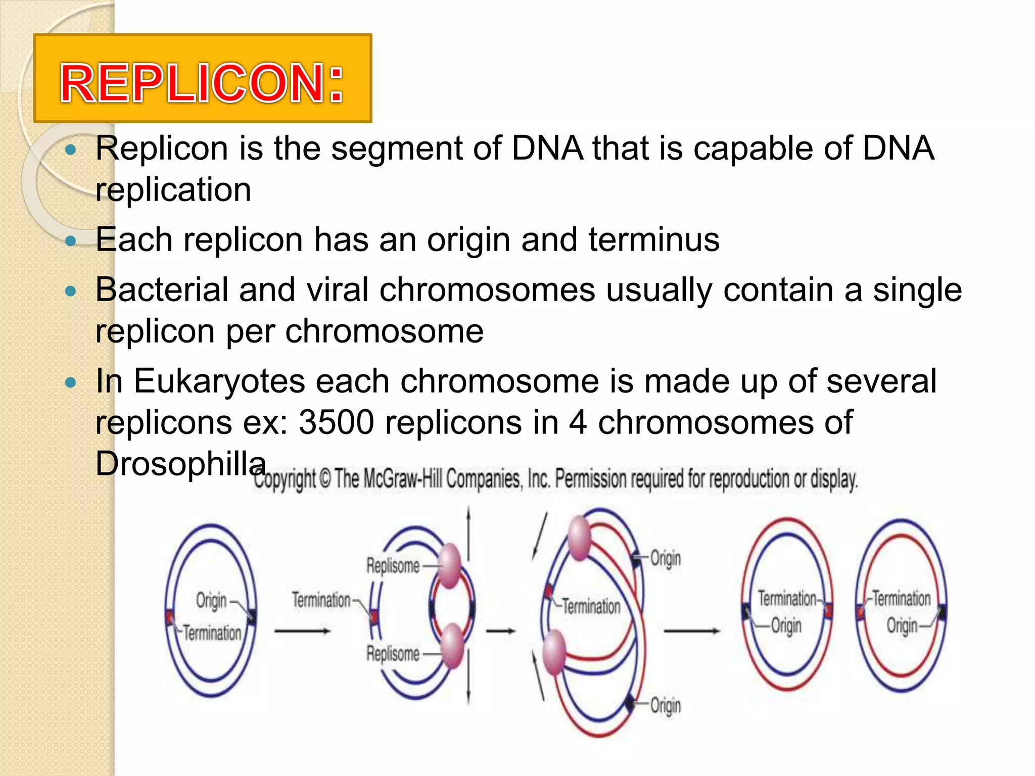
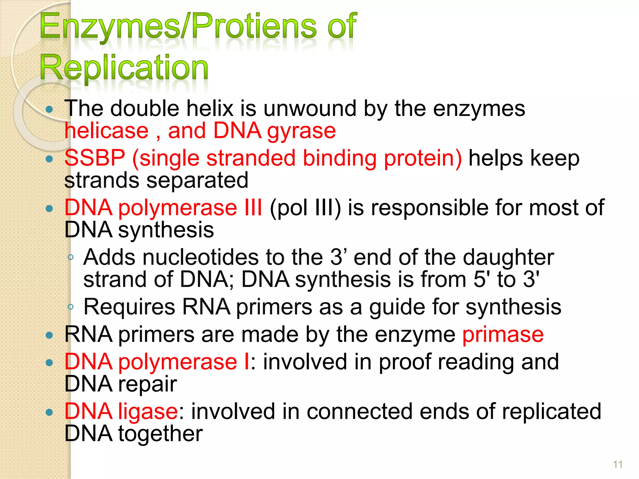
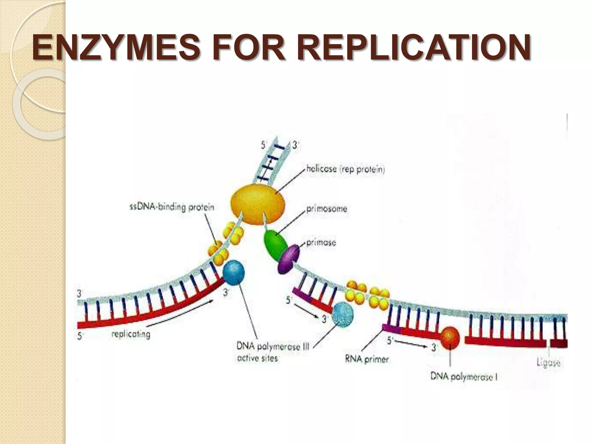
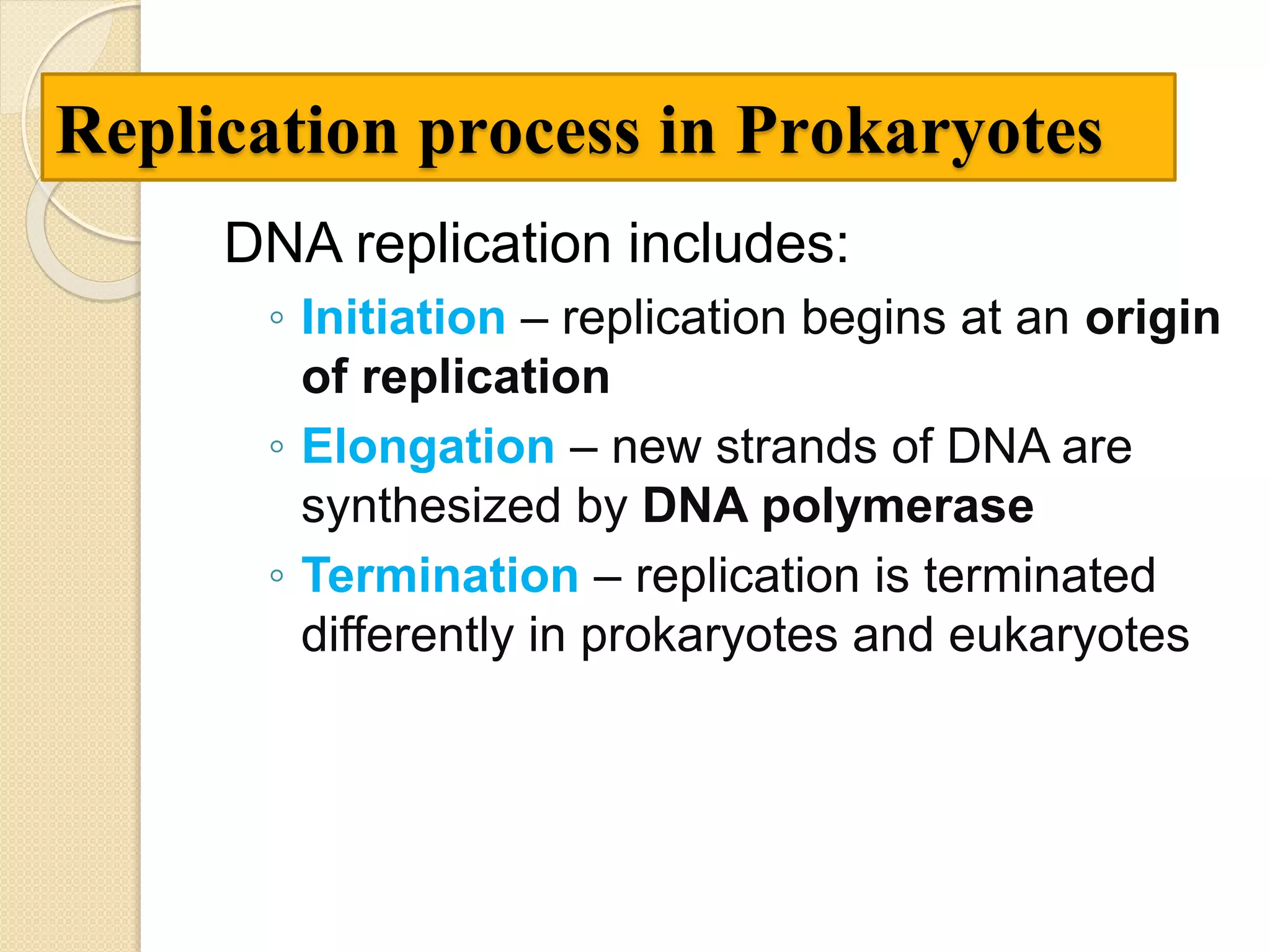


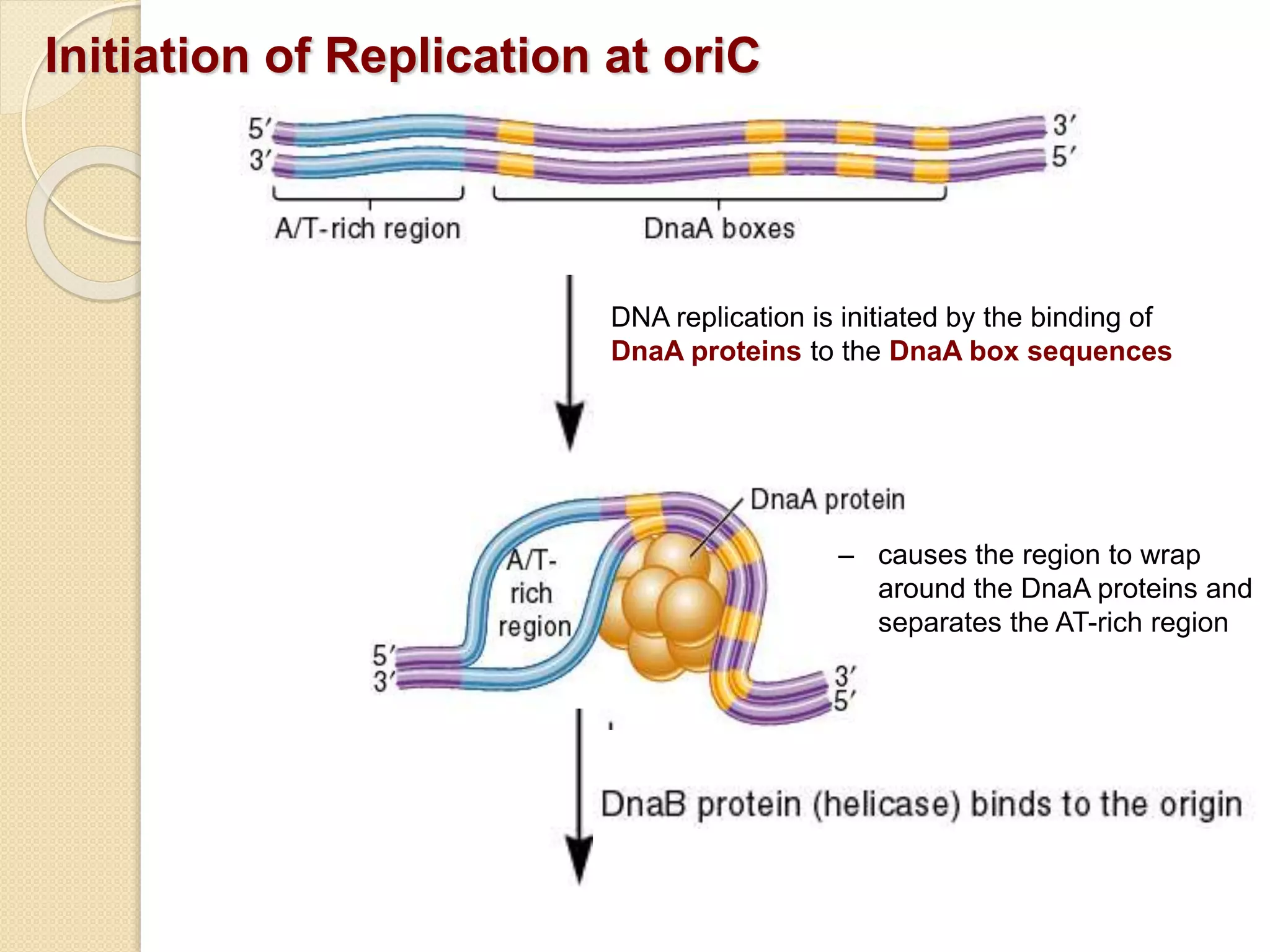
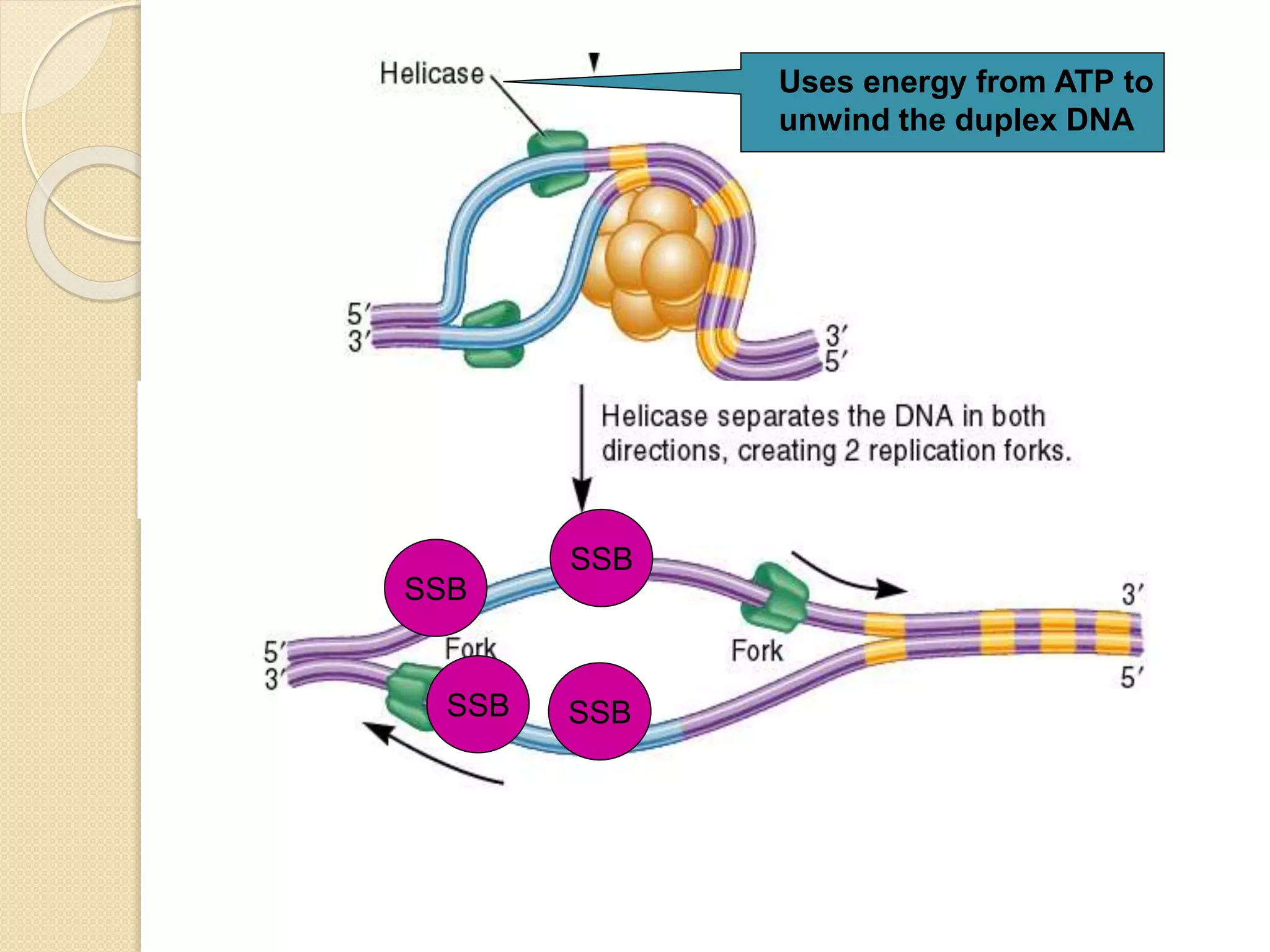

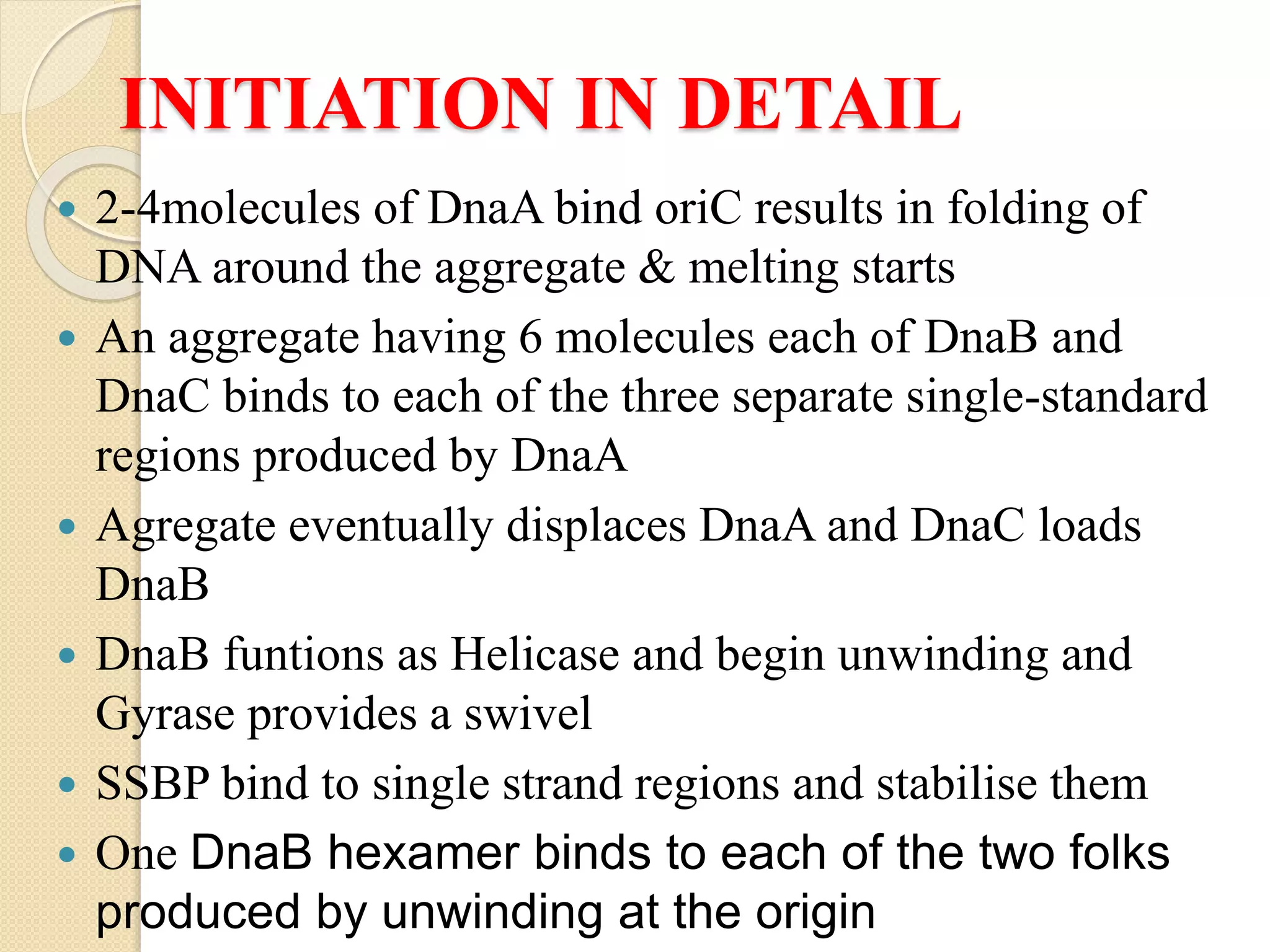


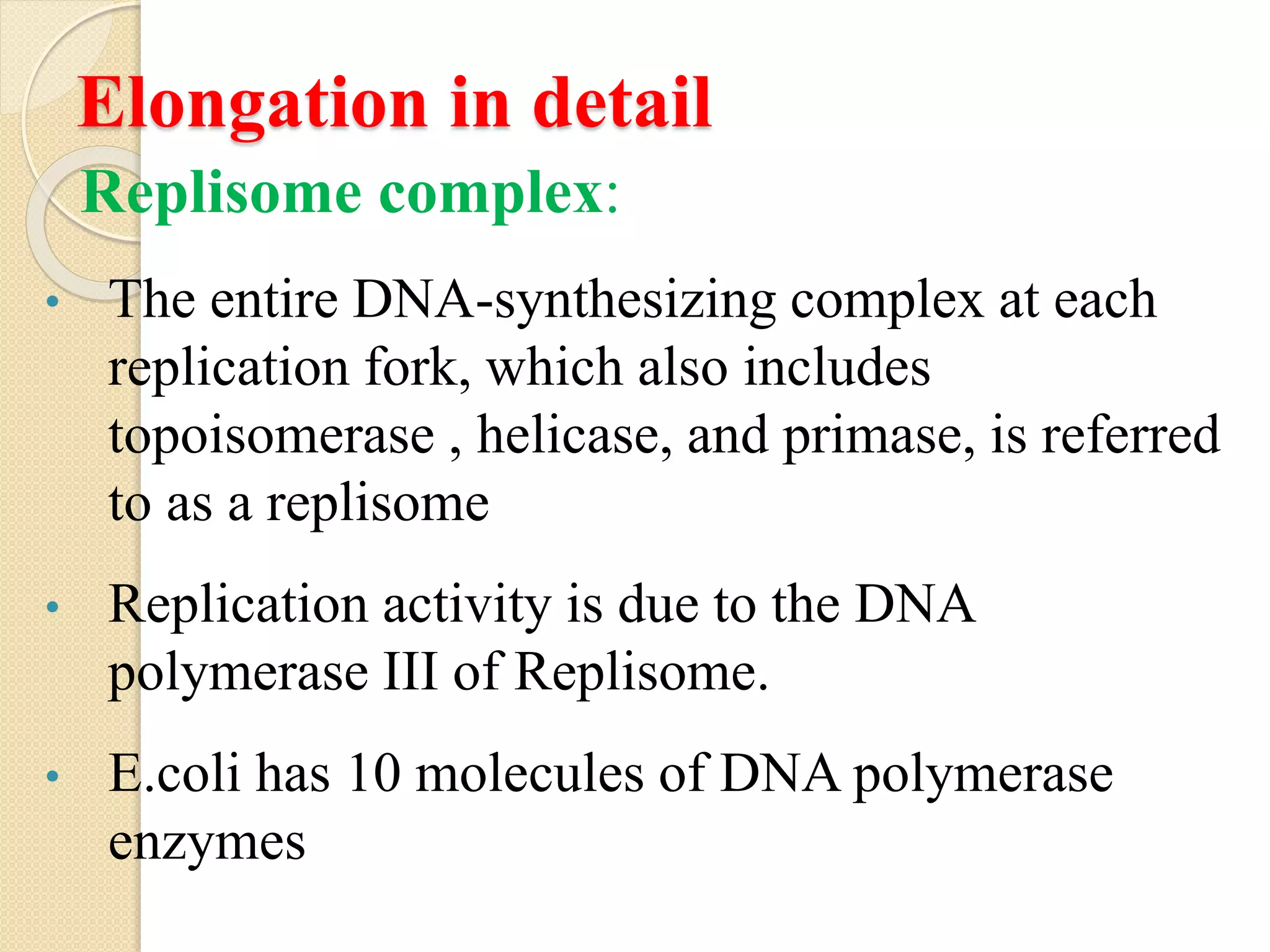

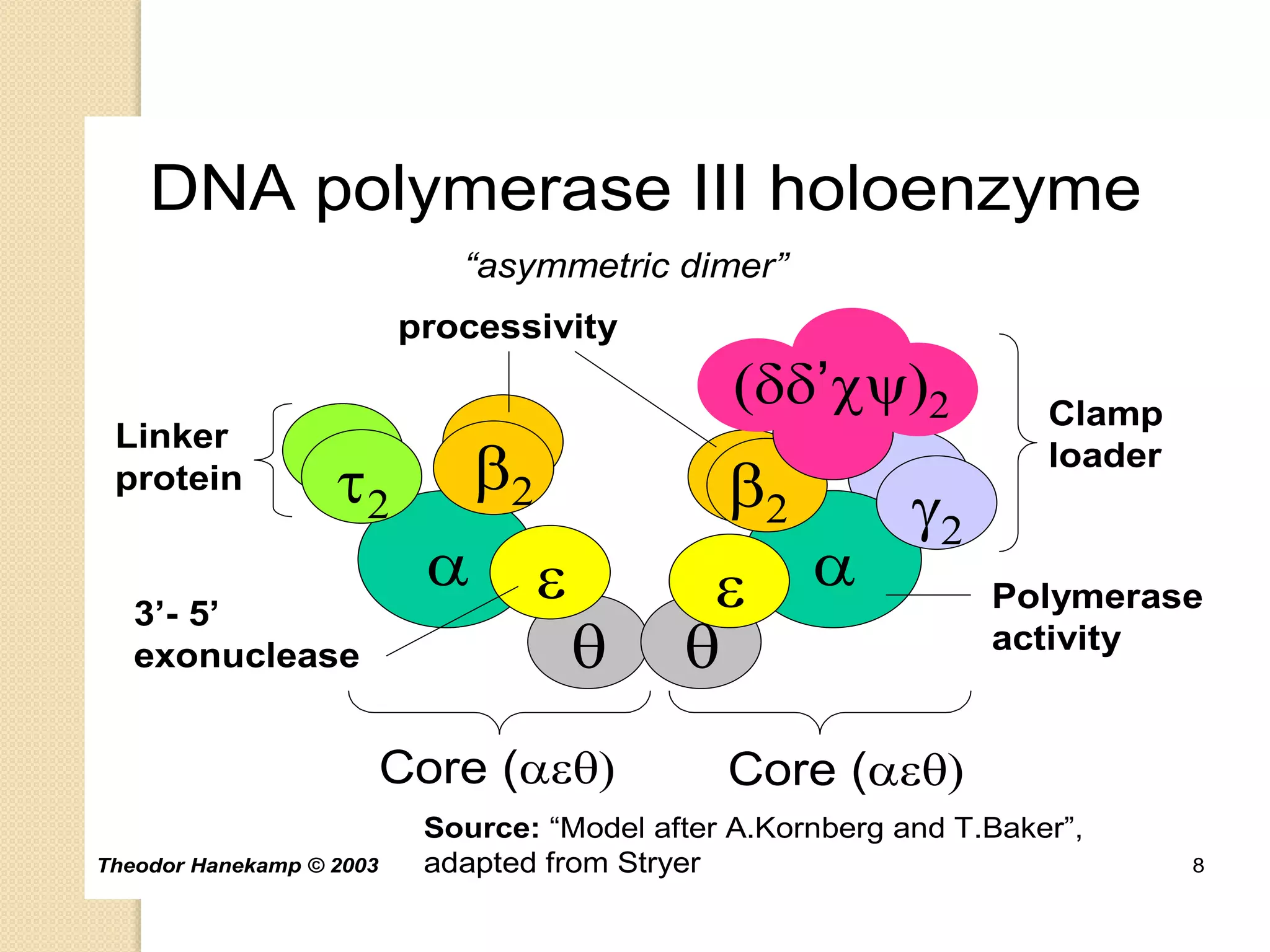

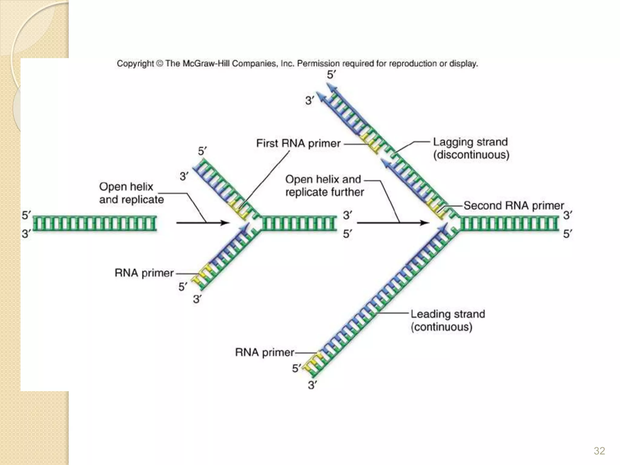


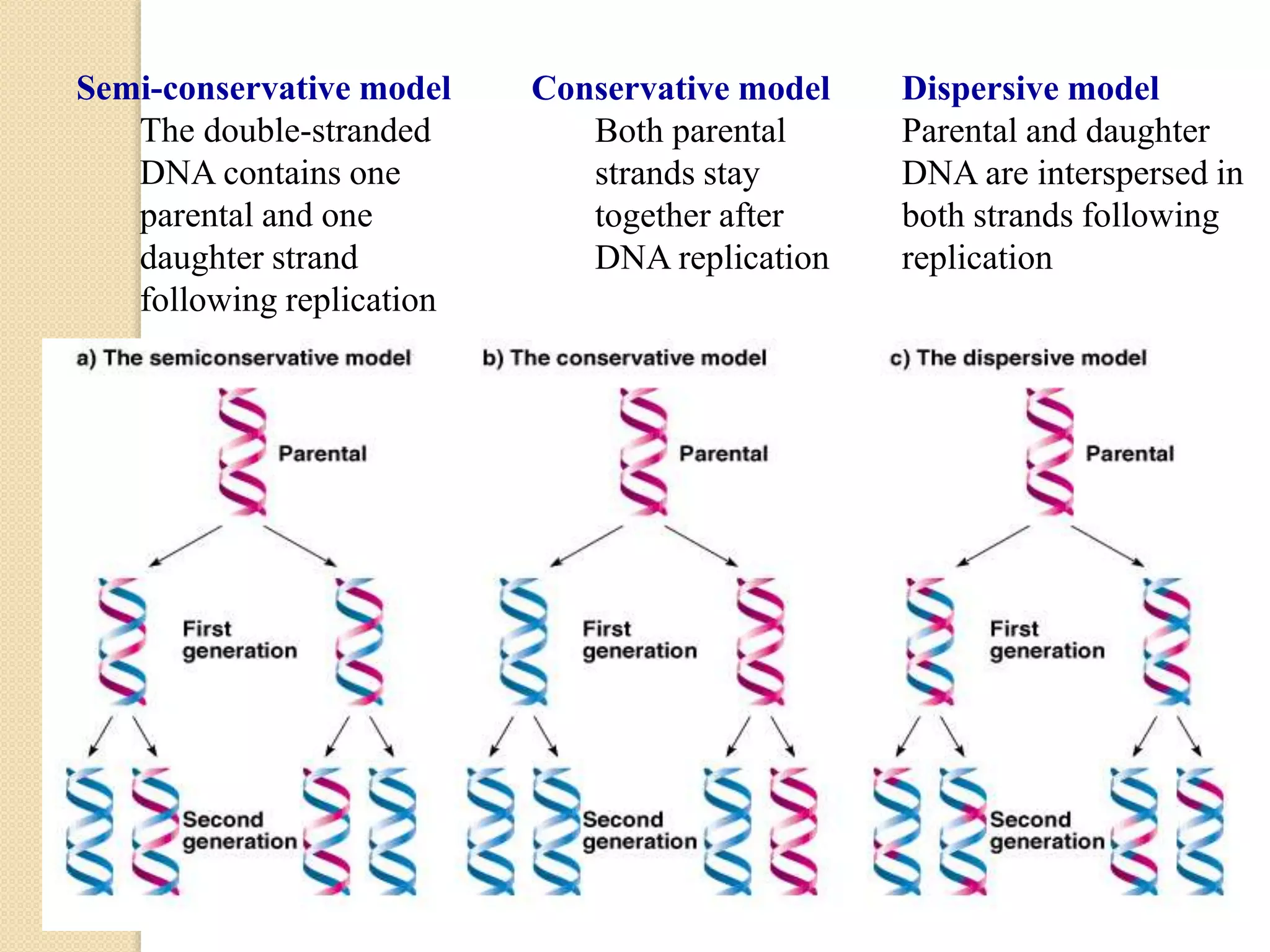


The document covers the process of DNA replication in prokaryotes, emphasizing its importance for biological inheritance, and details the stages of initiation, elongation, and termination. It describes key enzymes involved, such as DNA polymerase and helicase, as well as the semi-conservative replication model supported by Meselson and Stahl's experiment. Additionally, it discusses the structural features of DNA, including replicons and replication forks.


