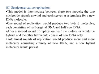
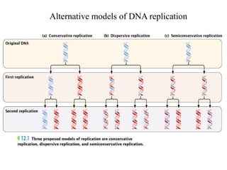
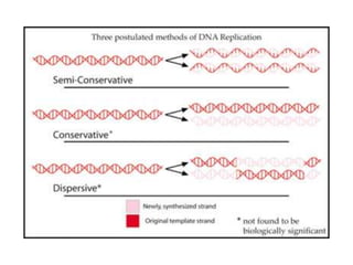
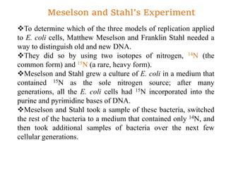
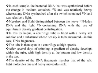
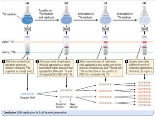
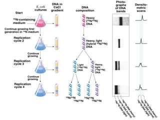
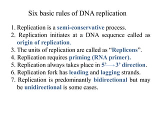
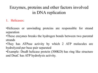
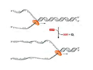
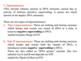
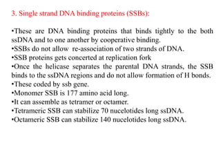
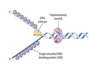
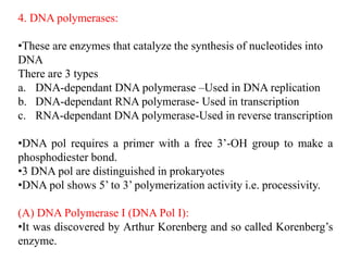
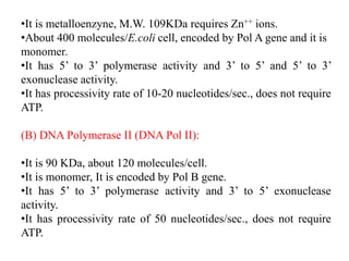
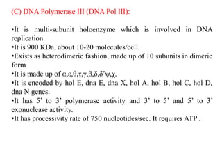
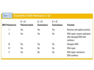
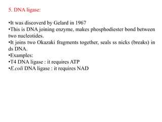
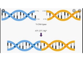
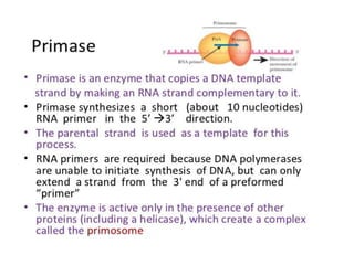
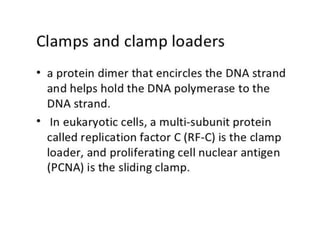
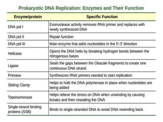
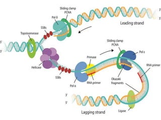
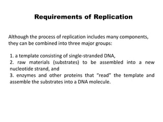
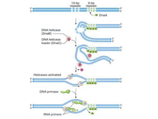
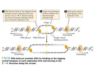
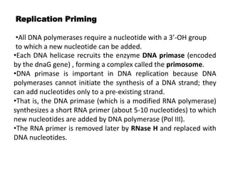
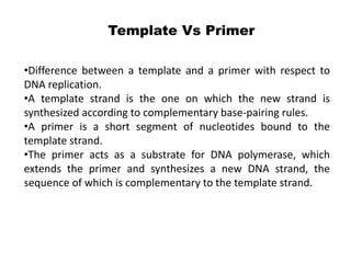
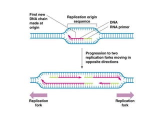
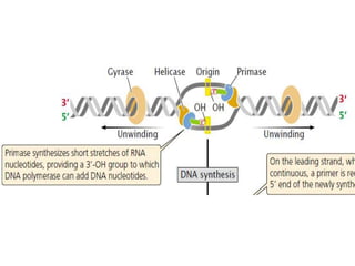
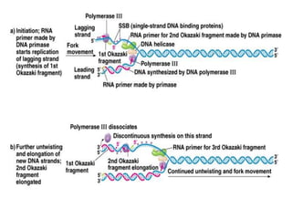
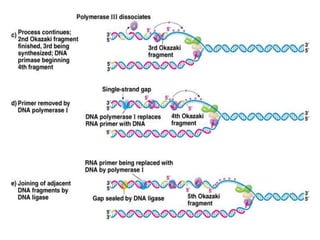
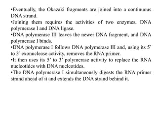
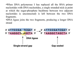
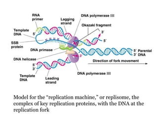
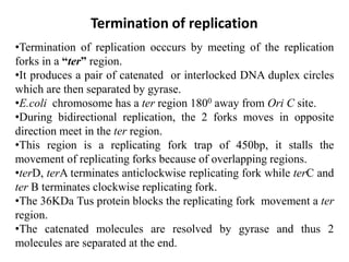
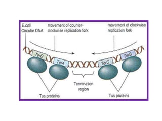
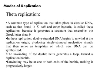
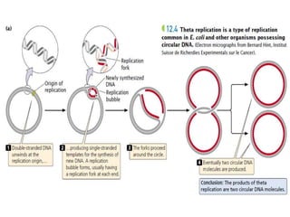
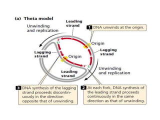
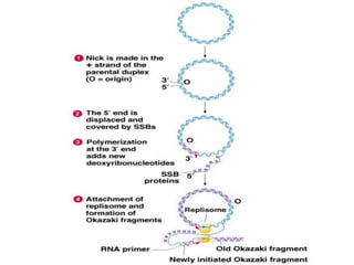
Prokaryotic DNA replication
Vishrut S. Ghare
(M.Sc Microbiology, SET)
Asst. Professor, S.B.B alias
A. Jedhe College, Pune
Prokaryotic DNA replication
i. Models of DNA replication. (Conservative, semiconservative,
and Dispersive)
ii. Meselson and Stahl’s experiment (semiconservative)
iii. Six basic rules of DNA replication
iv. Enzymes, proteins and other factors involved in DNA
replication.
v. Modes of DNA replication Rolling circle mechanism, theta
and linear DNA replication
Semiconservative Replication
From the three-dimensional structure of DNA that Watson and
Crick proposed in 1953, several important genetic implications
were immediately apparent.
The complementary nature of the two nucleotide strands in a
DNA molecule suggested that, during replication, each strand can
serve as a template for the synthesis of a new strand.
The specificity of base pairing (adenine with thymine; guanine
with cytosine) implied that only one sequence of bases can be
specified by each template, and so two DNA molecules built on
the pair of templates will be identical with the original.
This process is called semiconservative replication, because
each of the original nucleotide strands remains intact (conserved),
despite no longer being combined in the same molecule; the
original DNA molecule is half (semi) conserved during
replication.
i. Models of DNA replication.
(Conservative, semiconservative, and Dispersive)
Initially, three alternative models were proposed for DNA
replication.
(A) Conservative replication:
•In conservative replication, the entire double-stranded DNA
molecule serves as a template for a whole new molecule of DNA,
and the original DNA molecule is fully conserved during replication.
(B) Dispersive replication:
•In dispersive replication , both nucleotide strands break down
(disperse) into fragments, which serve as templates for the synthesis
of new DNA fragments, and then somehow reassemble into two
complete DNA molecules.
•In this model, each resulting DNA molecule is interspersed with
fragments of old and new DNA; none of the original molecule is
conserved.
(C) Semiconservative replication:
•This model is intermediate between these two models; the two
nucleotide strands unwind and each serves as a template for a new
DNA molecule.
•One round of replication would produce two hybrid molecules,
each consisting of half original DNA and half new DNA.
•After a second round of replication, half the molecules would be
hybrid, and the other half would consist of new DNA only.
• Additional rounds of replication would produce more and more
molecules consisting entirely of new DNA, and a few hybrid
molecules would persist.
Alternative models of DNA replication
Meselson and Stahl’s Experiment
To determine which of the three models of replication applied
to E. coli cells, Matthew Meselson and Franklin Stahl needed a
way to distinguish old and new DNA.
They did so by using two isotopes of nitrogen, 14N (the
common form) and 15N (a rare, heavy form).
Meselson and Stahl grew a culture of E. coli in a medium that
contained 15N as the sole nitrogen source; after many
generations, all the E. coli cells had 15N incorporated into the
purine and pyrimidine bases of DNA.
Meselson and Stahl took a sample of these bacteria, switched
the rest of the bacteria to a medium that contained only 14N, and
then took additional samples of bacteria over the next few
cellular generations.
In each sample, the bacterial DNA that was synthesized before
the change in medium contained 15N and was relatively heavy,
whereas any DNA synthesized after the switch contained 14N and
was relatively light.
Meselson and Stahl distinguished between the heavy 15N-laden
DNA and the light 14N-containing DNA with the use of
equilibrium density gradient centrifugation.
In this technique, a centrifuge tube is filled with a heavy salt
solution and a substance whose density is to be measured—in this
case, DNA fragments.
The tube is then spun in a centrifuge at high speeds.
After several days of spinning, a gradient of density develops
within the tube, with high density at the bottom and low density
at the top.
The density of the DNA fragments matches that of the salt:
light molecules rise and heavy molecules sink.
They found that DNA from bacteria grown only on medium
containing 15N produced a single band at the position expected of
DNA containing only 15N.
DNA from bacteria transferred to the medium with 14N and
allowed one round of replication also produced a single band, but
at a position intermediate between that expected of DNA with only
15N and that expected of DNA with only 14N.
After a second round of replication in medium with 14N, two
bands of equal intensity appeared, one in the intermediate position
and the other at the position expected of DNA that contained only
14N.
All samples taken after additional rounds of replication
produced two bands, and the band representing light DNA became
progressively stronger.
Meselson and Stahl’s results were exactly as expected for
semiconservative replication and are incompatible with those
predicated for both conservative and dispersive replication.
Six basic rules of DNA replication
1. Replication is a semi-conservative process.
2. Replication initiates at a DNA sequence called as
origin of replication.
3. The units of replication are called as “Replicons”.
4. Replication requires priming (RNA primer).
5. Replication always takes place in 5’ 3’ direction.
6. Replication fork has leading and lagging strands.
7. Replication is predominantly bidirectional but may
be unidirectional is some cases.
Enzymes, proteins and other factors involved
in DNA replication
1. Helicases:
•Helicases or unwinding proteins are responsible for strand
separation
•These enzymes breaks the hydrogen bonds between two parental
strands
•They has ATPase activity by which 2 ATP molecules are
hydrolyzed per base pair separated
•Example: DnaB helicase protein (300KD) has ring like structure
and DnaC has ATP hydrolysis activity.
2. Topoisomerases:
•This enzyme releases tension in DNA molecule caused due to
activity of helicase (positive supercoiling, it causes too much
tension in the duplex DNA structure)
There are two types of topoisomerases:
a. Type I topoisomerases: These are nicking and closing enzymes
which breaks and reseals one strand of DNA at a time, it
removes negative supercoiling in DNA.
studied byJames Wang in1971
b. Type II topoisomerases: These are nicking and closing enzymes
which breaks and reseals both the strands of DNA, it
introduces/creates negative supercoiling in DNA.
These are also called as “DNA gyrase” enzyme. Has two
subunits GyrA and B and depends on ATP.
studied by Martin Gellert in1976
3. Single strand DNA binding proteins (SSBs):
•These are DNA binding proteins that binds tightly to the both
ssDNA and to one another by cooperative binding.
•SSBs do not allow re-association of two strands of DNA.
•SSB proteins gets concerted at replication fork
•Once the helicase separates the parental DNA strands, the SSB
binds to the ssDNA regions and do not allow formation of H bonds.
•These coded by ssb gene.
•Monomer SSB is 177 amino acid long.
•It can assemble as tetramer or octamer.
•Tetrameric SSB can stabilize 70 nucelotides long ssDNA.
•Octameric SSB can stabilize 140 nucelotides long ssDNA.
4. DNA polymerases:
•These are enzymes that catalyze the synthesis of nucleotides into
DNA
There are 3 types
a. DNA-dependant DNA polymerase –Used in DNA replication
b. DNA-dependant RNA polymerase- Used in transcription
c. RNA-dependant DNA polymerase-Used in reverse transcription
•DNA pol requires a primer with a free 3’-OH group to make a
phosphodiester bond.
•3 DNA pol are distinguished in prokaryotes
•DNA pol shows 5’ to 3’ polymerization activity i.e. processivity.
(A) DNA Polymerase I (DNA Pol I):
•It was discovered by Arthur Korenberg and so called Korenberg’s
enzyme.
•It is metalloenzyne, M.W. 109KDa requires Zn++ ions.
•About 400 molecules/E.coli cell, encoded by Pol A gene and it is
monomer.
•It has 5’ to 3’ polymerase activity and 3’ to 5’ and 5’ to 3’
exonuclease activity.
•It has processivity rate of 10-20 nucleotides/sec., does not require
ATP.
(B) DNA Polymerase II (DNA Pol II):
•It is 90 KDa, about 120 molecules/cell.
•It is monomer, It is encoded by Pol B gene.
•It has 5’ to 3’ polymerase activity and 3’ to 5’ exonuclease
activity.
•It has processivity rate of 50 nucleotides/sec., does not require
ATP.
(C) DNA Polymerase III (DNA Pol III):
•It is multi-subunit holoenzyme which is involved in DNA
replication.
•It is 900 KDa, about 10-20 molecules/cell.
•Exists as heterodimeric fashion, made up of 10 subunits in dimeric
form
•It is made up of α,ε,θ,τ,γ,β,δ,δ’ψ,χ.
•It is encoded by hol E, dna E, dna X, hol A, hol B, hol C, hol D,
dna N genes.
•It has 5’ to 3’ polymerase activity and 3’ to 5’ and 5’ to 3’
exonuclease activity.
•It has processivity rate of 750 nucleotides/sec. It requires ATP .
5. DNA ligase:
•It was discoverd by Gelard in 1967
•This is DNA joining enzyme, makes phosphodiester bond between
two nucleotides.
•It joins two Okazaki fragments together, seals ss nicks (breaks) in
ds DNA.
•Examples:
•T4 DNA ligase : it requires ATP
•E.coli DNA ligase : it requires NAD
Requirements of Replication
Although the process of replication includes many components,
they can be combined into three major groups:
1. a template consisting of single-stranded DNA,
2. raw materials (substrates) to be assembled into a new
nucleotide strand, and
3. enzymes and other proteins that “read” the template and
assemble the substrates into a DNA molecule.
Replicons
•DNA molecule capable of replication is called as replicon.
•Rplicon is a DNA molecule which has origin of replication (ori)
and can be replicated autonomously in the cell.
•Replication initiates at ori site.
•The prokaryotic chromosome has single origin of replication site
and termination site, is called a single replicon.
•The stretch of DNA from the origin of replication to the two
termini of replication (where adjacent replication forks fuse) on
each side of the origin is called a replicon or replication unit.
The origin of replication
•The circular chromosome of E. coli has a single replication
origin (oriC).
•The minimal sequence required for oriC to function consists of
245 bp that contain several critical sites.
•The E. coli replicator is oriC, which spans 245 bp of DNA and
contains a cluster of three copies of a 13-bp AT-rich sequence and
four copies of a 9-bp sequence.
•For the initiation of replication, an initiator protein (DnaA) or
proteins bind to the replicator and denature the AT-rich region.
•DNA helicases (DnaB; encoded by the dnaB gene) are recruited
and are loaded onto the DNA by DNA helicase loader proteins
(DnaC; encoded by the dnaC gene).
•The helicases untwist the DNA in both directions from the origin
of replication by breaking the hydrogen bonds between the bases.
•The energy for the untwisting comes from the hydrolysis of ATP.
•To stabilize the single-stranded DNA long enough for replication
to take place, single-strand-binding (SSB) proteins attach tightly to
the exposed single-stranded DNA
•DNA gyrases removes the torsion strain caused due to Helicases,
removes positive supercoiling and introduces negative
supercoiling.
Replication Priming
•All DNA polymerases require a nucleotide with a 3’-OH group
to which a new nucleotide can be added.
•Each DNA helicase recruits the enzyme DNA primase (encoded
by the dnaG gene) , forming a complex called the primosome.
•DNA primase is important in DNA replication because DNA
polymerases cannot initiate the synthesis of a DNA strand; they
can add nucleotides only to a pre-existing strand.
•That is, the DNA primase (which is a modified RNA polymerase)
synthesizes a short RNA primer (about 5-10 nucleotides) to which
new nucleotides are added by DNA polymerase (Pol III).
•The RNA primer is removed later by RNase H and replaced with
DNA nucleotides.
•Difference between a template and a primer with respect to
DNA replication.
•A template strand is the one on which the new strand is
synthesized according to complementary base-pairing rules.
•A primer is a short segment of nucleotides bound to the
template strand.
•The primer acts as a substrate for DNA polymerase, which
extends the primer and synthesizes a new DNA strand, the
sequence of which is complementary to the template strand.
Template Vs Primer
Replication takes place in 5’ to 3’
direction
•The 2 strands of DNA duplex are anti-parallel, with one goes in
5’to 3’ direction and the other in 3’to 5’ direction.
•Because the two DNA strands run in opposite direction, the two
daughter strands being synthesized at the replication fork must also
run in opposite directions.
•Therefore overall chain growth must be in 5’ to3’direction for one
daughter strand and in the 3’ to 5’ direction for the other.
•However, all known DNA pol enzymes can extend the DNA
chains only in 5’ to3’direction , because incoming nucleotide can
react only with free 3’-OH end of the growing polynucleotide
chain,.
Leading and Lagging strand
synthesis
DNA polymerases can synthesize DNA only in the 5'-to-3'
direction, yet the two DNA strands are of opposite polarity.
To maintain the 5'-to-3' polarity of DNA synthesis on each
template, and to maintain one overall direction of replication fork
movement, DNA is made in opposite directions on the two
template strands.
The new strand being made in the same direction as the
movement of the replication fork is the leading strand.
and the new strand being made in the direction opposite that of
the movement of the replication fork is the lagging strand.
The leading strand needs a single RNA primer for its synthesis,
whereas the lagging strand needs a series of primers
Leading strands synthesis takes place continuously in the 5’ to
3’ end by the addition of nucleotides at the 3’ end in the direction
of unwinding of the replication fork.
It does not require clamping and unclamping of DNA pol III
from DNA.
 It requires about 1 or 2 DNA pol III per cell, this strand
synthesis is highly processive.
The other strand called lagging has its 3’ end facing away from
the replication fork.
It is polymerized in a discontinuous way in short pieces away
from the replication fork i.e. in the opposite direction of the
continuous strand.
This semi-discontinuous replication was studied by Okazaki
(Reiji and Tuneko Okazaki) et.al
It requires repetitive clamping and unclamping of DNA pol III
from DNA.
It requires about 10-20 molecules of enzyme per cell, synthesis
of this strand is less processive, many primers are needed.
The replication fork thus had 2 arms, a forward arm on which the
leading strand is synthesized continuously and a retrograde arm on
which the lagging strand is discontinuously synthesized via a
repeated synthesis of short fragments of DNA.
Each RNA primed nascent DNA fragment is referred as Okazaki
fragment.
E. coli DNA forms about 4000 Okazaki fragments in lagging
strand synthesis.
Each fragment is about 1000 bp long.
•Eventually, the Okazaki fragments are joined into a continuous
DNA strand.
•Joining them requires the activities of two enzymes, DNA
polymerase I and DNA ligase.
•DNA polymerase III leaves the newer DNA fragment, and DNA
polymerase I binds.
•DNA polymerase I follows DNA polymerase III and, using its 5’
to 3’ exonuclease activity, removes the RNA primer.
•It then uses its 5’ to 3’ polymerase activity to replace the RNA
nucleotides with DNA nucleotides.
•The DNA polymerase I simultaneously digests the RNA primer
strand ahead of it and extends the DNA strand behind it.
•When DNA polymerase I has replaced all the RNA primer
nucleotides with DNA nucleotides, a single stranded nick (a point
at which the sugar-phosphate backbone between two adjacent
nucleotides is unconnected) is left between the two DNA
fragments.
•DNA ligase joins the two fragments, producing a longer DNA
strand
Model for the “replication machine,” or replisome, the
complex of key replication proteins, with the DNA at the
replication fork
Termination of replication
•Termination of replication occcurs by meeting of the replication
forks in a “ter” region.
•It produces a pair of catenated or interlocked DNA duplex circles
which are then separated by gyrase.
•E.coli chromosome has a ter region 1800 away from Ori C site.
•During bidirectional replication, the 2 forks moves in opposite
direction meet in the ter region.
•This region is a replicating fork trap of 450bp, it stalls the
movement of replicating forks because of overlapping regions.
•terD, terA terminates anticlockwise replicating fork while terC and
ter B terminates clockwise replicating fork.
•The 36KDa Tus protein blocks the replicating fork movement a ter
region.
•The catenated molecules are resolved by gyrase and thus 2
molecules are separated at the end.
Modes of Replication
Theta replication:
• A common type of replication that takes place in circular DNA,
such as that found in E. coli and other bacteria, is called theta
replication, because it generates a structure that resembles the
Greek letter theta θ.
•In theta replication, double-stranded DNA begins to unwind at the
replication origin, producing single-stranded nucleotide strands
that then serve as templates on which new DNA can be
synthesized.
•The unwinding of the double helix generates a loop, termed a
replication bubble.
•Unwinding may be at one or both ends of the bubble, making it
progressively larger.
•The repliation in which one of the strand is synthesises
continuously and other discontinuously then it is referred as semi-
discontinuous replication.
•Since both the strands of the parental DNA acts a template,
synthesis of strand that uses 3’ to 5’ parental strand as template is
synthesized continuously and called as leading strand.
•The leading strand orientation is 5’ to 3’ of the replication fork
and is extending from origin of replication.
•The other template strand is oriented in 5’ to 3’ direction hence
copying it would results in synthesis of 3’ to 5’ direction relative
to the direction of fork movement.
•This new DNA strand is called lagging strand and is synthesised
discontinuously as a series of short DNA fragments (Okazaki
fragments).
•DNA ligase then joins the Okazaki fragments to make
uninterrupted daughter strand.
•Overwinding of unreplicated segment that is caused by unwinding
of the daughter branches is removed by nicking action of DNA
gyrase.
•The product of theta repication are two circular ds DNA
molcules.
•Theta replication occurs in E.coli, B.subtilis.
Rolling-circle replication
•Another form of replication, called rolling-circle replication
takes place in some viruses and in the F factor of E. coli.
•This form of replication is initiated by a break in one of the
nucleotide strands that creates a 3’-OH group and a 5’-phosphate
group.
•New nucleotides are added to the 3’ end of the broken strand,
with the inner (unbroken) strand used as a template.
•As new nucleotides are added to the 3’ end, the 5’ end of the
broken strand is displaced from the template, rolling out like
thread being pulled off a spool.
•The 3 end grows around the circle, giving rise to the name
rolling-circle model.
•The replication fork may continue around the circle a number of
times, producing several linked copies of the same sequence.
•With each revolution around the circle, the growing 3’ end
displaces the nucleotide strand synthesized in the preceding
revolution.
•Eventually, the linear DNA molecule is cleaved from the circle,
resulting in a double stranded circular DNA molecule and a
single-stranded linear DNA molecule.
•The linear molecule circularizes either before or after serving
as a template for the synthesis of a complementary strand.
Prokaryotic DNA replication

Prokaryotic DNA replication

  • 1.
    Prokaryotic DNA replication VishrutS. Ghare (M.Sc Microbiology, SET) Asst. Professor, S.B.B alias A. Jedhe College, Pune
  • 2.
    Prokaryotic DNA replication i.Models of DNA replication. (Conservative, semiconservative, and Dispersive) ii. Meselson and Stahl’s experiment (semiconservative) iii. Six basic rules of DNA replication iv. Enzymes, proteins and other factors involved in DNA replication. v. Modes of DNA replication Rolling circle mechanism, theta and linear DNA replication
  • 3.
    Semiconservative Replication From thethree-dimensional structure of DNA that Watson and Crick proposed in 1953, several important genetic implications were immediately apparent. The complementary nature of the two nucleotide strands in a DNA molecule suggested that, during replication, each strand can serve as a template for the synthesis of a new strand. The specificity of base pairing (adenine with thymine; guanine with cytosine) implied that only one sequence of bases can be specified by each template, and so two DNA molecules built on the pair of templates will be identical with the original. This process is called semiconservative replication, because each of the original nucleotide strands remains intact (conserved), despite no longer being combined in the same molecule; the original DNA molecule is half (semi) conserved during replication.
  • 4.
    i. Models ofDNA replication. (Conservative, semiconservative, and Dispersive) Initially, three alternative models were proposed for DNA replication. (A) Conservative replication: •In conservative replication, the entire double-stranded DNA molecule serves as a template for a whole new molecule of DNA, and the original DNA molecule is fully conserved during replication. (B) Dispersive replication: •In dispersive replication , both nucleotide strands break down (disperse) into fragments, which serve as templates for the synthesis of new DNA fragments, and then somehow reassemble into two complete DNA molecules. •In this model, each resulting DNA molecule is interspersed with fragments of old and new DNA; none of the original molecule is conserved.
  • 5.
    (C) Semiconservative replication: •Thismodel is intermediate between these two models; the two nucleotide strands unwind and each serves as a template for a new DNA molecule. •One round of replication would produce two hybrid molecules, each consisting of half original DNA and half new DNA. •After a second round of replication, half the molecules would be hybrid, and the other half would consist of new DNA only. • Additional rounds of replication would produce more and more molecules consisting entirely of new DNA, and a few hybrid molecules would persist.
  • 6.
    Alternative models ofDNA replication
  • 8.
    Meselson and Stahl’sExperiment To determine which of the three models of replication applied to E. coli cells, Matthew Meselson and Franklin Stahl needed a way to distinguish old and new DNA. They did so by using two isotopes of nitrogen, 14N (the common form) and 15N (a rare, heavy form). Meselson and Stahl grew a culture of E. coli in a medium that contained 15N as the sole nitrogen source; after many generations, all the E. coli cells had 15N incorporated into the purine and pyrimidine bases of DNA. Meselson and Stahl took a sample of these bacteria, switched the rest of the bacteria to a medium that contained only 14N, and then took additional samples of bacteria over the next few cellular generations.
  • 9.
    In each sample,the bacterial DNA that was synthesized before the change in medium contained 15N and was relatively heavy, whereas any DNA synthesized after the switch contained 14N and was relatively light. Meselson and Stahl distinguished between the heavy 15N-laden DNA and the light 14N-containing DNA with the use of equilibrium density gradient centrifugation. In this technique, a centrifuge tube is filled with a heavy salt solution and a substance whose density is to be measured—in this case, DNA fragments. The tube is then spun in a centrifuge at high speeds. After several days of spinning, a gradient of density develops within the tube, with high density at the bottom and low density at the top. The density of the DNA fragments matches that of the salt: light molecules rise and heavy molecules sink.
  • 10.
    They found thatDNA from bacteria grown only on medium containing 15N produced a single band at the position expected of DNA containing only 15N. DNA from bacteria transferred to the medium with 14N and allowed one round of replication also produced a single band, but at a position intermediate between that expected of DNA with only 15N and that expected of DNA with only 14N. After a second round of replication in medium with 14N, two bands of equal intensity appeared, one in the intermediate position and the other at the position expected of DNA that contained only 14N. All samples taken after additional rounds of replication produced two bands, and the band representing light DNA became progressively stronger. Meselson and Stahl’s results were exactly as expected for semiconservative replication and are incompatible with those predicated for both conservative and dispersive replication.
  • 15.
    Six basic rulesof DNA replication 1. Replication is a semi-conservative process. 2. Replication initiates at a DNA sequence called as origin of replication. 3. The units of replication are called as “Replicons”. 4. Replication requires priming (RNA primer). 5. Replication always takes place in 5’ 3’ direction. 6. Replication fork has leading and lagging strands. 7. Replication is predominantly bidirectional but may be unidirectional is some cases.
  • 16.
    Enzymes, proteins andother factors involved in DNA replication 1. Helicases: •Helicases or unwinding proteins are responsible for strand separation •These enzymes breaks the hydrogen bonds between two parental strands •They has ATPase activity by which 2 ATP molecules are hydrolyzed per base pair separated •Example: DnaB helicase protein (300KD) has ring like structure and DnaC has ATP hydrolysis activity.
  • 18.
    2. Topoisomerases: •This enzymereleases tension in DNA molecule caused due to activity of helicase (positive supercoiling, it causes too much tension in the duplex DNA structure) There are two types of topoisomerases: a. Type I topoisomerases: These are nicking and closing enzymes which breaks and reseals one strand of DNA at a time, it removes negative supercoiling in DNA. studied byJames Wang in1971 b. Type II topoisomerases: These are nicking and closing enzymes which breaks and reseals both the strands of DNA, it introduces/creates negative supercoiling in DNA. These are also called as “DNA gyrase” enzyme. Has two subunits GyrA and B and depends on ATP. studied by Martin Gellert in1976
  • 19.
    3. Single strandDNA binding proteins (SSBs): •These are DNA binding proteins that binds tightly to the both ssDNA and to one another by cooperative binding. •SSBs do not allow re-association of two strands of DNA. •SSB proteins gets concerted at replication fork •Once the helicase separates the parental DNA strands, the SSB binds to the ssDNA regions and do not allow formation of H bonds. •These coded by ssb gene. •Monomer SSB is 177 amino acid long. •It can assemble as tetramer or octamer. •Tetrameric SSB can stabilize 70 nucelotides long ssDNA. •Octameric SSB can stabilize 140 nucelotides long ssDNA.
  • 21.
    4. DNA polymerases: •Theseare enzymes that catalyze the synthesis of nucleotides into DNA There are 3 types a. DNA-dependant DNA polymerase –Used in DNA replication b. DNA-dependant RNA polymerase- Used in transcription c. RNA-dependant DNA polymerase-Used in reverse transcription •DNA pol requires a primer with a free 3’-OH group to make a phosphodiester bond. •3 DNA pol are distinguished in prokaryotes •DNA pol shows 5’ to 3’ polymerization activity i.e. processivity. (A) DNA Polymerase I (DNA Pol I): •It was discovered by Arthur Korenberg and so called Korenberg’s enzyme.
  • 22.
    •It is metalloenzyne,M.W. 109KDa requires Zn++ ions. •About 400 molecules/E.coli cell, encoded by Pol A gene and it is monomer. •It has 5’ to 3’ polymerase activity and 3’ to 5’ and 5’ to 3’ exonuclease activity. •It has processivity rate of 10-20 nucleotides/sec., does not require ATP. (B) DNA Polymerase II (DNA Pol II): •It is 90 KDa, about 120 molecules/cell. •It is monomer, It is encoded by Pol B gene. •It has 5’ to 3’ polymerase activity and 3’ to 5’ exonuclease activity. •It has processivity rate of 50 nucleotides/sec., does not require ATP.
  • 23.
    (C) DNA PolymeraseIII (DNA Pol III): •It is multi-subunit holoenzyme which is involved in DNA replication. •It is 900 KDa, about 10-20 molecules/cell. •Exists as heterodimeric fashion, made up of 10 subunits in dimeric form •It is made up of α,ε,θ,τ,γ,β,δ,δ’ψ,χ. •It is encoded by hol E, dna E, dna X, hol A, hol B, hol C, hol D, dna N genes. •It has 5’ to 3’ polymerase activity and 3’ to 5’ and 5’ to 3’ exonuclease activity. •It has processivity rate of 750 nucleotides/sec. It requires ATP .
  • 25.
    5. DNA ligase: •Itwas discoverd by Gelard in 1967 •This is DNA joining enzyme, makes phosphodiester bond between two nucleotides. •It joins two Okazaki fragments together, seals ss nicks (breaks) in ds DNA. •Examples: •T4 DNA ligase : it requires ATP •E.coli DNA ligase : it requires NAD
  • 32.
    Requirements of Replication Althoughthe process of replication includes many components, they can be combined into three major groups: 1. a template consisting of single-stranded DNA, 2. raw materials (substrates) to be assembled into a new nucleotide strand, and 3. enzymes and other proteins that “read” the template and assemble the substrates into a DNA molecule.
  • 33.
    Replicons •DNA molecule capableof replication is called as replicon. •Rplicon is a DNA molecule which has origin of replication (ori) and can be replicated autonomously in the cell. •Replication initiates at ori site. •The prokaryotic chromosome has single origin of replication site and termination site, is called a single replicon. •The stretch of DNA from the origin of replication to the two termini of replication (where adjacent replication forks fuse) on each side of the origin is called a replicon or replication unit. The origin of replication •The circular chromosome of E. coli has a single replication origin (oriC). •The minimal sequence required for oriC to function consists of 245 bp that contain several critical sites.
  • 34.
    •The E. colireplicator is oriC, which spans 245 bp of DNA and contains a cluster of three copies of a 13-bp AT-rich sequence and four copies of a 9-bp sequence. •For the initiation of replication, an initiator protein (DnaA) or proteins bind to the replicator and denature the AT-rich region. •DNA helicases (DnaB; encoded by the dnaB gene) are recruited and are loaded onto the DNA by DNA helicase loader proteins (DnaC; encoded by the dnaC gene). •The helicases untwist the DNA in both directions from the origin of replication by breaking the hydrogen bonds between the bases. •The energy for the untwisting comes from the hydrolysis of ATP. •To stabilize the single-stranded DNA long enough for replication to take place, single-strand-binding (SSB) proteins attach tightly to the exposed single-stranded DNA •DNA gyrases removes the torsion strain caused due to Helicases, removes positive supercoiling and introduces negative supercoiling.
  • 37.
    Replication Priming •All DNApolymerases require a nucleotide with a 3’-OH group to which a new nucleotide can be added. •Each DNA helicase recruits the enzyme DNA primase (encoded by the dnaG gene) , forming a complex called the primosome. •DNA primase is important in DNA replication because DNA polymerases cannot initiate the synthesis of a DNA strand; they can add nucleotides only to a pre-existing strand. •That is, the DNA primase (which is a modified RNA polymerase) synthesizes a short RNA primer (about 5-10 nucleotides) to which new nucleotides are added by DNA polymerase (Pol III). •The RNA primer is removed later by RNase H and replaced with DNA nucleotides.
  • 38.
    •Difference between atemplate and a primer with respect to DNA replication. •A template strand is the one on which the new strand is synthesized according to complementary base-pairing rules. •A primer is a short segment of nucleotides bound to the template strand. •The primer acts as a substrate for DNA polymerase, which extends the primer and synthesizes a new DNA strand, the sequence of which is complementary to the template strand. Template Vs Primer
  • 41.
    Replication takes placein 5’ to 3’ direction •The 2 strands of DNA duplex are anti-parallel, with one goes in 5’to 3’ direction and the other in 3’to 5’ direction. •Because the two DNA strands run in opposite direction, the two daughter strands being synthesized at the replication fork must also run in opposite directions. •Therefore overall chain growth must be in 5’ to3’direction for one daughter strand and in the 3’ to 5’ direction for the other. •However, all known DNA pol enzymes can extend the DNA chains only in 5’ to3’direction , because incoming nucleotide can react only with free 3’-OH end of the growing polynucleotide chain,.
  • 42.
    Leading and Laggingstrand synthesis DNA polymerases can synthesize DNA only in the 5'-to-3' direction, yet the two DNA strands are of opposite polarity. To maintain the 5'-to-3' polarity of DNA synthesis on each template, and to maintain one overall direction of replication fork movement, DNA is made in opposite directions on the two template strands. The new strand being made in the same direction as the movement of the replication fork is the leading strand. and the new strand being made in the direction opposite that of the movement of the replication fork is the lagging strand. The leading strand needs a single RNA primer for its synthesis, whereas the lagging strand needs a series of primers
  • 43.
    Leading strands synthesistakes place continuously in the 5’ to 3’ end by the addition of nucleotides at the 3’ end in the direction of unwinding of the replication fork. It does not require clamping and unclamping of DNA pol III from DNA.  It requires about 1 or 2 DNA pol III per cell, this strand synthesis is highly processive. The other strand called lagging has its 3’ end facing away from the replication fork. It is polymerized in a discontinuous way in short pieces away from the replication fork i.e. in the opposite direction of the continuous strand. This semi-discontinuous replication was studied by Okazaki (Reiji and Tuneko Okazaki) et.al It requires repetitive clamping and unclamping of DNA pol III from DNA.
  • 44.
    It requires about10-20 molecules of enzyme per cell, synthesis of this strand is less processive, many primers are needed. The replication fork thus had 2 arms, a forward arm on which the leading strand is synthesized continuously and a retrograde arm on which the lagging strand is discontinuously synthesized via a repeated synthesis of short fragments of DNA. Each RNA primed nascent DNA fragment is referred as Okazaki fragment. E. coli DNA forms about 4000 Okazaki fragments in lagging strand synthesis. Each fragment is about 1000 bp long.
  • 47.
    •Eventually, the Okazakifragments are joined into a continuous DNA strand. •Joining them requires the activities of two enzymes, DNA polymerase I and DNA ligase. •DNA polymerase III leaves the newer DNA fragment, and DNA polymerase I binds. •DNA polymerase I follows DNA polymerase III and, using its 5’ to 3’ exonuclease activity, removes the RNA primer. •It then uses its 5’ to 3’ polymerase activity to replace the RNA nucleotides with DNA nucleotides. •The DNA polymerase I simultaneously digests the RNA primer strand ahead of it and extends the DNA strand behind it.
  • 48.
    •When DNA polymeraseI has replaced all the RNA primer nucleotides with DNA nucleotides, a single stranded nick (a point at which the sugar-phosphate backbone between two adjacent nucleotides is unconnected) is left between the two DNA fragments. •DNA ligase joins the two fragments, producing a longer DNA strand
  • 49.
    Model for the“replication machine,” or replisome, the complex of key replication proteins, with the DNA at the replication fork
  • 50.
    Termination of replication •Terminationof replication occcurs by meeting of the replication forks in a “ter” region. •It produces a pair of catenated or interlocked DNA duplex circles which are then separated by gyrase. •E.coli chromosome has a ter region 1800 away from Ori C site. •During bidirectional replication, the 2 forks moves in opposite direction meet in the ter region. •This region is a replicating fork trap of 450bp, it stalls the movement of replicating forks because of overlapping regions. •terD, terA terminates anticlockwise replicating fork while terC and ter B terminates clockwise replicating fork. •The 36KDa Tus protein blocks the replicating fork movement a ter region. •The catenated molecules are resolved by gyrase and thus 2 molecules are separated at the end.
  • 52.
    Modes of Replication Thetareplication: • A common type of replication that takes place in circular DNA, such as that found in E. coli and other bacteria, is called theta replication, because it generates a structure that resembles the Greek letter theta θ. •In theta replication, double-stranded DNA begins to unwind at the replication origin, producing single-stranded nucleotide strands that then serve as templates on which new DNA can be synthesized. •The unwinding of the double helix generates a loop, termed a replication bubble. •Unwinding may be at one or both ends of the bubble, making it progressively larger.
  • 53.
    •The repliation inwhich one of the strand is synthesises continuously and other discontinuously then it is referred as semi- discontinuous replication. •Since both the strands of the parental DNA acts a template, synthesis of strand that uses 3’ to 5’ parental strand as template is synthesized continuously and called as leading strand. •The leading strand orientation is 5’ to 3’ of the replication fork and is extending from origin of replication. •The other template strand is oriented in 5’ to 3’ direction hence copying it would results in synthesis of 3’ to 5’ direction relative to the direction of fork movement. •This new DNA strand is called lagging strand and is synthesised discontinuously as a series of short DNA fragments (Okazaki fragments). •DNA ligase then joins the Okazaki fragments to make uninterrupted daughter strand.
  • 54.
    •Overwinding of unreplicatedsegment that is caused by unwinding of the daughter branches is removed by nicking action of DNA gyrase. •The product of theta repication are two circular ds DNA molcules. •Theta replication occurs in E.coli, B.subtilis.
  • 57.
    Rolling-circle replication •Another formof replication, called rolling-circle replication takes place in some viruses and in the F factor of E. coli. •This form of replication is initiated by a break in one of the nucleotide strands that creates a 3’-OH group and a 5’-phosphate group. •New nucleotides are added to the 3’ end of the broken strand, with the inner (unbroken) strand used as a template. •As new nucleotides are added to the 3’ end, the 5’ end of the broken strand is displaced from the template, rolling out like thread being pulled off a spool. •The 3 end grows around the circle, giving rise to the name rolling-circle model. •The replication fork may continue around the circle a number of times, producing several linked copies of the same sequence.
  • 58.
    •With each revolutionaround the circle, the growing 3’ end displaces the nucleotide strand synthesized in the preceding revolution. •Eventually, the linear DNA molecule is cleaved from the circle, resulting in a double stranded circular DNA molecule and a single-stranded linear DNA molecule. •The linear molecule circularizes either before or after serving as a template for the synthesis of a complementary strand.