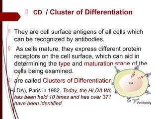
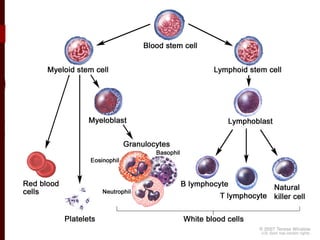
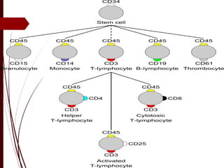
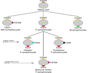
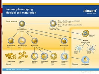
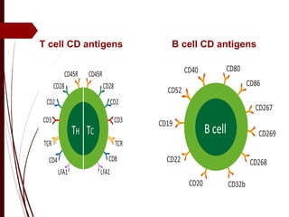
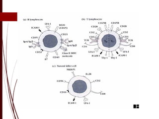
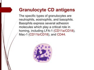
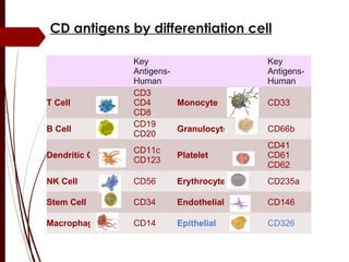
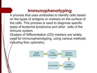
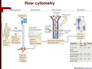
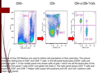
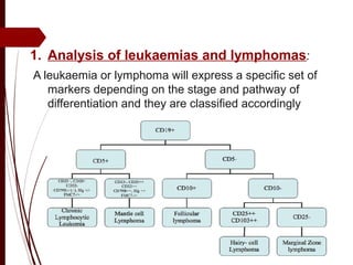
(CD) s
Clusters of Differentiation
Fawzeia Abo Ali
Ain shams faculty of
medicine
  CD / Cluster of Differentiation
 They are cell surface antigens of all cells which 
can be recognized by antibodies.
  As cells mature, they express different protein 
receptors on the cell surface, which can aid in 
determining the type and maturation stage of the 
cells being examined.
 are called Clusters of Differentiation
 HLDA), Paris in 1982. Today, the HLDA Workshop meeting
has been held 10 times and has over 371 CD markers
have been identified
 Properties:
 They are proteins or antigen markers
 A surface marker was given a CD number 
when two monoclonal antibodies bind to it.
 Numbers are assigned arbitrarily
  A small letter w before the number 
designation stands for "workshop". 
 CD antigens serve as receptors and ligands, 
and regulate cell signalling .
 In some cases CD antigens are expressed 
only at certain stages of development or 
under certain conditions
T cell CD antigens B cell CD antigens
Granulocyte CD antigens
.
The specific types of granulocytes are
neutrophils, eosinophils, and basophils.
Basophils express several adhesion
molecules which play a critical role in
homing, including LFA-1 (CD11a/CD18),
Mac-1 (CD11b/CD18), and CD44.
Key
Antigens-
Human
Key
Antigens-
Human
T Cell
CD3
CD4
CD8
Monocyte CD33
B Cell
CD19
CD20
Granulocyte CD66b
Dendritic Cell
CD11c
CD123
Platelet
CD41
CD61
CD62
NK Cell CD56 Erythrocyte CD235a
Stem Cell CD34 Endothelial CD146
Macrophage CD14 Epithelial CD326
CD antigens by differentiation cell
Immunophenotyping
A process that uses antibodies to identify cells based
on the types of antigens or markers on the surface of
the cells. This process is used to diagnose specific
types of leukemia lymphoma and other cells of the
immune system.
Clusters of Differentiation (CD) markers are widely
used for immunophenotyping, using various methods
including flow cytometry.
Flow cytometry
Example of how CD Markers are used to define cell populations in flow cytometry. The picture
shows the Gating tree of CD4+
and CD8+
T cells. In the left panel leukocytes (CD45+
cells) are
gated into gate 1. In the middle panel only shows cells of gate 1 which are all the leukocytes of the
sample. In this panel T cells (CD3+
) are gated into Gate 2. The right panel shows CD3+
T cells and
seperates CD4+
and CD8-
T Helper cells (upper left quadrant) and C8+
and Cd4-
cytotoxic cells
(lower right panel)
1. Analysis of leukaemias and lymphomas:
A leukaemia or lymphoma will express a specific set of
markers depending on the stage and pathway of
differentiation and they are classified accordingly
2. Detection of minimal residual disease
Minimal residual disease (MRD) was
defined as disease beyond the limit of
morphological detection using conventional
microscopy.
Patients with acute leukaemia were considered
to be in remission when bone marrow
samples contained <5% neoplastic cells.
Flow cytometric methods can detect far
lower levels of disease,
3. Stem cell enumeration
 Haemopoietic stem cells in the bone marrow can be
identified by their expression of CD34. Normally, the
number of such cells in bone marrow is low and is
negligible in peripheral blood. However, if mobilisation of
CD34 positive cells from the bone marrow is stimulated,
stem cells can be harvested from the peripheral blood
as well as the marrow.
4. Solid organ transplantation
 T cell cross-match: Flow cytometry can be used to
crossmatch a recipient's serum with donor lymphocytes.
 analysis of the peripheral blood lymphocytes may help
to indicate early rejection
5.  Immunodeficiency diseases
 Diseases resulting from primary immunodeficiencies,
can be a result of defects in T-cells, B-cells,
granulocytes or monocytes.
 In many of these diseases surface or cytoplasmic
proteins are missing or have impaired function. They
are characterized by immunophenotyping, the
selection of antibodies being based on the clinical
presentation.
6. HIV infection.
 Determination of the numbers of CD4 +ve
lymphocytes in the peripheral blood is used to
monitor patients with HIV infections .
 The percentage of CD4 +ve cells can be obtained in
a single tube by staining for CD45/CD3/CD4.
7. Paroxysmal nocturnal haemoglobinuria
 The white cells and red cells produced are
dysfunctional and are susceptible to lysis. Analysis of
this clonal abnormality by flow cytometry, in general,
can be accomplished by analysis of CD55 and CD59
on red cells .
8. Immunotherapy
 Rituxan (Rituximab) - a monoclonal antibody
against CD20.
 Gazyva (Obinutuzumab): targets CD20 antigen, used in
initial treatment for small lymphocytic
lymphoma/chronic lymphocytic leukemia.
 Arzerra (Ofatumumab): targets CD 20 antigen, used in
SLL/CLL.
 Campath (Alemtuzumab): targets CD52 antigen in
SLL/CLL and peripheral T-cell lymphomas.
 Adcetris (Brentuximab vedotin): targets CD30 and is
attached to a chemotherapy drug. Used in anaplastic
large cell lymphoma.
Clusters of differentiation (CDs)

Clusters of differentiation (CDs)

  • 1.
    (CD) s Clusters ofDifferentiation Fawzeia Abo Ali Ain shams faculty of medicine
  • 2.
      CD /Cluster of Differentiation  They are cell surface antigens of all cells which  can be recognized by antibodies.   As cells mature, they express different protein  receptors on the cell surface, which can aid in  determining the type and maturation stage of the  cells being examined.  are called Clusters of Differentiation  HLDA), Paris in 1982. Today, the HLDA Workshop meeting has been held 10 times and has over 371 CD markers have been identified
  • 3.
     Properties:  They are proteins or antigen markers A surface marker was given a CD number  when two monoclonal antibodies bind to it.  Numbers are assigned arbitrarily   A small letter w before the number  designation stands for "workshop".   CD antigens serve as receptors and ligands,  and regulate cell signalling .  In some cases CD antigens are expressed  only at certain stages of development or  under certain conditions
  • 8.
    T cell CDantigens B cell CD antigens
  • 10.
    Granulocyte CD antigens . Thespecific types of granulocytes are neutrophils, eosinophils, and basophils. Basophils express several adhesion molecules which play a critical role in homing, including LFA-1 (CD11a/CD18), Mac-1 (CD11b/CD18), and CD44.
  • 11.
    Key Antigens- Human Key Antigens- Human T Cell CD3 CD4 CD8 Monocyte CD33 BCell CD19 CD20 Granulocyte CD66b Dendritic Cell CD11c CD123 Platelet CD41 CD61 CD62 NK Cell CD56 Erythrocyte CD235a Stem Cell CD34 Endothelial CD146 Macrophage CD14 Epithelial CD326 CD antigens by differentiation cell
  • 12.
    Immunophenotyping A process thatuses antibodies to identify cells based on the types of antigens or markers on the surface of the cells. This process is used to diagnose specific types of leukemia lymphoma and other cells of the immune system. Clusters of Differentiation (CD) markers are widely used for immunophenotyping, using various methods including flow cytometry.
  • 13.
  • 14.
    Example of howCD Markers are used to define cell populations in flow cytometry. The picture shows the Gating tree of CD4+ and CD8+ T cells. In the left panel leukocytes (CD45+ cells) are gated into gate 1. In the middle panel only shows cells of gate 1 which are all the leukocytes of the sample. In this panel T cells (CD3+ ) are gated into Gate 2. The right panel shows CD3+ T cells and seperates CD4+ and CD8- T Helper cells (upper left quadrant) and C8+ and Cd4- cytotoxic cells (lower right panel)
  • 16.
    1. Analysis ofleukaemias and lymphomas: A leukaemia or lymphoma will express a specific set of markers depending on the stage and pathway of differentiation and they are classified accordingly
  • 17.
    2. Detection ofminimal residual disease Minimal residual disease (MRD) was defined as disease beyond the limit of morphological detection using conventional microscopy. Patients with acute leukaemia were considered to be in remission when bone marrow samples contained <5% neoplastic cells. Flow cytometric methods can detect far lower levels of disease,
  • 18.
    3. Stem cellenumeration  Haemopoietic stem cells in the bone marrow can be identified by their expression of CD34. Normally, the number of such cells in bone marrow is low and is negligible in peripheral blood. However, if mobilisation of CD34 positive cells from the bone marrow is stimulated, stem cells can be harvested from the peripheral blood as well as the marrow. 4. Solid organ transplantation  T cell cross-match: Flow cytometry can be used to crossmatch a recipient's serum with donor lymphocytes.  analysis of the peripheral blood lymphocytes may help to indicate early rejection
  • 19.
    5.  Immunodeficiency diseases Diseases resulting from primary immunodeficiencies, can be a result of defects in T-cells, B-cells, granulocytes or monocytes.  In many of these diseases surface or cytoplasmic proteins are missing or have impaired function. They are characterized by immunophenotyping, the selection of antibodies being based on the clinical presentation.
  • 20.
    6. HIV infection. Determination of the numbers of CD4 +ve lymphocytes in the peripheral blood is used to monitor patients with HIV infections .  The percentage of CD4 +ve cells can be obtained in a single tube by staining for CD45/CD3/CD4. 7. Paroxysmal nocturnal haemoglobinuria  The white cells and red cells produced are dysfunctional and are susceptible to lysis. Analysis of this clonal abnormality by flow cytometry, in general, can be accomplished by analysis of CD55 and CD59 on red cells .
  • 21.
    8. Immunotherapy  Rituxan(Rituximab) - a monoclonal antibody against CD20.  Gazyva (Obinutuzumab): targets CD20 antigen, used in initial treatment for small lymphocytic lymphoma/chronic lymphocytic leukemia.  Arzerra (Ofatumumab): targets CD 20 antigen, used in SLL/CLL.  Campath (Alemtuzumab): targets CD52 antigen in SLL/CLL and peripheral T-cell lymphomas.  Adcetris (Brentuximab vedotin): targets CD30 and is attached to a chemotherapy drug. Used in anaplastic large cell lymphoma.