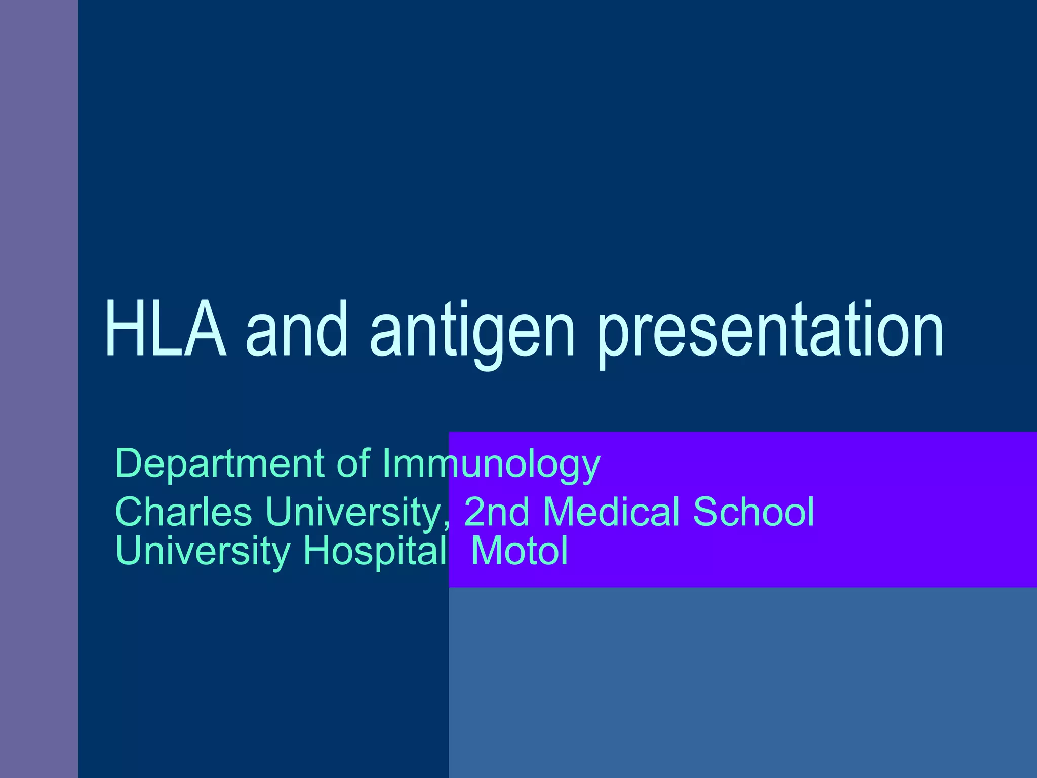
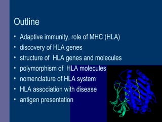
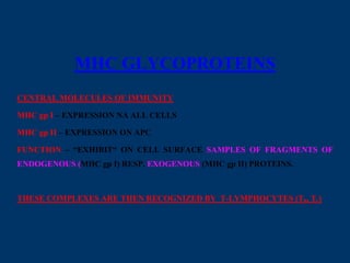
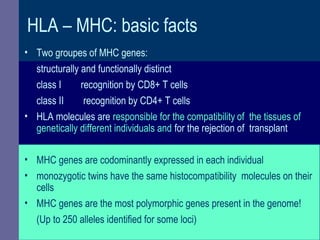
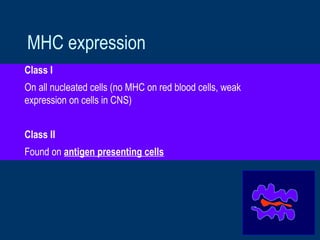
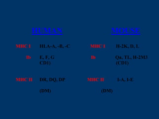
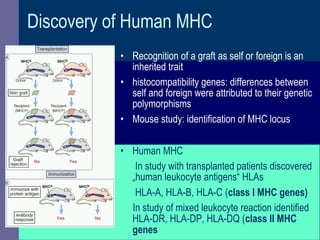
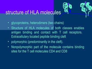
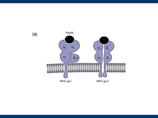
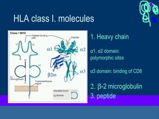
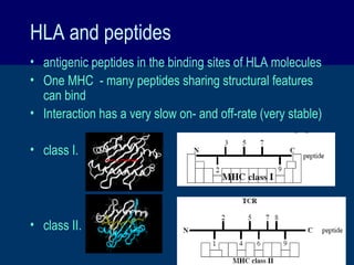
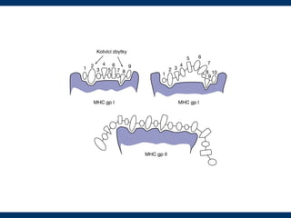
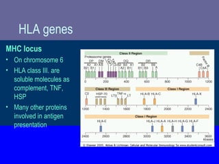
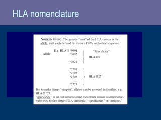
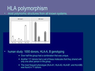
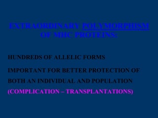
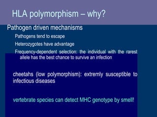
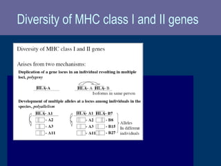
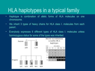
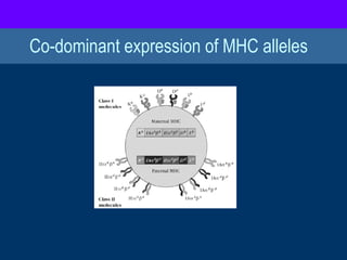
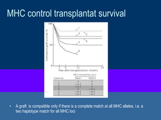
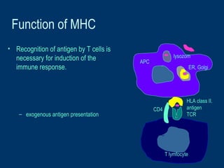
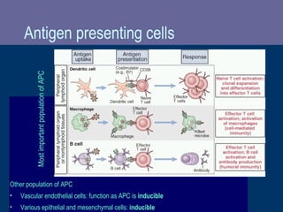
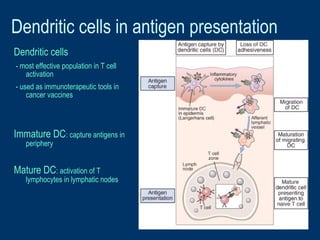
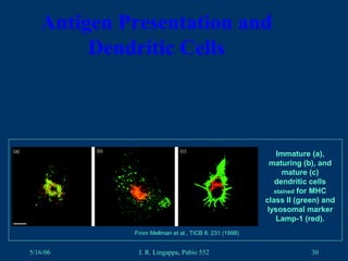
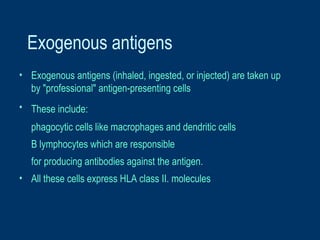
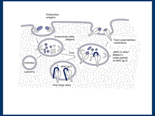
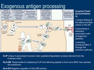
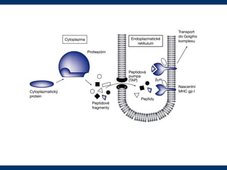
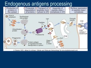
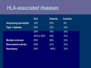
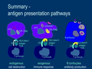
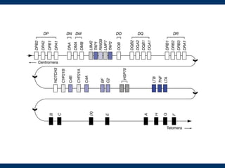
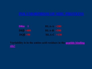
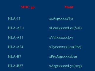
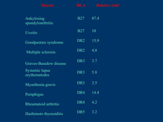
HLA and antigen presentation
Department of Immunology
Charles University, 2nd Medical School
University Hospital Motol
MHC in adaptive immunity
Innate Adaptive
Specificity For structures shared by groups
of related microbes
For antigens of microbes and for
nonmicrobial antigens
Diversity Limited; germline-encoded Very large; receptors are produced by
somatic recombination of gene
segments
Memory None Yes
Nonreactivity to self Yes Yes
Physical and
chemical barriers
Skin, mucosal epithelia;
antimicrobial chemicals
Lymphocytes in epithelia; antibodies
secreted at epithelial surfaces
Blood proteins Complement Antibodies
Cells Phagocytes (macrophages,
neutrophils), natural killer cells
T and B Lymphocytes
Characteristics
Components
T cells recognise cell-associated
antigens displayed on
MHC = major histocompatibility complex
Outline
• Adaptive immunity, role of MHC (HLA)
• discovery of HLA genes
• structure of HLA genes and molecules
• polymorphism of HLA molecules
• nomenclature of HLA system
• HLA association with disease
• antigen presentation
MHC GLYCOPROTEINS
CENTRAL MOLECULES OF IMMUNITY
MHC gp I – EXPRESSION NA ALL CELLS
MHC gp II – EXPRESSION ON APC
FUNCTION – “EXHIBIT“ ON CELL SURFACE SAMPLES OF FRAGMENTS OF
ENDOGENOUS (MHC gp I) RESP. EXOGENOUS (MHC gp II) PROTEINS.
THESE COMPLEXES ARE THEN RECOGNIZED BY T-LYMPHOCYTES (Th, Tc)
HLA – MHC: basic facts
• Two groupes of MHC genes:
structurally and functionally distinct
class I recognition by CD8+ T cells
class II recognition by CD4+ T cells
• HLA molecules are responsible for the compatibility of the tissues of
genetically different individuals and for the rejection of transplant
• MHC genes are codominantly expressed in each individual
• monozygotic twins have the same histocompatibility molecules on their
cells
• MHC genes are the most polymorphic genes present in the genome!
(Up to 250 alleles identified for some loci)
MHC expression
Class I
On all nucleated cells (no MHC on red blood cells, weak
expression on cells in CNS)
Class II
Found on antigen presenting cells
HUMAN MOUSE
MHC I HLA-A, -B, -C MHC I H-2K, D, L
Ib E, F, G Ib Qa, TL, H-2M3
CD1) (CD1)
MHC II DR, DQ, DP MHC II I-A, I-E
(DM) (DM)
Discovery of Human MHC
• Recognition of a graft as self or foreign is an
inherited trait
• histocompatibility genes: differences between
self and foreign were attributed to their genetic
polymorphisms
• Mouse study: identification of MHC locus
• Human MHC
In study with transplanted patients discovered
„human leukocyte antigens“ HLAs
HLA-A, HLA-B, HLA-C (class I MHC genes)
In study of mixed leukocyte reaction identified
HLA-DR, HLA-DP, HLA-DQ (class II MHC
genes
structure of HLA molecules
• glycoproteins, heterodimers (two chains)
• Structure of HLA molecules of both classes enables
antigen binding and contact with T cell receptors.
Extracellulary located peptide binding cleft
• polymorphic (predominantly in the cleft).
• Nonpolymorphic part of the molecule contains binding
sites for the T cell molecules CD4 and CD8
HLA class I. molecules
1. Heavy chain
α1, α2 domain:
polymorphic sites
α3 domain: binding of CD8
2. β-2 microglobulin
3. peptide
Structure of HLA class II. molecules
1. α chain
α1: polymorphic sites
α2: binding of CD4
2. β chain
β1: polymorphic sites
β2: binding of CD4
3. peptide
HLA and antigens
• Most T lymphocytes recognize only peptides
• T cells are specific for amino acid sequences of peptides - TCR
• Intracellular antigens are presented in connection with HLA class I. -
CD8+ T cells recognition
• Extracellular antigens are presented in connection with HLA class II.
– CD4+ T cells recognition
Experiment:
• T cell response – only when peptide is attached to APC
HLA and peptides
• antigenic peptides in the binding sites of HLA molecules
• One MHC - many peptides sharing structural features
can bind
• Interaction has a very slow on- and off-rate (very stable)
• class I.
• class II.
HLA genes
MHC locus
• On chromosome 6
• HLA class III. are
soluble molecules as
complement, TNF,
HSP
• Many other proteins
involved in antigen
presentation
HLA nomenclature
HLA polymorphism
• most polymorhic structures from all known systems.
• human study: 1000 donors, HLA-A, B genotyping
• Over half the group had a combination that was unique.
• Another 111 donors had a set of these molecules that they shared with
only one other person in the group.
• The most frequent phenotype (HLA-A1, HLA-A3, HLA-B7, and HLA-B8)
was found in 11 donors.
EXTRAORDINARY POLYMORPHISM
OF MHC PROTEINS:
HUNDREDS OF ALLELIC FORMS
IMPORTANT FOR BETTER PROTECTION OF
BOTH AN INDIVIDUAL AND POPULATION
(COMPLICATION – TRANSPLANTATIONS)
HLA polymorphism – why?
Pathogen driven mechanisms
Pathogens tend to escape
Heterozygotes have advantage
Frequency-dependent selection: the individual with the rarest
allele has the best chance to survive an infection
cheetahs (low polymorphism): extremly susceptible to
infectious diseases
vertebrate species can detect MHC genotype by smell!
Diversity of MHC class I and II genes
HLA haplotypes in a typical family
• Haplotype is combination of allelic forms of HLA molecules on one
chromosome.
• We inherit 3 types of heavy chains for HLA class I. molecules from each
parent .
• Everybody expresses 6 different types of HLA class I. molecules unless
honmozygous status for some of the types was inherited.
Co-dominant expression of MHC alleles
MHC control transplantat survival
• A graft is compatible only if there is a complete match at all MHC alleles, i.e. a
two haplotype match for all MHC loci
Function of MHC
• Recognition of antigen by T cells is
necessary for induction of the
immune response.
– exogenous antigen presentation
HLA class II.
antigen
TCR
CD4
T lymfocyte
APC
lysozom
ER, Golgi
Antigen presenting cells
Other population of APC
• Vascular endothelial cells: function as APC is inducible
• Various epithelial and mesenchymal cells: inducible
MostimportantpopulationofAPC
Dendritic cells in antigen presentation
Dendritic cells
- most effective population in T cell
activation
- used as immunoterapeutic tools in
cancer vaccines
Immature DC: capture antigens in
periphery
Mature DC: activation of T
lymphocytes in lymphatic nodes
5/16/06 J. R. Lingappa, Pabio 552 30
Antigen Presentation and
Dendritic Cells
From Mellman et al., TICB 8: 231 (1998)
Immature (a),
maturing (b), and
mature (c)
dendritic cells
stained for MHC
class II (green) and
lysosomal marker
Lamp-1 (red).
Exogenous antigens
• Exogenous antigens (inhaled, ingested, or injected) are taken up
by "professional" antigen-presenting cells
• These include:
phagocytic cells like macrophages and dendritic cells
B lymphocytes which are responsible
for producing antibodies against the antigen.
• All these cells express HLA class II. molecules
Exogenous antigen processing
CLIP (Class II-associated invariant chain peptide)-Degradation product derived from the
Invariant chain.
HLA-DM Responsible for displacing CLIP and allowing peptide to bind once MHC has reached
the endosome.
HLA-DO Negative regulator of HLA-DM activity
Invariant Chain
(Ii)-Contributes
to:
1.proper folding of
the alpha and beta
chains in the ER
2.preventing in
advertent
association with
endogenous
peptides
3.directing
immature MHC II
through the
secretory pathway
Endogenous antigens processing
TAP (transporter associated with antigen presentation)
• Transport associated
protein - TAP is
responsible for the peptide
transport from cytoplasm to
ER.
• Proteins are degraded to
peptide in proteasome.
• The peptides are picked up
by TAP proteins and
transported from the cytosol
into the RER where they
assemble with
– the transmembrane polypeptide and beta-2 microglobulin.
– this trimolecular complex then moves through the Golgi apparatus and is
inserted in the plasma membrane
Imunodeficiences - MHC defect
• Bare lymphocyte syndrome:
mutation in genes regulating class II MHC transcription
- reduced number of CD4+ T cells in periphery
- defective activation of CD4+ T cells
- fatal, treatment: BM transplantation
• Class I MHC deficiencies:
- decreased number of CD4+ T cells in periphery
- caused by TAP1, TAP2
- patients suffer from respiratory tract bacterial infection
HLA-associated diseases
HLA Patients Controls
Ankylosing spondylitis B27 90% 9%
Type 1 diabetes DR3 52% 23%
DR4 74% 24%
DR3 or DR4 93% 43%
Multiple sclerosis DR2 86% 33%
Rheumatoid arthritis DR4 81% 24%
Narcolepsy DR2 >95% 33%
Summary -
antigen presentation pathways
CD4
T lymfocyte
B lymfocyte
HLA class I.
antigen
TCR
CD8
T lymfocyte
APC
ER, Golgi
HLA class II.
antigen
TCR
CD4
T lymfocyte
APC
lysozome
ER, Golgi
HLA class II.
antigen
TCR
endogenous exogenous B lymfocytes
cell destruction immune response antibody production
POLYMORPHISM OF MHC PROTEINS
DRα 1 HLA-A >280
DRβ >400 HLA-B >500
DQβ >50 HLA-C >130
Variability is in the amino acid residues in the peptide binding
site!
MHC gp Motif
HLA-11 xxAspxxxxxTyr
HLA-A2,1 xLeuxxxxxxLeu(VaI)
HLA-A11 xVaIxxxxxxLys
HLA-A24 xTyrxxxxxxLeu(Phe)
HLA-B7 xProArgxxxxxLeu
HLA-B27 xArgxxxxxxLys(Arg)
Disease HLA Relative risk*
Ankylosing
spondyloarthritis
B27 87.4
Uveitis B27 10
Goodpasture syndrome DR2 15.9
Multiple sclerosis DR2 4.8
Graves-Basedow disease
DR3 3.7
Systemic lupus
erythematodes
DR3 5.8
Myasthenia gravis DR3 2.5
Pemphigus DR4 14.4
Rheumatoid arthritis DR4 4.2
Hashimoto thyreoiditis DR5 3.2

HLA and antigen presentation

  • 1.
    HLA and antigenpresentation Department of Immunology Charles University, 2nd Medical School University Hospital Motol
  • 2.
    MHC in adaptiveimmunity Innate Adaptive Specificity For structures shared by groups of related microbes For antigens of microbes and for nonmicrobial antigens Diversity Limited; germline-encoded Very large; receptors are produced by somatic recombination of gene segments Memory None Yes Nonreactivity to self Yes Yes Physical and chemical barriers Skin, mucosal epithelia; antimicrobial chemicals Lymphocytes in epithelia; antibodies secreted at epithelial surfaces Blood proteins Complement Antibodies Cells Phagocytes (macrophages, neutrophils), natural killer cells T and B Lymphocytes Characteristics Components T cells recognise cell-associated antigens displayed on MHC = major histocompatibility complex
  • 3.
    Outline • Adaptive immunity,role of MHC (HLA) • discovery of HLA genes • structure of HLA genes and molecules • polymorphism of HLA molecules • nomenclature of HLA system • HLA association with disease • antigen presentation
  • 4.
    MHC GLYCOPROTEINS CENTRAL MOLECULESOF IMMUNITY MHC gp I – EXPRESSION NA ALL CELLS MHC gp II – EXPRESSION ON APC FUNCTION – “EXHIBIT“ ON CELL SURFACE SAMPLES OF FRAGMENTS OF ENDOGENOUS (MHC gp I) RESP. EXOGENOUS (MHC gp II) PROTEINS. THESE COMPLEXES ARE THEN RECOGNIZED BY T-LYMPHOCYTES (Th, Tc)
  • 5.
    HLA – MHC:basic facts • Two groupes of MHC genes: structurally and functionally distinct class I recognition by CD8+ T cells class II recognition by CD4+ T cells • HLA molecules are responsible for the compatibility of the tissues of genetically different individuals and for the rejection of transplant • MHC genes are codominantly expressed in each individual • monozygotic twins have the same histocompatibility molecules on their cells • MHC genes are the most polymorphic genes present in the genome! (Up to 250 alleles identified for some loci)
  • 6.
    MHC expression Class I Onall nucleated cells (no MHC on red blood cells, weak expression on cells in CNS) Class II Found on antigen presenting cells
  • 7.
    HUMAN MOUSE MHC IHLA-A, -B, -C MHC I H-2K, D, L Ib E, F, G Ib Qa, TL, H-2M3 CD1) (CD1) MHC II DR, DQ, DP MHC II I-A, I-E (DM) (DM)
  • 8.
    Discovery of HumanMHC • Recognition of a graft as self or foreign is an inherited trait • histocompatibility genes: differences between self and foreign were attributed to their genetic polymorphisms • Mouse study: identification of MHC locus • Human MHC In study with transplanted patients discovered „human leukocyte antigens“ HLAs HLA-A, HLA-B, HLA-C (class I MHC genes) In study of mixed leukocyte reaction identified HLA-DR, HLA-DP, HLA-DQ (class II MHC genes
  • 9.
    structure of HLAmolecules • glycoproteins, heterodimers (two chains) • Structure of HLA molecules of both classes enables antigen binding and contact with T cell receptors. Extracellulary located peptide binding cleft • polymorphic (predominantly in the cleft). • Nonpolymorphic part of the molecule contains binding sites for the T cell molecules CD4 and CD8
  • 11.
    HLA class I.molecules 1. Heavy chain α1, α2 domain: polymorphic sites α3 domain: binding of CD8 2. β-2 microglobulin 3. peptide
  • 13.
    Structure of HLAclass II. molecules 1. α chain α1: polymorphic sites α2: binding of CD4 2. β chain β1: polymorphic sites β2: binding of CD4 3. peptide
  • 15.
    HLA and antigens •Most T lymphocytes recognize only peptides • T cells are specific for amino acid sequences of peptides - TCR • Intracellular antigens are presented in connection with HLA class I. - CD8+ T cells recognition • Extracellular antigens are presented in connection with HLA class II. – CD4+ T cells recognition Experiment: • T cell response – only when peptide is attached to APC
  • 16.
    HLA and peptides •antigenic peptides in the binding sites of HLA molecules • One MHC - many peptides sharing structural features can bind • Interaction has a very slow on- and off-rate (very stable) • class I. • class II.
  • 18.
    HLA genes MHC locus •On chromosome 6 • HLA class III. are soluble molecules as complement, TNF, HSP • Many other proteins involved in antigen presentation
  • 19.
  • 20.
    HLA polymorphism • mostpolymorhic structures from all known systems. • human study: 1000 donors, HLA-A, B genotyping • Over half the group had a combination that was unique. • Another 111 donors had a set of these molecules that they shared with only one other person in the group. • The most frequent phenotype (HLA-A1, HLA-A3, HLA-B7, and HLA-B8) was found in 11 donors.
  • 21.
    EXTRAORDINARY POLYMORPHISM OF MHCPROTEINS: HUNDREDS OF ALLELIC FORMS IMPORTANT FOR BETTER PROTECTION OF BOTH AN INDIVIDUAL AND POPULATION (COMPLICATION – TRANSPLANTATIONS)
  • 22.
    HLA polymorphism –why? Pathogen driven mechanisms Pathogens tend to escape Heterozygotes have advantage Frequency-dependent selection: the individual with the rarest allele has the best chance to survive an infection cheetahs (low polymorphism): extremly susceptible to infectious diseases vertebrate species can detect MHC genotype by smell!
  • 23.
    Diversity of MHCclass I and II genes
  • 24.
    HLA haplotypes ina typical family • Haplotype is combination of allelic forms of HLA molecules on one chromosome. • We inherit 3 types of heavy chains for HLA class I. molecules from each parent . • Everybody expresses 6 different types of HLA class I. molecules unless honmozygous status for some of the types was inherited.
  • 25.
  • 26.
    MHC control transplantatsurvival • A graft is compatible only if there is a complete match at all MHC alleles, i.e. a two haplotype match for all MHC loci
  • 27.
    Function of MHC •Recognition of antigen by T cells is necessary for induction of the immune response. – exogenous antigen presentation HLA class II. antigen TCR CD4 T lymfocyte APC lysozom ER, Golgi
  • 28.
    Antigen presenting cells Otherpopulation of APC • Vascular endothelial cells: function as APC is inducible • Various epithelial and mesenchymal cells: inducible MostimportantpopulationofAPC
  • 29.
    Dendritic cells inantigen presentation Dendritic cells - most effective population in T cell activation - used as immunoterapeutic tools in cancer vaccines Immature DC: capture antigens in periphery Mature DC: activation of T lymphocytes in lymphatic nodes
  • 30.
    5/16/06 J. R.Lingappa, Pabio 552 30 Antigen Presentation and Dendritic Cells From Mellman et al., TICB 8: 231 (1998) Immature (a), maturing (b), and mature (c) dendritic cells stained for MHC class II (green) and lysosomal marker Lamp-1 (red).
  • 31.
    Exogenous antigens • Exogenousantigens (inhaled, ingested, or injected) are taken up by "professional" antigen-presenting cells • These include: phagocytic cells like macrophages and dendritic cells B lymphocytes which are responsible for producing antibodies against the antigen. • All these cells express HLA class II. molecules
  • 33.
    Exogenous antigen processing CLIP(Class II-associated invariant chain peptide)-Degradation product derived from the Invariant chain. HLA-DM Responsible for displacing CLIP and allowing peptide to bind once MHC has reached the endosome. HLA-DO Negative regulator of HLA-DM activity Invariant Chain (Ii)-Contributes to: 1.proper folding of the alpha and beta chains in the ER 2.preventing in advertent association with endogenous peptides 3.directing immature MHC II through the secretory pathway
  • 35.
  • 36.
    TAP (transporter associatedwith antigen presentation) • Transport associated protein - TAP is responsible for the peptide transport from cytoplasm to ER. • Proteins are degraded to peptide in proteasome. • The peptides are picked up by TAP proteins and transported from the cytosol into the RER where they assemble with – the transmembrane polypeptide and beta-2 microglobulin. – this trimolecular complex then moves through the Golgi apparatus and is inserted in the plasma membrane
  • 39.
    Imunodeficiences - MHCdefect • Bare lymphocyte syndrome: mutation in genes regulating class II MHC transcription - reduced number of CD4+ T cells in periphery - defective activation of CD4+ T cells - fatal, treatment: BM transplantation • Class I MHC deficiencies: - decreased number of CD4+ T cells in periphery - caused by TAP1, TAP2 - patients suffer from respiratory tract bacterial infection
  • 40.
    HLA-associated diseases HLA PatientsControls Ankylosing spondylitis B27 90% 9% Type 1 diabetes DR3 52% 23% DR4 74% 24% DR3 or DR4 93% 43% Multiple sclerosis DR2 86% 33% Rheumatoid arthritis DR4 81% 24% Narcolepsy DR2 >95% 33%
  • 41.
    Summary - antigen presentationpathways CD4 T lymfocyte B lymfocyte HLA class I. antigen TCR CD8 T lymfocyte APC ER, Golgi HLA class II. antigen TCR CD4 T lymfocyte APC lysozome ER, Golgi HLA class II. antigen TCR endogenous exogenous B lymfocytes cell destruction immune response antibody production
  • 43.
    POLYMORPHISM OF MHCPROTEINS DRα 1 HLA-A >280 DRβ >400 HLA-B >500 DQβ >50 HLA-C >130 Variability is in the amino acid residues in the peptide binding site!
  • 44.
    MHC gp Motif HLA-11xxAspxxxxxTyr HLA-A2,1 xLeuxxxxxxLeu(VaI) HLA-A11 xVaIxxxxxxLys HLA-A24 xTyrxxxxxxLeu(Phe) HLA-B7 xProArgxxxxxLeu HLA-B27 xArgxxxxxxLys(Arg)
  • 45.
    Disease HLA Relativerisk* Ankylosing spondyloarthritis B27 87.4 Uveitis B27 10 Goodpasture syndrome DR2 15.9 Multiple sclerosis DR2 4.8 Graves-Basedow disease DR3 3.7 Systemic lupus erythematodes DR3 5.8 Myasthenia gravis DR3 2.5 Pemphigus DR4 14.4 Rheumatoid arthritis DR4 4.2 Hashimoto thyreoiditis DR5 3.2

Editor's Notes

  • #26 Humans synthesize three different types of class II. molecules designated HLA-A, HLA-B, and HLA-C (heterozygote – 6 different class I molecules on every cell). These differ only in their heavy chain, all sharing the same type of beta-2 microglobulin. HLA class I. molecules are expressed on all nucleated cells of the body. HLA class I. molecules are not expressed on red blood cells and only minor level of expression is detected on the cells of central nervous system. The genes encoding the different heavy chains are clustered on chromosome 6 in the major histocompatibility complex.