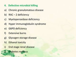
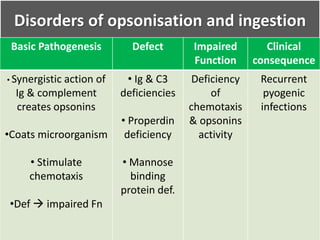
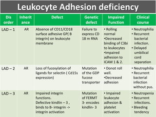
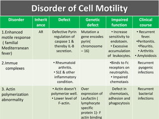
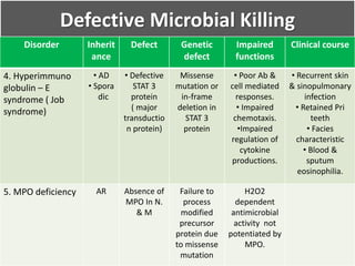
LEUKOCYTE FUNCTIONS
DEFECTS
Dr Nidhi Rai
Introduction
• Essential cellular components of blood.
• Develop from the pluripotential hematopoietic stem
cells in the bone marrow.
• Leukocytes include
 Granulocytes:
– Neutrophils
– Basophils
– Eosinophils
 Monocyte
 Lymphocyte
Primary function: To protect the host from
infectious agents or pathogens.
Innate Immune Response
Acquired/Adaptive /Specific
immune system
• 1st response to
common class of
pathogens.
• Neutrophils &
Monocytes – Play
a major role.
• Rapid but limited.
• Initiated in
lymphoid tissue
when pathogens
encounter
lymphocytes.
• Lymphocytes –
plays a major role.
Neutrophils
• Neutrophils are very mobile cells
• Two types of granules:
– Primary or azurophilic
– Secondary or specific
• All granules contain enzymes which are involved in killing
and digesting bacteria and fungi.
Functions of Neutrophils
• Innate immune response: 4 steps
1. Adherence
2. Migration ( chemotaxis)
3. Phagocytosis
4. Bacterial killing
1. Adherence
• Activation of leukocytes & vascular endothelial cells (VEC)
by inflammatory mediators(cytokines).
• Activation result is activity of 3 classes of cytoadhesion
mol. (CAM) & their ligands.
• CAM & their ligands play a major role.
1. Selectins & their ligands
2. Intercellular adhesion molecule (ICAM)
3. Beta 2 (CD18) family of leukocyte integrin & their
ligands.
2. Migration- pseudopodia forms
ICAM
E & P Selectins
3. Phagocytosis
Some pathogens are
recognized by opsonization
(IgG & C3b)
PAMP
PRR
Degranulation
4.Bacterial Killing & Digestion
• Phagosomes  fuse with the lysosome( phagolysosome)
 discharge their granules  Bacterial killing &
digestion.
• Microbicidal mechanism – 2 types
1. Oxygen dependent
2. Oxygen independent
Oxygen Dependent
• Neutrophils activation is accompanied by increase in O2
use- Respiratory burst/ oxidative burst.
• Respiratory burst – caused by activated NADPH oxidase.
 Increase in O2 consumption.
 Increase glycolysis.
 Production of ROS ( superoxide)
 Increase in glucose oxidation by HMP shunt
• Activated NADPH – detected by NBT, Cytochrome oxidase
or chemiluminescence test.
• MPO independent:
 H2O2
 Superoxide ion
 Hydroxyl ion
 Singlet oxygen
• MPO dependent: Present in primary granules
Oxygen Independent
• Mediated by the granules present in primary,gelatinase,specific
granules.
• Acid pH of phagosomes  Increase activity of granular protein
 Microbicidal activity
• Alone insufficient to kill microbes.
• Include :
 Lysozyme
 Lactoferrin
 BPI( Bacterial permeability inducing proteins)
 Defensins
 Collagenase
Monocytes Functions
• Active in both innate and adaptive IR.
• Phagocytosis.
• Ingest activated clotting factors thus limiting the
coagulation process.
• Secrete a variety of substance affects the functions of
other cells especially lymphocytes.
Eosinophils
• Eosinophils : 2 - 8 % leukocytes
• Derived from hematopoietic stem cells
• bilobed nucleus with numerous bright orange
cytoplasmic granules
• Originates from IL -5 responsive CD 34 + myeloid
progenitor cells
• IL -5 has lineage specificity for eosinophils
Function of Eosinophils
• Cationic proteins, cytokines and leukotriens- in
eosinophil granules mediate
 Parasite defense
 Allergic response
 Tissue infiltration
 Immune modulation
Basophils
• Basophils are least of the circulating leucocytes
• They constitute less than 1% of the circulating leucocytes
or absolute counts of 0.02-0.08x10⁹/lt
• Normal range on BMA is 0-0.5%
• Circulates in blood and normally not found in tissues
• Recruited to tissues during inflammatory or immunologic
response
Functions of Basophils
• Basophils – Key player in Allergy
• Degranulation
• Late Phase Reaction
Lymphocytes
• Specific component of immune system.
• Different types :
 B-Lymphocytes (“B cells)
 T- Lymphocytes (“T cells”)
Natural Killer Cells (NK cells)
Memory Cells
Suppressor Cells
• They have different functions in specific immunity.
B-LYMPHOCYTES
• Mature in bone marrow, then carried to lymphoid tissue
via blood stream and lymphatic circulation.
• This process of maturation and migration takes place
throughout life.
• Other lymphocytes can be generated via mitosis of B
lymphocytes resident in lymphoid tissues
T-LYMPHOCYTES
• Immature lymphocytes leave bone marrow during fetal
and early neonatal life.
• Go to thymus gland.
• Mature there before they go on to other lymphoid
tissues.
Role in Specific Immunity
• The body must be able to recognize the difference
between “self” and “nonself.”
• Any lymphocytes with antibodies that recognize one’s
own body tissue as an antigen are killed during fetal life.
• Stages of specific immune response-
1) Antigen encounter and recognition by lymphocytes.
2) Lymphocyte activation.
3) Attack
1) Antigen encounter and recognition by lymphocytes
• Specific lymphocytes are programmed to recognize a
specific antigen.
• Usually happens in a lymphoid organ, bloodstream, or
lymph vessel.
2) Lymphocyte activation:
• Once a lymphocyte has recognized an antigen, it
undergoes numerous cycles of mitotic divisions, making
more of the same.
• Some of the newly produced cells carry out the attack;
others influence the activation and function of the attack
cells
3) Attack:
B-lymphocytes have specific receptors on their cell
membrane – ANTIBODIES – that bind with invading
materials/organisms
Function of T Lymphocytes
• Do not produce antibodies.
• Function in “cell-mediated immunity.”
• “NATURAL KILLER” cells destroy viruses.
• Secrete “lymphokines” which attract phagocytic cells.
• Secrete “perforin” which eats the cells membrane or
viral coat of invaders.
• Helper T cells:
• Induce macrophages to destroy other antigens
• STIMULATE B-LYMPHOCYTES TO PRODUCE
ANTIBODIES.
• “Suppressor T Cells” prevent overreaction of the system.
(Inhibit B-lymphocye production.)
Disorders of Leukocyte Function
1. Disorders of opsonisation and ingestion
2. Defective adhesion of Leukocytes.
a) Leukocyte adhesion deficiency
b) Drug induced
3. Defective locomotion and chemotaxis
a) Actin polymerization abnormalities.
b) Neonatal neutrophils
c) Interleukin 2 administration
d) Cardiopulmonary bypass
e) Enhanced motile response
4. Defective microbial killing
a) Chronic granulomatous disease
b) RAC – 2 deficiency
c) Myeloperoxidase deficiency
d) Hyper immunoglobulin syndrome
e) G6PD deficiency
f) Extensive burns
g) Glycogen storage disease
h) Ethanol toxicity
i) End stage renal disease
j) Diabetes mellitus
5. Abnormal structure of the nucleus or of an organelle
a) Hereditary macropolycytes
b) Hereditary hyper segmentation
c) Pelger- Huet anomaly
d) Alder- Reilly anomaly
e) May – Hegglin anomaly
6. Degranulation defect
a) Chediak – Higashi disease
b) Specific granule deficiency
Disorders of opsonisation and ingestion
Basic Pathogenesis Defect Impaired
Function
Clinical
consequence
• Synergistic action of
Ig & complement
creates opsonins
•Coats microorganism
• Stimulate
chemotaxis
•Def  impaired Fn
• Ig & C3
deficiencies
• Properdin
deficiency
• Mannose
binding
protein def.
Deficiency
of
chemotaxis
& opsonins
activity
Recurrent
pyogenic
infections
Defective Adhesion
Main functional defect
• Unable to successfully activate and "grab" or adhere to
the blood vessel wall because they lack the necessary
podium & if present but may be defective and not work
properly.
• Since these leukocytes cannot adhere, experience an
"adhesion deficiency",
Leukocyte Adhesion deficiency
Dis
order
Inherit
ance
Defect Genetic
defect
Impaired
Function
Clinical
course
LAD – 1 AR Absence of CD11/CD18
surface adhesive GP( B
integrin) on leukocyte
membrane
Failure to
express CD
18 m RNA
• Rolling
normal
•Decreased
binding of C3bi
to leukocytes
•Impaired
adhesion to
ICAM 1 & 2.
• Neutrophilia
• Recurrent
bacterial
infection.
• Delayed
umbilical
cord
separation
LAD – 2 AR Loss of fucosylation of
ligands for selectin ( Cd15s
expression)
Mutation
of the GDP
fucose
transporter
• Donot roll
well.
•Decreased
adhesion
• Neutrophilia
• Recurrent
bacterial
infection
without pus.
LAD – 3 AR Impaired integrin
functions.
Defective kindlin – 3 ,
binds to B- integrin ->
integrin activation
Mutation
of FERMT
3- encodes
kindlin- 3
• Impaired
leukocyte
adhesion &
platelet
activation
• Neutropenia
• Recurrent
infections.
• Bleeding
tendency
Disorder of Cell Motility
Disorder Inherit
ance
Defect Genetic
defect
Impaired
function
Clinical
course
1.Enhanced
motile response
( familial
Mediterranean
fever)
AR Defective Pyrin
regulation of
caspase 1 &
thereby IL-B
secretion.
Mutation a
gene encodes
pyrin(
chromosome
– 16)
• Increase
sensitivity to
endotoxin.
• Excessive
accumulation
of leukocytes.
• Recurrent
fever.
•Peritonitis.
•Pleuritis.
• Arthritis
• Amyloidosis
2.Immue
complexes
• Rheumatoid
arthritis.
• SLE & other
inflammatory
condition.
•Binds to Fc
receptors on
neutrophils.
• Impaired
chemotaxis
Recurrent
pyogenic
infections
3. Actin
polymerization
abnormality
• Actin doesn’t
polymerize well.
• Lower level of
F-actin.
Over
expression of
Leufactin (
lymphocyte
specific
protein 1)- F
actin binding
Defect in
chemotaxis,
adhesion and
phagocytosis
Recurrent
bacterial
infections
Defective microbial killing
• The ultimate step in the elimination of infectious agents
and necrotic cells is their killing and degradation within
neutrophils and macrophages.
• Any defect or deficiency causes recurrent infections
Chronic Granulomatous Disease
• Affecting the functions of neutrophils & monocytes.
• Defect : Absent or Reduced function of the respiratory
burst.
• Genetic defect: Congenital defects in the five components
of the enzyme NADPH oxidase.
• Clinical features: Rucurrent catalase +ve infections
• Hallmark : presence of granulomas caused by thechronic
inflammatory response to the pathogen
• Functional defects: Defects in opsonisation, phagocytosis,
or intracellular killing.
• Lab diagnosis:
1. Bacterial killing test
2. Phorbol myristate acetate (PMA)
3. Nitroblue tetrazolium (NBT) test
4. Flow cytometric analysis (Most sensitive)
5. Prenatal diagnosis of CGD :Analysis of DNA from
chorionic villous sampling or amniotic fluid cells
Defective Microbial Killing
Disorder Inheri
tance
Defect Genetic
defect
Impaired
functions
Clinical
course
1. G6PD
Deficiency
< 5 % of normal
activity of G6PD
• Failure to
activate NADPH
oxidase( required
for phagocytosis).
• Hemolytic
anemia
Infection with
catalase +ve
organism
2. RAC – 2
Deficiency
AD Negative
inhibition by
mutated protein
of Rac- 2
mediated
function
Mutation in
Rac- 2
Failure to activate
receptor mediated
O2 generation &
chemotaxis.
• Neutrophilia.
• Recurrent
bacterial
infections
3. Deficiencies
of glutathione
reductase &
synthetase
AR Due to def.
Failure to
detoxify H2O2
Excessive
formation of
H2O2
Minimal
problem with
recurrent
pyogenic
infections
Defective Microbial Killing
Disorder Inherit
ance
Defect Genetic
defect
Impaired
functions
Clinical course
4. Hyperimmuno
globulin – E
syndrome ( Job
syndrome)
• AD
• Spora
dic
• Defective
STAT 3
protein
( major
transductio
n protein)
Missense
mutation or
in-frame
deletion in
STAT 3
protein
• Poor Ab &
cell mediated
responses.
• Impaired
chemotaxis.
•Impaired
regulation of
cytokine
productions.
• Recurrent skin
& sinopulmonary
infection
• Retained Pri
teeth
• Facies
characteristic
• Blood &
sputum
eosinophilia.
5. MPO deficiency AR Absence of
MPO In N.
& M
Failure to
process
modified
precursor
protein due
to missense
mutation
H2O2
dependent
antimicrobial
activity not
potentiated by
MPO.
Degranulation Abnormalities
• Normally in stimulated cells signal transduction cascade
activates G proteins enhanced intracellular Ca2+ ,
protein kinase activation.
• Culminate in secretion ( Fusion of granule membrane
with phagosomes of the plasma membrane) –
Degranulation.
• Defect in degranulation- Reduce bactericidal activity
( Defective Leukocytic function)
Chediak- Higashi Syndrome
• Congenital gigantism of peroxidase positive granules.
• Inheritance – AR
• Basic defect : Increased fusion of cytoplasmic granules 
Abnormal large peroxidase positive granules.
• Giant granules are formed in most granules containing cells
through out the body.
• Genetic defect: CHS1/LYST gene –encodes a protein , regulate
granule fusion.
Pathogenesis:
Early stage of Myelopoiesis
Azurophilic granules fuse to form
giant granules
Large granules- Reduced hydrolytic
enzymes
Precursors -
phagocytosed in the
marrow
Moderate Neutropenia
In PB- Normal ingestion of
particles & O2 met. By
Leukocyte
Slow & inconsistence delivery of
hydrolytic enzymes
Slow Microbial killing
Bacterial
Infection
CHS Cell Mb.-
More fluid than
normal.
1. Fusion of
granules.
2. Reducing
expression of
CD11b/CD18(M
ac-1)
3. Increase C-AMP
Decrease
chemotaxis
• Monocytes – same functional derangement.
• Perforin deficient NK cells impair cytotoxic activity 
unable to kill microbes.
Clinical Course:
• Characteristically present with partial albinism , silvery
hair & photophobia.
• Recurrent pyogenic infections.
• Increase bleeding tendencies.
• Usually quiescent.
• >85% of patients Accelerated phase
 Lymphadenopathy, neutropenia,hepatosplenomegaly as
a manifestation of hemophagocytic syndrome.
Lab Findings:
• Characteristic microscopic findings
1. Large, often multiple, peroxidase +ve granules in the
granulocyte of blood and BM.
2. Large melanosomes in the hair.
• Abnormal platelet aggregation (def. of storage pool of
ADP & serotonin).
• Early phase- blood counts normal
• Late stage – cytopenias.
Specific Granules Deficiency
Inheritance Functional
defect
Genetic defect Impaired
Function
Lab . Findings
• AR
• Acquired
form
 Thermally
injured Pt.
 MDS
• Absence of
specific granules
(ALP+)
• MPO +ve
primary granules
present.
• Confined to
myeloid cells
only.
• Functional loss
of myeloid
transcription
factor due to
Mutation or
reduced
expression of
growth factor
independence
(Gfi) and C/EBPE
which regulates
specific granules
formation
• Impaired
chemotaxis &
bactericidal
activity.
• Bilobed nuclei
in neutrophils.
•Def. of
Defensin,gelatina
se, collgenase, vit
B12 binding
protein &
lectoferrin.
(Present in sec
granules.)
• Presence of
neutrophils
devoid of specific
granules but
containing
azurophilic
granules on PB.
• Confirmatory
test- def. Of
lactoferrin or vit
B12 binding
protein.
Abnormal Structure of the Nucleus or
Organelle
Disorder Morphological defect Impaired
function
Clinical course or
associated condition
1. Pelger –
Huet
Anomaly
• Distinct shapes of the nuclei of
leukocyte with decrease number of
nuclear segment.
• Nuclei –Rod like , dumb bell shaped,
pinch nez with small, round or oval
individual lobes.
Function
normally
2. Alder –
Reilly
Anomaly
• Large, dark azurophilic & basophilic
granules in all leukocytes.
•Distinguish from toxic granules by
staining metachrmoatically with
toluidine blue.
• Lymphocytes – occur in clusters
surrounded by vacuoles & shaped like
a dot or comma ( Gasser’s cell)
Associated with
Mucopolysaccharidosis
Abnormal Structure of the Nucleus or
Organelle
Disorder Morphological Defect Impaired
Functions
Clinical course /
associated
conditions
3. May Hegglin
Anomaly
( Autosomal
dominant)
•Giant fused , well defined
granules in granulocytes &
lymphocytes.
• Inclusion similar to Dohle
bodies – distinguished from
it as these are more large &
rounded.
Engulf but
donot kill
microorganism.
• Serious, often fatal.
• Repeated pyogenic
infections
4. Hereditary
Hypersegmentation
( Autosomal
dominant)
Increase number of
neutrophilc segments
5. Hereditary
hypersegmentation of
eosinophils and –ve
staining for
peroxidase
Lack of sudanophilia &
peroxidase activity
Basophilic inclusions
May Hegglin anomaly
Note the granules
Alder Reilly anomaly-
hurler’s syndrome
Pelger Huet Anomaly
Workup of the Patient with
Recurrent Infections
1..Initial evaluation
• History, physical
examination.
• Family history
• Blood counts
•Bacterial counts
Functional asplenia
Wiskot- Aldrich syndrome
Neutropenia work up
( BM,ANA,CBC)
Howell Jolly Bodies
Thrombocytopenia,
eczema
Neutrophil <1500
Neutrophil G6PD def.
Chediak – Higashi
syndrome
Specific granules Def.
Hemolytic anemia
Partial albinism,
abnormal granules
Abnormal granules
Pelger-Huet anomaly
If normal
•2. Ig/Complement
work up- Ig Levels.
•Ab titers
• Delayed
hypersensitivity
•Total T cells
Hypogammaglobulin
syndromes
e.g. linked
agammaglobulinemia
Severe combined immuno
def, AIDS, Mucocutaneous
candidiasis,ataxia-
telegiectasia
Hypocomplementemia
syndrome
IgE.- 2000 IU/ml
Hyper- IgE
Decreased Ig
Cellular Immuno
def.
Decreased complement
Stat 3 mutation
If normal
3. Phagocyte
evaluation.
•NBT
•DHR assay
• Chemotaxis assay
If normal
Neutrophil G6PD def.
LAD.Chediak-Higashi
syndrome, Specific granules
Def,Rac-2 def.
Chronic granulomatous ds
Complement def, humoral
defects
Absent O2-,
Abnormal NBT,DHR
Only abnormal chemotaxis
Abbreviated O2- production
- GSH pathway
Abnormal response to
activated control serum
4.Further phagocyte
evaluation.
• MPO stain
•Flow
cytometry(CD11/18)
•Quantitative
ingestion assay
MPO Def
Opsonins Defect
Neutrophil actin dysfn
MPO absent
Decrease ingestion
with Pt’s serum
Decreases ingestion
with control serum
LAD-1.
LAD-2
LAD-3.
Absent Cd11/18
Decreased sLe
expression by flow
Failure to generate O2-
when challenged with
unopsonized zymosam
References
• Wintrobes clinical hematology
• William’s hematology
• Postgraduate hematology
• McKenzie Clinical lab hematology
• Robbins basic pathology
Thank You

Leukocyte functions disorder

  • 1.
  • 2.
    Introduction • Essential cellularcomponents of blood. • Develop from the pluripotential hematopoietic stem cells in the bone marrow. • Leukocytes include  Granulocytes: – Neutrophils – Basophils – Eosinophils  Monocyte  Lymphocyte
  • 3.
    Primary function: Toprotect the host from infectious agents or pathogens. Innate Immune Response Acquired/Adaptive /Specific immune system • 1st response to common class of pathogens. • Neutrophils & Monocytes – Play a major role. • Rapid but limited. • Initiated in lymphoid tissue when pathogens encounter lymphocytes. • Lymphocytes – plays a major role.
  • 4.
    Neutrophils • Neutrophils arevery mobile cells • Two types of granules: – Primary or azurophilic – Secondary or specific • All granules contain enzymes which are involved in killing and digesting bacteria and fungi.
  • 5.
    Functions of Neutrophils •Innate immune response: 4 steps 1. Adherence 2. Migration ( chemotaxis) 3. Phagocytosis 4. Bacterial killing
  • 6.
    1. Adherence • Activationof leukocytes & vascular endothelial cells (VEC) by inflammatory mediators(cytokines). • Activation result is activity of 3 classes of cytoadhesion mol. (CAM) & their ligands. • CAM & their ligands play a major role. 1. Selectins & their ligands 2. Intercellular adhesion molecule (ICAM) 3. Beta 2 (CD18) family of leukocyte integrin & their ligands.
  • 8.
    2. Migration- pseudopodiaforms ICAM E & P Selectins
  • 9.
    3. Phagocytosis Some pathogensare recognized by opsonization (IgG & C3b) PAMP PRR Degranulation
  • 10.
    4.Bacterial Killing &Digestion • Phagosomes  fuse with the lysosome( phagolysosome)  discharge their granules  Bacterial killing & digestion. • Microbicidal mechanism – 2 types 1. Oxygen dependent 2. Oxygen independent
  • 11.
    Oxygen Dependent • Neutrophilsactivation is accompanied by increase in O2 use- Respiratory burst/ oxidative burst. • Respiratory burst – caused by activated NADPH oxidase.  Increase in O2 consumption.  Increase glycolysis.  Production of ROS ( superoxide)  Increase in glucose oxidation by HMP shunt • Activated NADPH – detected by NBT, Cytochrome oxidase or chemiluminescence test.
  • 12.
    • MPO independent: H2O2  Superoxide ion  Hydroxyl ion  Singlet oxygen • MPO dependent: Present in primary granules
  • 13.
    Oxygen Independent • Mediatedby the granules present in primary,gelatinase,specific granules. • Acid pH of phagosomes  Increase activity of granular protein  Microbicidal activity • Alone insufficient to kill microbes. • Include :  Lysozyme  Lactoferrin  BPI( Bacterial permeability inducing proteins)  Defensins  Collagenase
  • 14.
    Monocytes Functions • Activein both innate and adaptive IR. • Phagocytosis. • Ingest activated clotting factors thus limiting the coagulation process. • Secrete a variety of substance affects the functions of other cells especially lymphocytes.
  • 15.
    Eosinophils • Eosinophils :2 - 8 % leukocytes • Derived from hematopoietic stem cells • bilobed nucleus with numerous bright orange cytoplasmic granules • Originates from IL -5 responsive CD 34 + myeloid progenitor cells • IL -5 has lineage specificity for eosinophils
  • 16.
    Function of Eosinophils •Cationic proteins, cytokines and leukotriens- in eosinophil granules mediate  Parasite defense  Allergic response  Tissue infiltration  Immune modulation
  • 17.
    Basophils • Basophils areleast of the circulating leucocytes • They constitute less than 1% of the circulating leucocytes or absolute counts of 0.02-0.08x10⁹/lt • Normal range on BMA is 0-0.5% • Circulates in blood and normally not found in tissues • Recruited to tissues during inflammatory or immunologic response
  • 18.
    Functions of Basophils •Basophils – Key player in Allergy • Degranulation • Late Phase Reaction
  • 19.
    Lymphocytes • Specific componentof immune system. • Different types :  B-Lymphocytes (“B cells)  T- Lymphocytes (“T cells”) Natural Killer Cells (NK cells) Memory Cells Suppressor Cells • They have different functions in specific immunity.
  • 20.
    B-LYMPHOCYTES • Mature inbone marrow, then carried to lymphoid tissue via blood stream and lymphatic circulation. • This process of maturation and migration takes place throughout life. • Other lymphocytes can be generated via mitosis of B lymphocytes resident in lymphoid tissues
  • 21.
    T-LYMPHOCYTES • Immature lymphocytesleave bone marrow during fetal and early neonatal life. • Go to thymus gland. • Mature there before they go on to other lymphoid tissues.
  • 22.
    Role in SpecificImmunity • The body must be able to recognize the difference between “self” and “nonself.” • Any lymphocytes with antibodies that recognize one’s own body tissue as an antigen are killed during fetal life. • Stages of specific immune response- 1) Antigen encounter and recognition by lymphocytes. 2) Lymphocyte activation. 3) Attack
  • 23.
    1) Antigen encounterand recognition by lymphocytes • Specific lymphocytes are programmed to recognize a specific antigen. • Usually happens in a lymphoid organ, bloodstream, or lymph vessel. 2) Lymphocyte activation: • Once a lymphocyte has recognized an antigen, it undergoes numerous cycles of mitotic divisions, making more of the same. • Some of the newly produced cells carry out the attack; others influence the activation and function of the attack cells
  • 24.
    3) Attack: B-lymphocytes havespecific receptors on their cell membrane – ANTIBODIES – that bind with invading materials/organisms
  • 25.
    Function of TLymphocytes • Do not produce antibodies. • Function in “cell-mediated immunity.” • “NATURAL KILLER” cells destroy viruses. • Secrete “lymphokines” which attract phagocytic cells. • Secrete “perforin” which eats the cells membrane or viral coat of invaders.
  • 26.
    • Helper Tcells: • Induce macrophages to destroy other antigens • STIMULATE B-LYMPHOCYTES TO PRODUCE ANTIBODIES. • “Suppressor T Cells” prevent overreaction of the system. (Inhibit B-lymphocye production.)
  • 27.
    Disorders of LeukocyteFunction 1. Disorders of opsonisation and ingestion 2. Defective adhesion of Leukocytes. a) Leukocyte adhesion deficiency b) Drug induced 3. Defective locomotion and chemotaxis a) Actin polymerization abnormalities. b) Neonatal neutrophils c) Interleukin 2 administration d) Cardiopulmonary bypass e) Enhanced motile response
  • 28.
    4. Defective microbialkilling a) Chronic granulomatous disease b) RAC – 2 deficiency c) Myeloperoxidase deficiency d) Hyper immunoglobulin syndrome e) G6PD deficiency f) Extensive burns g) Glycogen storage disease h) Ethanol toxicity i) End stage renal disease j) Diabetes mellitus
  • 29.
    5. Abnormal structureof the nucleus or of an organelle a) Hereditary macropolycytes b) Hereditary hyper segmentation c) Pelger- Huet anomaly d) Alder- Reilly anomaly e) May – Hegglin anomaly 6. Degranulation defect a) Chediak – Higashi disease b) Specific granule deficiency
  • 30.
    Disorders of opsonisationand ingestion Basic Pathogenesis Defect Impaired Function Clinical consequence • Synergistic action of Ig & complement creates opsonins •Coats microorganism • Stimulate chemotaxis •Def  impaired Fn • Ig & C3 deficiencies • Properdin deficiency • Mannose binding protein def. Deficiency of chemotaxis & opsonins activity Recurrent pyogenic infections
  • 31.
    Defective Adhesion Main functionaldefect • Unable to successfully activate and "grab" or adhere to the blood vessel wall because they lack the necessary podium & if present but may be defective and not work properly. • Since these leukocytes cannot adhere, experience an "adhesion deficiency",
  • 32.
    Leukocyte Adhesion deficiency Dis order Inherit ance DefectGenetic defect Impaired Function Clinical course LAD – 1 AR Absence of CD11/CD18 surface adhesive GP( B integrin) on leukocyte membrane Failure to express CD 18 m RNA • Rolling normal •Decreased binding of C3bi to leukocytes •Impaired adhesion to ICAM 1 & 2. • Neutrophilia • Recurrent bacterial infection. • Delayed umbilical cord separation LAD – 2 AR Loss of fucosylation of ligands for selectin ( Cd15s expression) Mutation of the GDP fucose transporter • Donot roll well. •Decreased adhesion • Neutrophilia • Recurrent bacterial infection without pus. LAD – 3 AR Impaired integrin functions. Defective kindlin – 3 , binds to B- integrin -> integrin activation Mutation of FERMT 3- encodes kindlin- 3 • Impaired leukocyte adhesion & platelet activation • Neutropenia • Recurrent infections. • Bleeding tendency
  • 33.
    Disorder of CellMotility Disorder Inherit ance Defect Genetic defect Impaired function Clinical course 1.Enhanced motile response ( familial Mediterranean fever) AR Defective Pyrin regulation of caspase 1 & thereby IL-B secretion. Mutation a gene encodes pyrin( chromosome – 16) • Increase sensitivity to endotoxin. • Excessive accumulation of leukocytes. • Recurrent fever. •Peritonitis. •Pleuritis. • Arthritis • Amyloidosis 2.Immue complexes • Rheumatoid arthritis. • SLE & other inflammatory condition. •Binds to Fc receptors on neutrophils. • Impaired chemotaxis Recurrent pyogenic infections 3. Actin polymerization abnormality • Actin doesn’t polymerize well. • Lower level of F-actin. Over expression of Leufactin ( lymphocyte specific protein 1)- F actin binding Defect in chemotaxis, adhesion and phagocytosis Recurrent bacterial infections
  • 34.
    Defective microbial killing •The ultimate step in the elimination of infectious agents and necrotic cells is their killing and degradation within neutrophils and macrophages. • Any defect or deficiency causes recurrent infections
  • 35.
    Chronic Granulomatous Disease •Affecting the functions of neutrophils & monocytes. • Defect : Absent or Reduced function of the respiratory burst. • Genetic defect: Congenital defects in the five components of the enzyme NADPH oxidase. • Clinical features: Rucurrent catalase +ve infections • Hallmark : presence of granulomas caused by thechronic inflammatory response to the pathogen
  • 36.
    • Functional defects:Defects in opsonisation, phagocytosis, or intracellular killing. • Lab diagnosis: 1. Bacterial killing test 2. Phorbol myristate acetate (PMA) 3. Nitroblue tetrazolium (NBT) test 4. Flow cytometric analysis (Most sensitive) 5. Prenatal diagnosis of CGD :Analysis of DNA from chorionic villous sampling or amniotic fluid cells
  • 37.
    Defective Microbial Killing DisorderInheri tance Defect Genetic defect Impaired functions Clinical course 1. G6PD Deficiency < 5 % of normal activity of G6PD • Failure to activate NADPH oxidase( required for phagocytosis). • Hemolytic anemia Infection with catalase +ve organism 2. RAC – 2 Deficiency AD Negative inhibition by mutated protein of Rac- 2 mediated function Mutation in Rac- 2 Failure to activate receptor mediated O2 generation & chemotaxis. • Neutrophilia. • Recurrent bacterial infections 3. Deficiencies of glutathione reductase & synthetase AR Due to def. Failure to detoxify H2O2 Excessive formation of H2O2 Minimal problem with recurrent pyogenic infections
  • 38.
    Defective Microbial Killing DisorderInherit ance Defect Genetic defect Impaired functions Clinical course 4. Hyperimmuno globulin – E syndrome ( Job syndrome) • AD • Spora dic • Defective STAT 3 protein ( major transductio n protein) Missense mutation or in-frame deletion in STAT 3 protein • Poor Ab & cell mediated responses. • Impaired chemotaxis. •Impaired regulation of cytokine productions. • Recurrent skin & sinopulmonary infection • Retained Pri teeth • Facies characteristic • Blood & sputum eosinophilia. 5. MPO deficiency AR Absence of MPO In N. & M Failure to process modified precursor protein due to missense mutation H2O2 dependent antimicrobial activity not potentiated by MPO.
  • 39.
    Degranulation Abnormalities • Normallyin stimulated cells signal transduction cascade activates G proteins enhanced intracellular Ca2+ , protein kinase activation. • Culminate in secretion ( Fusion of granule membrane with phagosomes of the plasma membrane) – Degranulation. • Defect in degranulation- Reduce bactericidal activity ( Defective Leukocytic function)
  • 40.
    Chediak- Higashi Syndrome •Congenital gigantism of peroxidase positive granules. • Inheritance – AR • Basic defect : Increased fusion of cytoplasmic granules  Abnormal large peroxidase positive granules. • Giant granules are formed in most granules containing cells through out the body. • Genetic defect: CHS1/LYST gene –encodes a protein , regulate granule fusion.
  • 41.
    Pathogenesis: Early stage ofMyelopoiesis Azurophilic granules fuse to form giant granules Large granules- Reduced hydrolytic enzymes Precursors - phagocytosed in the marrow Moderate Neutropenia In PB- Normal ingestion of particles & O2 met. By Leukocyte Slow & inconsistence delivery of hydrolytic enzymes Slow Microbial killing Bacterial Infection CHS Cell Mb.- More fluid than normal. 1. Fusion of granules. 2. Reducing expression of CD11b/CD18(M ac-1) 3. Increase C-AMP Decrease chemotaxis
  • 42.
    • Monocytes –same functional derangement. • Perforin deficient NK cells impair cytotoxic activity  unable to kill microbes.
  • 44.
    Clinical Course: • Characteristicallypresent with partial albinism , silvery hair & photophobia. • Recurrent pyogenic infections. • Increase bleeding tendencies. • Usually quiescent. • >85% of patients Accelerated phase  Lymphadenopathy, neutropenia,hepatosplenomegaly as a manifestation of hemophagocytic syndrome.
  • 45.
    Lab Findings: • Characteristicmicroscopic findings 1. Large, often multiple, peroxidase +ve granules in the granulocyte of blood and BM. 2. Large melanosomes in the hair. • Abnormal platelet aggregation (def. of storage pool of ADP & serotonin). • Early phase- blood counts normal • Late stage – cytopenias.
  • 46.
    Specific Granules Deficiency InheritanceFunctional defect Genetic defect Impaired Function Lab . Findings • AR • Acquired form  Thermally injured Pt.  MDS • Absence of specific granules (ALP+) • MPO +ve primary granules present. • Confined to myeloid cells only. • Functional loss of myeloid transcription factor due to Mutation or reduced expression of growth factor independence (Gfi) and C/EBPE which regulates specific granules formation • Impaired chemotaxis & bactericidal activity. • Bilobed nuclei in neutrophils. •Def. of Defensin,gelatina se, collgenase, vit B12 binding protein & lectoferrin. (Present in sec granules.) • Presence of neutrophils devoid of specific granules but containing azurophilic granules on PB. • Confirmatory test- def. Of lactoferrin or vit B12 binding protein.
  • 47.
    Abnormal Structure ofthe Nucleus or Organelle Disorder Morphological defect Impaired function Clinical course or associated condition 1. Pelger – Huet Anomaly • Distinct shapes of the nuclei of leukocyte with decrease number of nuclear segment. • Nuclei –Rod like , dumb bell shaped, pinch nez with small, round or oval individual lobes. Function normally 2. Alder – Reilly Anomaly • Large, dark azurophilic & basophilic granules in all leukocytes. •Distinguish from toxic granules by staining metachrmoatically with toluidine blue. • Lymphocytes – occur in clusters surrounded by vacuoles & shaped like a dot or comma ( Gasser’s cell) Associated with Mucopolysaccharidosis
  • 48.
    Abnormal Structure ofthe Nucleus or Organelle Disorder Morphological Defect Impaired Functions Clinical course / associated conditions 3. May Hegglin Anomaly ( Autosomal dominant) •Giant fused , well defined granules in granulocytes & lymphocytes. • Inclusion similar to Dohle bodies – distinguished from it as these are more large & rounded. Engulf but donot kill microorganism. • Serious, often fatal. • Repeated pyogenic infections 4. Hereditary Hypersegmentation ( Autosomal dominant) Increase number of neutrophilc segments 5. Hereditary hypersegmentation of eosinophils and –ve staining for peroxidase Lack of sudanophilia & peroxidase activity
  • 49.
    Basophilic inclusions May Hegglinanomaly Note the granules Alder Reilly anomaly- hurler’s syndrome Pelger Huet Anomaly
  • 50.
    Workup of thePatient with Recurrent Infections
  • 51.
    1..Initial evaluation • History,physical examination. • Family history • Blood counts •Bacterial counts Functional asplenia Wiskot- Aldrich syndrome Neutropenia work up ( BM,ANA,CBC) Howell Jolly Bodies Thrombocytopenia, eczema Neutrophil <1500 Neutrophil G6PD def. Chediak – Higashi syndrome Specific granules Def. Hemolytic anemia Partial albinism, abnormal granules Abnormal granules Pelger-Huet anomaly If normal •2. Ig/Complement work up- Ig Levels. •Ab titers • Delayed hypersensitivity •Total T cells Hypogammaglobulin syndromes e.g. linked agammaglobulinemia Severe combined immuno def, AIDS, Mucocutaneous candidiasis,ataxia- telegiectasia Hypocomplementemia syndrome IgE.- 2000 IU/ml Hyper- IgE Decreased Ig Cellular Immuno def. Decreased complement Stat 3 mutation If normal 3. Phagocyte evaluation. •NBT •DHR assay • Chemotaxis assay If normal Neutrophil G6PD def. LAD.Chediak-Higashi syndrome, Specific granules Def,Rac-2 def. Chronic granulomatous ds Complement def, humoral defects Absent O2-, Abnormal NBT,DHR Only abnormal chemotaxis Abbreviated O2- production - GSH pathway Abnormal response to activated control serum 4.Further phagocyte evaluation. • MPO stain •Flow cytometry(CD11/18) •Quantitative ingestion assay MPO Def Opsonins Defect Neutrophil actin dysfn MPO absent Decrease ingestion with Pt’s serum Decreases ingestion with control serum LAD-1. LAD-2 LAD-3. Absent Cd11/18 Decreased sLe expression by flow Failure to generate O2- when challenged with unopsonized zymosam
  • 52.
    References • Wintrobes clinicalhematology • William’s hematology • Postgraduate hematology • McKenzie Clinical lab hematology • Robbins basic pathology
  • 53.

Editor's Notes

  • #15 If granulocyte activation persists,neutrophils release substances such asmonocytic chemotactic protein that attract monocytes to the area. These in turn release monokines that enhance lymphocytic infiltration. Monocytes phagoctosed cellular debris , effete cells &n other particulate matter.
  • #32  leukocytes are unable to successfully activate and "grab" or adhere to the blood vessel wall because they lack the necessary "hooks". Sometimes the "hooks" are present but may be defective and not work properly. Since these leukocytes cannot adhere, because of missing or defective "hooks", they experience an "adhesion deficiency“,
  • #33 LAD 2- CD 18 expression is normal/selectin and their ligand ( carbohydrate whose structure is related to sialyl lex (CD 15s) – Important role in neutrophils adhesion so lack of Cd 15 s expression didn’t bind to surface.  The Kindlin-1, -2, and -3, perform an essential role in activation of integrin adhesion receptors,
  • #34  Pyrin is produced in certain white blood cells (neutrophils, eosinophils and monocytes) that play a role in inflammation and in fighting infection.  pyrin helps regulate inflammation by interacting with the cytoskeleton. Pyrin may direct the migration of white blood cells to sites of inflammation 
  • #36 X linked recessive disease caused by a mutation in the glycoprotein 91phagocyte oxidase gene (gp91 phox gene), which encodes a membrane bound subunit of the enzyme; the remaining one third of cases are inherited in an autosomal recessive manner and are caused by defects in the cytosol components p47phox (25%), p67phox (5%),or the smaller membrane bound subunit,p22phox (5%). In general, patients with p22phox and p67phox deficiency CGD have a similar clinical phenotype to those with X linked 91phox deficiency, but patients with 47 phox deficiency follow a milder course.85–88 Patients with X linked CGD can, on the basis of genotypic analysis, be divided into the X910 (complete eficiency), X91− (partial deficiency), or X91+ (stable but inactive gp91 phox) subgroups.89
  • #37 The bacterial killing test is a comprehensive screening test for defects in opsonisation,phagocytosis, or intracellular killing. It looks at the ability of phagocytes to kill catalase positive bacteria, The phorbol myristate acetate (PMA) nitroblue tetrazolium (NBT) slide test is an extremely easy and reliable screening test for disorders of oxidative metabolism 3. Neutrophils are exposed to a stimulus,such as PMA, incubated with NBT, and staining assessed by counting 100 neutrophils on a smear. After stimulation, more than 95% of neutrophils from healthy individuals stain positive, with a blue black precipitate of formazan granules, whereas < 5% of neutrophils are stained in individuals with CGD. Carriers of X linked CGD usually show 30–70% positive cells Flow cytometry:Neutrophils are stimulated in vitro using reagents such as PMA and the superoxide or hydrogen peroxide generated is measured.
  • #38  Rac proteins are members of the Rho family of GTPases and are key mediators of phagocyte functions, through their involvement in the control of migration to the site of infection, phagocytosis and reactive oxygen species (ROS) production by the nicotinamide adenine dinucleotide phosphate (NADPH) oxidase. Rac-GTP is a component of the membrane-assembled NADPH oxidase complex, 
  • #39 Stat 3 is a major transduction protein affecting pathways involving would healing. Angiogenesis, immunity and cancer. Mpo Catalyses the production of HOCL in phagosome Microbicidal def if leukocyte early after ingestion of microorganism.
  • #45 Accelerated phase due to lyphocytic infiltration in liver, spleen, arrow & CNS.
  • #47 Impaired chemotaxis is due to def of intracellular pool of molecules exhibit in the tertiary or sec granules.
  • #52 The flow cytometric dihydrorhodamine 123 (DHR) assay -The DHR assay measured change in fluorescence of DHR-loaded granulocytes after phorbol myristate acetate (PMA) stimulation. . Chemotaxis assay is based on the premise of creating a gradient of the chemotactic agent and allowing cells to migrate through a membrane towards the chemotactic agent. If the agent is not chemotactic for the cell, then the majority of the cells will remain on the membrane. If the agent is chemotactic, then the cells will migrate through the membrane and settle on the bottom of the well of the chemotaxis plate.