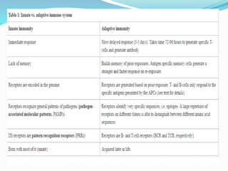
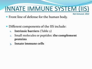
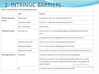
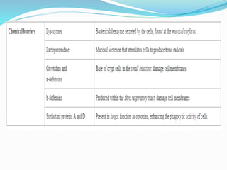
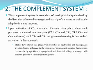
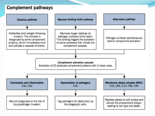
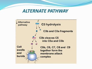
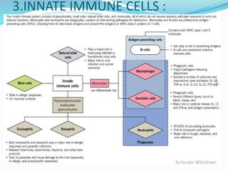
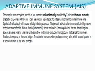
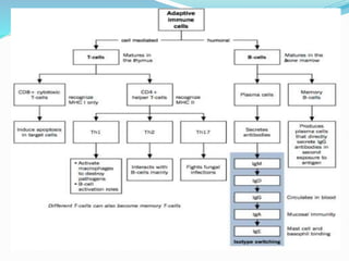
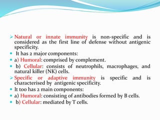
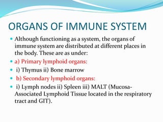
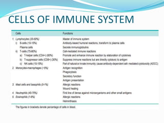
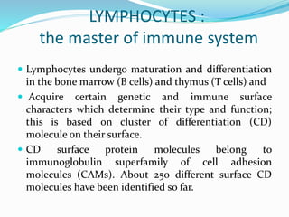
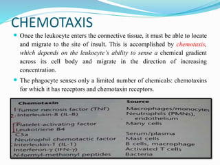
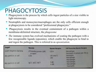
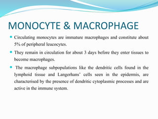
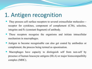
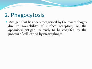
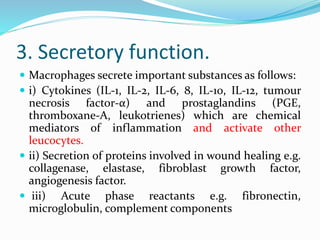
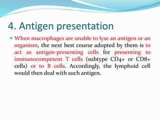
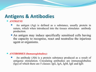
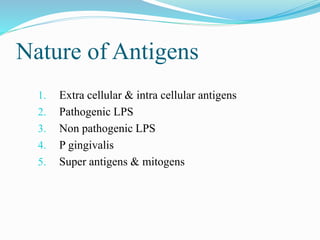
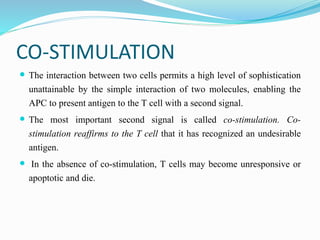
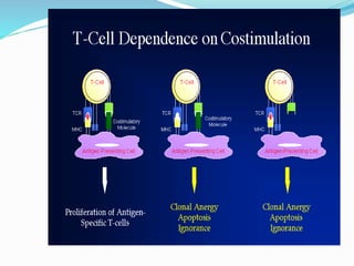
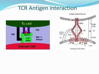
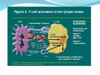
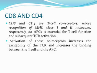
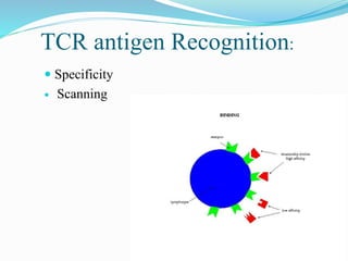
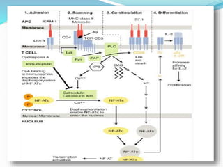
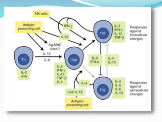
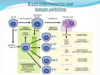
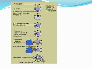
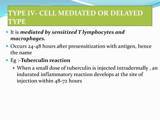
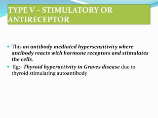
SlideShare a Scribd company logo
PRESENTED BY:
MANPREET KAUR BEHL
MDS STUDENT
INTRODUCTION
•Immunity is a biological term that describes a state of
having sufficient biological defences to avoid infection,
disease or other unwanted biological invasion.
Immunity involves both specific and non-specific
components.
•It is defined as the resistance of a host to
pathogens and their toxic products.
HISTORICAL BACKGROUND
 The prehistoric view of disease was that it was caused
by supernatural forces, and that illness was a form of
punishment for “bad deeds”.
 Between the time of Hippocrates and the 19th century,
diseases were attributed to an alteration or imbalance
in one of the four humors (blood, phlegm, yellow bile
or black bile).
•Miasma Theory
•Chinese physicians in the eleventh century observed that
the inhalation of small pox crusts presented the
subsequent occurrence of the disease.
 However, it was with Louis Pasteur’s Germ theory of
disease that the fledgling science of immunology began to
explain how bacteria caused disease, and how, following
infection, the human body gained the ability to resist
further insults.
 Further development of preventive immunization were
made possible by Louis Pasteur (1881), who coined the term
“vaccine”.
 Later Robert Koch discovered the tubercle bacillus and
developed his studies of bacterial etiology of infectious
diseases.
 E. Metchin Koff (1880’s) elucidated the importance of
phagocytosis by leukocytes.
 Paul Ehrlich (1908) proposed the humoral theory of
antibody formation.
INNATE IMMUNE SYSTEM (IIS)
Nat Immunol. 2012
 Front line of defense for the human body.
 Different components of the IIS include:
1. Intrinsic barriers (Table 2)
2. Small molecules or peptides: the complement
proteins
3. Innate immune cells
1. INTRINSIC BARRIERS :
2. THE COMPLEMENT SYSTEM :
 The complement system is comprised of small proteins synthesized by
the liver that enhance the strength and activity of an innate as well as the
adaptive immune response.
 Upon activation of C3, a cascade of events takes place where each
precursor is cleaved into two parts (C3 à C3a and C3b, C4 à C4a and
C4b and so on) until C9a and C9b are generated (naming is due to their
activation in the sequence).
 Studies have shown that phagocytic properties of neutrophils and macrophages
are significantly enhanced in the presence of complement proteins. Furthermore,
chemotaxis by cytokines is upregulated and bacterial killing is stronger with
different proteins of the complement system.
ALTERNATE PATHWAY
3.INNATE IMMUNE CELLS :
ADAPTIVE IMMUNE SYSTEM (AIS)
 Natural or innate immunity is non-specific and is
considered as the first line of defense without antigenic
specificity.
 It has 2 major components:
 a) Humoral: comprised by complement.
 b) Cellular: consists of neutrophils, macrophages, and
natural killer (NK) cells.
 Specific or adaptive immunity is specific and is
characterised by antigenic specificity.
 It too has 2 main components:
 a) Humoral: consisting of antibodies formed by B cells.
 b) Cellular: mediated by T cells.
ORGANS OF IMMUNE SYSTEM
 Although functioning as a system, the organs of
immune system are distributed at different places in
the body. These are as under:
 a) Primary lymphoid organs:
 i) Thymus ii) Bone marrow
 b) Secondary lymphoid organs:
 i) Lymph nodes ii) Spleen iii) MALT (Mucosa-
Associated Lymphoid Tissue located in the respiratory
tract and GIT).
CELLS OF IMMUNE SYSTEM
LYMPHOCYTES :
the master of immune system
 Lymphocytes undergo maturation and differentiation
in the bone marrow (B cells) and thymus (T cells) and
 Acquire certain genetic and immune surface
characters which determine their type and function;
this is based on cluster of differentiation (CD)
molecule on their surface.
 CD surface protein molecules belong to
immunoglobulin superfamily of cell adhesion
molecules (CAMs). About 250 different surface CD
molecules have been identified so far.
T-lymphocytes (T-cell) :
 Produced in the bone marrow but mature in the thymus.
 Have a unique antigen receptor, generated during development in the thymus.
 This T-cell receptor (TCR) recognizes a specific peptide sequences bound to the major
histocompatibility complex (MHC) on APCs and the infected cells. This TCR and MHC
interaction allows T-cells to activate and proliferate.
 MHC is a cell surface molecule that mediates interaction of immune cells with other immune
cells or body cells. In humans, MHC is also called human leukocyte antigen (HLA).
 Each person inherits up to 6 class I molecules and 12 class II molecules. Each different
type of HLA molecule binds a different antigen sequence for presentation.
 MHC class I molecules are found on all nucleated cells of the body. They present
cytosolic protein sequences, and cytotoxic T cells are main responding cell.
 In contrast, MHC class II molecules are only found on lymphoid tissue cells. They
present proteins found in lysosomes and endosomes or extracellular pathogens, and T
helper cells are the responding cells.
T-CELL
 These cells are implicated in inciting cell-mediated
immunity and delayed type of hypersensitivity.
 T cells in circulation comprise 75-80% of
lymphocytes.
 Pan T cell markers are CD3, CD7 and CD2. Besides, T
cells also carry receptor (TCR) for recognition of MHC
molecules.
 Depending upon functional activity, T cells have two
major subtypes: T helper cells and T suppressor cells.
T cells
TH0
TH1
 TH cells – CD4 Positive TH2
TH3
TH17
TC1
 TC cells – CD8 Positive TC2
 Double Negative T cells – CD4 Negative CD8 Negative
 NK T Cells
 Regulatory T Cells
T-HELPER CELL
 Abbreviated as TH cells,
 these cells promote and enhance the immune reaction and are also termed as T-
regulatory cells.
 They carry CD4 molecule on their surface and hence are also called CD4+ cells.
CD4+ cells in circulation are about twice the number of CD8+ cells (CD4+/CD8 ratio
2:1).
 These cells act by elaboration of variety of cytokines.
 Depending upon the type of cytokines elaborated, these TH cells are further of two
subclasses:
 TH 1 and TH 2.
 TH 1 cells elaborate IL-2 and interferon (IFN)-γ.
 TH 2 cells elaborate IL-4, IL-5, IL-6, and IL-10.
 CD4+ cells are predominantly involved in cell-mediated reactions to viral infections
(e.g. in HIV), tissue transplant reactions and tumour lysis.
Effector functions of TH cells:
TH cell activation and maturation will lead to release of
various cytokine and that can lead to:
1. B cell differentiation to plasma cell (TH2 response)
2. Activation of TC cells (TH1 response)
3. Activation or suppression of other cells such as
macrophages and NK cells.
T-SUPPRESSOR CELLS
 Abbreviated as TS cells,
 they suppress immune reactions but are cytotoxic and actually destroy the
invading antigen; hence are also termed as cytotoxic T lymphocytes (CTL).
 These cells carry CD8 molecule on their surface and hence are also called
CD8+ cells.
 CD8+ cells in circulation are about half the number of CD4+ cells.
 Compared to CD4+ cells which act by elaboration of cytokines, CD8+ cells
are directly cytotoxic to the antigen.
 CD8+ cells are particularly involved in destroying cells infected with viruses,
foreign cells and tumour cells.
NK CELLS
 NK cells comprise about 10-15% of circulating lymphocytes.
 These lymphocytes do not have B or T cell markers, nor are these cells
dependent upon thymus for development unlike CD4+ and CD8+ T cells.
 NK cells carry surface molecules of CD2, CD16 and CD56, but negative for T
cell marker CD3.
 NK cells are morphologically distinct from B and T cells in being large
granular lymphocytes.
 NK cells are part of the natural or innate immunity. These cells recognise
antibody-coated target cells and bring about killing of the target directly; this
process is termed as antibody dependent cell-mediated cytotoxicity (ADCC).
 This mechanism is particularly operative against viruses and tumour cells
B-lymphocytes (B cells):
 Produced and mature in the bone marrow.
 Play the lead role in humoral immunity and mainly functions to produce various types of
antibody. Several different types of B-cells exist but only the major ones are discussed
here: memory B-cells and plasma cells.
 Memory cells are formed from the activated B-cells specific for a certain antigen
encountered during an active immune response. These cells remain in the circulation for
long periods of time and respond immediately upon the exposure of the same antigen.
 Plasma cells produce and secrete copious amounts of various antibodies. Each plasma cell
is specific for a specific antibody (one plasma cell cannot secrete all antibodies).
 B-cell activation can be T-cell dependent or it can also be T-cell independent. Since MHC can
present peptide sequences only, carbohydrate sequences directly bind B-cells and cross-link
antibody receptors (antibodies can act as B-cell receptors also) leading to B-cell activation.
 While B cells differentiate into plasma cells which form specific
antibodies.
 T cells get functionally activated on coming in contact with appropriate
antigen. Upon coming in contact with antigen, it is the macrophage, i.e.
specialised antigen-presenting cell such as dendritic cell, and the major
histocompatibilty complex (MHC) in the macrophage, which
determines whether the invading antigen is to be presented to B cells or
T cells. Some strong antigens that cannot be dealt by antibody response
from B cells such as certain microorganisms (e.g. viruses, mycobacteria
M. tuberculosis and M. leprae), cancer cells, tissue transplantation
antigen etc, are presented to T cells.
B-CELL
 On coming in contact with antigen (e.g. invading microorganims), B
cells are activated to proliferate and transform into plasmacytoid
lymphocytes and then into plasma cells.
 Depending upon the maturation stage of B cells, specific CD molecules
appear on the cell surface which can be identified by CD markers;
common B cell markers include: CD 19, 20, 21, 23.
 These cells also possess B cell receptors (BCR) for surface
immunoglobulins (IgM and IgG) and Fc receptor for attaching to
antibody molecule. T cell help is provided to B cells by a subset of T
helper cells, TH 2, by elaborated interleukins (IL-4, IL-5, IL-10, IL-13).
LEUKOCYTE FUNCTIONS:
 CHEMOTAXIS
 PHAGOCYTOSIS
 KILLING
 ANTIGEN PROCESSING & PRESENTATION
CHEMOTAXIS
 Once the leukocyte enters the connective tissue, it must be able to locate
and migrate to the site of insult. This is accomplished by chemotaxis,
which depends on the leukocyte’s ability to sense a chemical gradient
across its cell body and migrate in the direction of increasing
concentration.
 The phagocyte senses only a limited number of chemicals: chemotaxins
for which it has receptors and chemotaxin receptors.
PHAGOCYTOSIS Phagocytosis is the process by which cells ingest particles of a size visible to
light microscopy.
 Neutrophils and monocytes/macrophages are the only cells efficient enough
at phagocytosis to be considered “professional phagocytes.”
 Phagocytosis results in the eventual containment of a pathogen within a
membrane-delimited structure, the phagosome
 The immune system has evolved mechanisms of coating the pathogen with a
few recognizable ligands (opsonins), which enable the phagocyte to bind to
and ingest the pathogen. This is referred to as opsonization.
 Phagocytes kill bacteria through two broad categories
of killing mechanisms.
 One category is based on the reduction of oxygen and
is referred to as “oxidative.”
 Oxidative mechanisms require
 (1) the presence of oxygen and
 (2) an oxidationreduction potential, Eh, at or above -
160 mV.
 Microbicidal activity occurs by forming toxic, reduced-oxygen
metabolites such as superoxide anion (O2-) using the NADPH oxidase
system.
 The superoxide anion also contributes to the formation of hydrogen
peroxide (H2O2), which is capable of diffusing across membranes.
 In the presence of H2O2 and chloride, MPO catalyzes the formation of
hypochlorous acid (HOCl).
 Second category of killing mechanisms, the nonoxidative mechanisms.
 It requires phagosome-lysosome fusion, phagolysosome.
 It results in the secretion of lysosomal components into the phagolysosome.
 Less than 30 seconds after phagocytosis, neutrophils secrete specific granule
components into the phagolysosome.
 Specific granules contain several microbiocidal components, including
lysozyme and lactoferrin.
 Lysozyme is an enzyme that possesses enzymedependent bactericidal activity
and enzyme-independent bactericidal and fungicidal activity.
 Lactoferrin is a bacteriostatic compound that contains a bactericidal peptide
domain, lactoferricin.
MONOCYTE & MACROPHAGE
 Circulating monocytes are immature macrophages and constitute about
5% of peripheral leucocytes.
 They remain in circulation for about 3 days before they enter tissues to
become macrophages.
 The macrophage subpopulations like the dendritic cells found in the
lymphoid tissue and Langerhans’ cells seen in the epidermis, are
characterised by the presence of dendritic cytoplasmic processes and are
active in the immune system.
Salient features and
important immune
functions of macrophages
1. Antigen recognition
 They possess cell surface receptors to several extracellular molecules—
receptor for cytokines, component of complement (C3b), selectins,
integrins and Fc (constant fragment) of antibody.
 These receptors recognise the organisms and initiate intracellular
mechanism in macrophages.
 Antigen to become recognisable can also get coated by antibodies or
complement, the process being termed as opsonisation.
 Macrophages have capacity to distinguish self from non-self by
presence of human leucocyte antigens (HLA) or major histocompatibilty
complex (MHC).
2. Phagocytosis
 Antigen that has been recognised by the macrophages
due to availability of surface receptors, or the
opsonised antigen, is ready to be engulfed by the
process of cell-eating by macrophages
3. Secretory function.
 Macrophages secrete important substances as follows:
 i) Cytokines (IL-1, IL-2, IL-6, 8, IL-10, IL-12, tumour
necrosis factor-α) and prostaglandins (PGE,
thromboxane-A, leukotrienes) which are chemical
mediators of inflammation and activate other
leucocytes.
 ii) Secretion of proteins involved in wound healing e.g.
collagenase, elastase, fibroblast growth factor,
angiogenesis factor.
 iii) Acute phase reactants e.g. fibronectin,
microglobulin, complement components
4. Antigen presentation
 When macrophages are unable to lyse an antigen or an
organism, the next best course adopted by them is to
act as antigen-presenting cells for presenting to
immunocompetent T cells (subtype CD4+ or CD8+
cells) or to B cells. Accordingly, the lymphoid cell
would then deal with such antigen.
Antigens & Antibodies
 ANTIGENS
 An antigen (Ag) is defined as a substance, usually protein in
nature, which when introduced into the tissues stimulates antibody
production.
 An antigen may induce specifically sensitised cells having
the capacity to recognise, react and neutralise the injurious
agent or organisms.
 ANTIBODIES (Immunoglobulins):
 An antibody (Ab) is a protein substance produced as a result of
antigenic stimulation. Circulating antibodies are immunoglobulins
(Igs) of which there are 5 classes: IgG, IgA, IgM, IgE and IgD.
Nature of Antigens
1. Extra cellular & intra cellular antigens
2. Pathogenic LPS
3. Non pathogenic LPS
4. P gingivalis
5. Super antigens & mitogens
Antigen Presentation:
Antigen Processing and Presentation
 MHC is a locus on short arm of chromosome 6 (6p21.3)
that encodes MHC classes I,II, III, which are involved in
antigen uptake, processing and presentation.
 MHC class I molecules: present intracellular antigens to
CD8+ T cells and NK cells.
 MHC class II molecules: present extracellular antigens to
CD4+ Tcells.
 MHC class III molecules include complement factors B,
C2 and C4.
Antigen Presenting Cells
Includes B cells, macrophages, dermal dendritic cells
and Langerhan cells
These cells naturally express MHC-II molecules to
activate specific effector T cells.
Generate antigen specific immune response to
periodontal pathogens.
HLA SYSTEM AND
MAJOR HISTOCOMPATIBILITY
COMPLEX
 HLA stands for Human Leucocyte Antigens because these antigens or
genetic proteins in the body which determine one’s own tissue from
non-self (histocompatibility) were first discovered on the surface of
leucocytes.
 Subsequently, it was found that HLA are actually gene complexes of
proteins on the surface of all nucleated cells of the body and platelets.
 Since these complexes are of immense importance in matching donor
and recipient for organ transplant, they are called major
histocompatibility complex (MHC) or HLA complex
 Out of various genes for histocompatibility,
most of the transplantation antigens or MHC
are located on a portion of chromosome 6 of
all nucleated cells of the body and platelets.
 These genes occupy four regions or loci—A,
B, C and D, on the short (p) arm of
chromosome 6 and exhibit marked variation in
allelic genes at each locus.
 Therefore, the product of HLA antigens is
highly polymorphic.
 HLA system is part of immunoglobulin
superfamily of CAMs.
3 CLASSES OF HLA :
 Class I MHC antigens have loci as HLA-A, HLA-B and HLA-C. CD8+ (i.e. T
suppressor) lymphocytes carry receptors for class I MHC and these cells are used to
identify class I antigen on them.
 Class II MHC antigens have single locus as HLA-D. These antigens have further 3
loci: DR, DQ and DP.
 Class II MHC is identified by B cells and CD4+ (i.e. T helper) cells.
 Class III MHC antigens are some components of the complement system (C2 and C4)
coded on HLA complex but are not associated with HLA expression and are not used
in antigen identification.
 In view of high polymorphism of class I and class II genes, they have a number of
alleles on loci numbered serially like HLA-A 1, HLA-A 2, HLA-A 3 etc.
 MHC antigens present on the cell surface help the macrophage in its function of
bacterial antigen recognition i.e. they help to identify self from foreign, and
accordingly present the foreign antigen to T cells (CD4+ or CD8+) or to B cells.
 MHC classes I, II, and III molecules, which are involved with antigen uptake,
processing, and presentation.
 All cells process and present self derived antigens (intracellular antigens) in
association with MHC class I molecules.
 MHC class I molecules are used to present intracellular antigens to CD8+ T cells and
NK cells.
 MHC class III molecules include complement factors B, C2, and C4.
 Antigens derived from extracellular sources are presented by professional antigen-
presenting cells (APCs) in association with MHC class II molecules.
 The three main professional APCs are
 peripheral DCs,
 monocyte derivatives, and
 B cells.
CO-STIMULATION
 The interaction between two cells permits a high level of sophistication
unattainable by the simple interaction of two molecules, enabling the
APC to present antigen to the T cell with a second signal.
 The most important second signal is called co-stimulation. Co-
stimulation reaffirms to the T cell that it has recognized an undesirable
antigen.
 In the absence of co-stimulation, T cells may become unresponsive or
apoptotic and die.
Activation – Signal transduction
Co-stimulation – B71 & B72 interaction
with CD28.
TOLL LIKE RECEPTORS
 A receptor molecule named “toll,” first identified in fruit flies
(Drosophila spp.), was shown to be important in certain responses to
injury or infection.
 The human toll-like receptors (TLRs) are stimulated by highly
conserved bacterial components such as LPS and are important in
dictating the adaptations found in the innate immune system.
 TLRs cause APCs to upregulate the co-stimulatory B7 molecules.
Although T cells may constantly interact with antigen, co-stimulation
enables this interaction to progress to T-cell proliferation.
SPECIFIC IMMUNE RESPONSES:
 Chronic inflammation, if protracted, can result in an adaptation called the
specific immune response.
 The specific immune response requires lymphocytes, which use two types of
receptors, to generate
specific immune responses, the B-cell antigen receptor (BCR) and the T-cell
antigen receptor (TCR).
 Four phases are involved in the generation of specific immunity:
 (1) clonal selection, the selection of lymphocytes that bear receptors (BCRs or
TCRs) recognizing the specific antigen;
 (2) clonal expansion, the proliferation of those lymphocytes;
 (3) clonal contraction, the death of “effector” lymphocytes; and
 (4) memory, the maintenance of an expanded clone of cells that bears the
specific receptors (BCRs or TCRs) recognizing the antigen .
 As long as a sufficient number of lymphocytes are maintained to provide
protection against a specific antigen, the individual is said to be “immune.”
Protective immunity is achieved if an adequate number of the lymphocytes are maintained in the memory
phase, and the individual is said to be immune.
this does not always occur because an individual (1) may not undergo sufficient clonal expansion, (2) may
undergo excessive clonal contraction, or (3) may be unable to maintain memory.
(Modified from Ahmed R, Gray D: Science 272(5258):54, 1996.)
Clonal Selection:
Specific Immune Responses
Four phases are involved:
 Clonal Selection: selection of lymphocytes that bear
receptors (BCR’s or TCR’s) recognizing the specific antigen.
 Clonal Expansion: proliferation of those lymphocytes.
 Clonal contraction: death of “effector” lymphocytes.
 Memory: the maintainence of an expanded clone of cells.
T-CELL RESPONSES
 T cells may express 3000 to 50,000 TCRs on their
surface.
 Antigens are presented to the TCR by MHC class I or II
molecules on the APC.
 The TCR recognizes and binds the MHC-peptide
complex.
 Antigen (Ag) is contacted by the TCR using variable
domains found at the Nterminus of the TCRα and β
subunits.
TCR Antigen interaction
CD8 AND CD4
 CD8 and CD4 are T-cell co-receptors, whose
recognition of MHC class I and II molecules,
respectively, on APCs is essential for T-cell function
and subsequent TCR activation.
 Activation of these co-receptors increases the
excitability of the TCR and increases the binding
between the T cell and the APC.
 The low affinity of the TCR enables the T cell to bind
APCs in a reversible manner, which occurs between
multiple TCRs and one or a few antigens over time.
 This time-dependent interaction of many TCRs with a
few antigens is referred to as scanning.
 Scanning that leads to T-cell activation is called serial
triggering.
 To fully activate T-cells, multiple TCR engagement
must be sustained for 2 to 20 hours.
TCR antigen Recognition:
 Specificity
 Scanning
ACTIVATION OF T-CELL
proliferative differentiation.
begins with activation of the
protein tyrosine kinases lck, TCR
fyn, PLC
ZAP CD28
Activation of CD28
prepares the T cell to
receive the
costimulatory
signals, which are
important in T-cell
survival and function
Diacyl
glycerol
(DAG)
1,4,5-
inositol
triphosph
ate (IP3).
DAG signals the
activation of a
transcription-
activating
cofactor, NF-ATn
Ca+2
activation of a
calmodulin-
calcineurin
A/B
phosphatase.
dephosp
horylates
NF-ATc
active nuclear factor of activated T
cells
(NF-AT).
upregulates transcription of
genes, such as those for interleukin-2 (IL-2) and
the IL-2 receptor α subunit (IL-2Rα),
stimulate
proliferative differentiation
B-cell Differentiation and
Isotype switching
ROLE OF HLA COMPLEX
 1. Organ transplantation. The greater the genetic disparity between
donor and recipient in HLA system, the stronger and more rapid will be
the rejection reaction.
 2. Regulation of the immune system.
 Class I and II histocompatibility antigens play a role in regulating both
cellular and humoral immunity:
 Class I MHC antigens regulate the function of cytotoxic T cells (CD8+
subpopulation) e.g. in virus infections.
 Class II MHC antigens regulate the function of helper T cells (CD4+
subpopulation).
3. Association of diseases with HLA.
An increasing number of diseases have been found to have association
with some specific histocompatibility antigens. These disorders include
the following:
i) Inflammatory disorders e.g. ankylosing spondylitis.
ii) Autoimmune disorders e.g. rheumatoid arthritis, insulindependent
diabetes mellitus.
iii) Inherited disorders of metabolism e.g. idiopathic haemochromatosis.
Basophils and Mast Cells
 Basophils are a type of circulating granulocytes (0-1%) while
mast cells are their counterparts seen in tissues, especially in
connective tissue around blood vessels and in submucosal
location.
 Basophils and mast cells have IgE surface receptor; thus on
coming in contact with antigen binding to IgE (e.g. allergic
reaction to parasites), these cells get activated and release
granules i.e. degranulate.
 These granules contain substances such as: histamine, platelet
activating factor, heparin and certain chemical mediators (e.g.
prostaglandins, leukotrienes).
 Mast cells and basophils are thus involved in mediating
inflammation in allergic reactions and have a role in wound
healing
Neutrophils
 Polymorphonuclear neutrophils (PMNs) are normally the most
numerous of the circulating leucocytes (40-75%).
 The cytoplasm of PMNs contains lysosomal granules of three types:
primary (azurophilic), secondary, and tertiary.
 PMNs have similar function to those of macrophages and are therefore
appropriately referred to as ‘microphages’ owing to their role as first
line of defense against an invading foreign organism in the body.
 However, these cells have limitation of size and type of organisms to be
engulfed e.g. while they are capable of acting against bacteria and small
foreign particulate material but not against viruses and large particles.
Eosinophils
 Eosinophils are also circulating granulocytes (1-6%).
 These cells play a role in allergic reactions and in
intestinal helminthiasis.
 The granules of eosinophils contain lysosomal
enzymes, peroxidases, and chemical mediators of
inflammation (e.g. prostaglandins, leukotrienes).
 On coming in contact with IgE opsonised antigen
(e.g. helminths), eosinophils degranulate and release
the chemicals stored in granules and incite
inflammation
Hypersensitivity
 It is defined as state of exaggerated immune
response to an antigen.
 The lesions of hypersensitivity are produced due
to interaction between antigen and products
of immune response.
HYPERSENSITIVITY REACTIONS
TYPE I – Anaphylactic type
TYPE II – Cytotoxic type
TYPE III – Immune complex type
TYPE IV – Cell mediated or delayed type
TYPE V- Stimulatory or antireceptor
TYPE I – Anaphylactic type
 It is defined as state of rapid developing immune response to
an antigen to which the individual is exposed.
 It is mediated by IgE antibody and is due to powerful effects of
histamine.
 It may be local / general depending upon the amount of histamine
released.
 Local :- hay fever, bronchial asthma
 Systemic :- bee stings, penicillin, horse serum
TYPE II – Cytotoxic type
 These are defined as reactions which cause injury to
the cell by combining antibodies with cell surface
antigens.
 IgG and IgM involved
 Eg :-
• Mismatched transfusion
reaction
• Autoimmune haemolytic
anaemia
• Erythroblastosis foetalis
TYPE III – Immune complex type
 These result from formation of immune complexes
by direct antigen antibody combination, causing
cell injury.
 The formation of antigen-antibody complexes in
the serum , followed by deposition in tissues, is the
key event in causing these reactions .
 2 types of reactions occur:
a) Local: Arthus reaction- when rabbits were repeatedly
injected subcutaneously with horse serum, the initial
injections were without any local effect, but later injections
caused local oedema, induration and haemorrhage.
a) Systemic :Serum sickness-seen in persons who receive high
dose of horse antitoxin against tetanus, gas gangrene
After 7-12 days, there occurs fever,
lymphadenopathy, splenomegaly,
glomerulonephritis, endocarditis, urticarial
rash.
TYPE IV- CELL MEDIATED OR DELAYED
TYPE
 It is mediated by sensitized T lymphocytes and
macrophages.
 Occurs 24-48 hours after presensitization with antigen, hence
the name
 Eg :-Tuberculin reaction
 When a small dose of tuberculin is injected intradermally , an
indurated inflammatory reaction develops at the site of
injection within 48-72 hours
TYPE V – STIMULATORY OR
ANTIRECEPTOR
 This an antibody mediated hypersensitivity where
antibody reacts with hormone receptors and stimulates
the cells.
 Eg:- Thyroid hyperactivity in Graves disease due to
thyroid stimulating autoantibody
IMMUNITY : the basic concepts

More Related Content

What's hot

Immune response
Immune responseImmune response
Immune response
raghunathp
 
Basic immunology
Basic immunologyBasic immunology
Basic immunology
Hema Duddukuri
 
B-cell and humoral immunity
B-cell and humoral immunityB-cell and humoral immunity
B-cell and humoral immunity
Santosh Kumar Yadav
 
ANTIGEN PRESENTING CELL
ANTIGEN PRESENTING CELLANTIGEN PRESENTING CELL
ANTIGEN PRESENTING CELL
Dr Sreelakshmi
 
Cells of immune response
Cells of immune responseCells of immune response
Cells of immune response
Esraa Gamal
 
Immunology
ImmunologyImmunology
Immunology
Aditi Singh
 
Mhc
MhcMhc
Innate Immunity
Innate ImmunityInnate Immunity
Antigens, hapteins, immunogens lectures 10.1.06
Antigens, hapteins, immunogens lectures 10.1.06Antigens, hapteins, immunogens lectures 10.1.06
Antigens, hapteins, immunogens lectures 10.1.06Bruno Mmassy
 
Immunology 1, 2, 3
Immunology 1, 2, 3Immunology 1, 2, 3
Immunology 1, 2, 3
Ahmed Elshebiny
 
Antigen antibody interaction
Antigen antibody interactionAntigen antibody interaction
Antigen antibody interaction
Bipul Das
 
Autoimmunity and Tolerance
Autoimmunity and ToleranceAutoimmunity and Tolerance
Autoimmunity and Tolerance
Atifa Ambreen
 
Introduction to immunology
Introduction to immunologyIntroduction to immunology
Introduction to immunology
promotemedical
 
Antigen
AntigenAntigen
Antigen
Natasha Sayal
 
Cell mediated immunity
Cell mediated immunityCell mediated immunity
Cell mediated immunity
Garima Thakur
 
Immunological tolerance
Immunological toleranceImmunological tolerance
Tumor immunology
Tumor immunologyTumor immunology
Tumor immunology
Promila Sheoran
 
Immunity - The basic concept
Immunity - The basic conceptImmunity - The basic concept
Immunity - The basic concept
Dr Sudeep Madhusudan Chaudhari
 
Adaptive immunity
Adaptive immunity  Adaptive immunity
Adaptive immunity
Abdullah Abobakr
 

What's hot (20)

Immune response
Immune responseImmune response
Immune response
 
Basic immunology
Basic immunologyBasic immunology
Basic immunology
 
B-cell and humoral immunity
B-cell and humoral immunityB-cell and humoral immunity
B-cell and humoral immunity
 
ANTIGEN PRESENTING CELL
ANTIGEN PRESENTING CELLANTIGEN PRESENTING CELL
ANTIGEN PRESENTING CELL
 
Cells of immune response
Cells of immune responseCells of immune response
Cells of immune response
 
Immunology
ImmunologyImmunology
Immunology
 
Mhc
MhcMhc
Mhc
 
Innate Immunity
Innate ImmunityInnate Immunity
Innate Immunity
 
Antigens, hapteins, immunogens lectures 10.1.06
Antigens, hapteins, immunogens lectures 10.1.06Antigens, hapteins, immunogens lectures 10.1.06
Antigens, hapteins, immunogens lectures 10.1.06
 
Immunology 1, 2, 3
Immunology 1, 2, 3Immunology 1, 2, 3
Immunology 1, 2, 3
 
Antigen antibody interaction
Antigen antibody interactionAntigen antibody interaction
Antigen antibody interaction
 
Autoimmunity and Tolerance
Autoimmunity and ToleranceAutoimmunity and Tolerance
Autoimmunity and Tolerance
 
Introduction to immunology
Introduction to immunologyIntroduction to immunology
Introduction to immunology
 
Antigen
AntigenAntigen
Antigen
 
Basic Immunology
Basic ImmunologyBasic Immunology
Basic Immunology
 
Cell mediated immunity
Cell mediated immunityCell mediated immunity
Cell mediated immunity
 
Immunological tolerance
Immunological toleranceImmunological tolerance
Immunological tolerance
 
Tumor immunology
Tumor immunologyTumor immunology
Tumor immunology
 
Immunity - The basic concept
Immunity - The basic conceptImmunity - The basic concept
Immunity - The basic concept
 
Adaptive immunity
Adaptive immunity  Adaptive immunity
Adaptive immunity
 

Similar to IMMUNITY : the basic concepts

Immunology
ImmunologyImmunology
1.7...............humoral & cell mediated immunity.pdf
1.7...............humoral & cell mediated immunity.pdf1.7...............humoral & cell mediated immunity.pdf
1.7...............humoral & cell mediated immunity.pdf
DarHanan
 
Cell mediated immunity
Cell mediated immunityCell mediated immunity
Cell mediated immunity
Amen Ullah
 
cellmediatedimmunity-181225200148.pdf
cellmediatedimmunity-181225200148.pdfcellmediatedimmunity-181225200148.pdf
cellmediatedimmunity-181225200148.pdf
AnukrittiMehra
 
Basic immunology from the dermatologic point of view (adaptive immunity)
Basic immunology from the dermatologic point of view (adaptive immunity)Basic immunology from the dermatologic point of view (adaptive immunity)
Basic immunology from the dermatologic point of view (adaptive immunity)
Hima Farag
 
BRANCHES OF ADAPTIVE IMMUNITY.docx
BRANCHES OF ADAPTIVE IMMUNITY.docxBRANCHES OF ADAPTIVE IMMUNITY.docx
BRANCHES OF ADAPTIVE IMMUNITY.docx
MARGARET OTEYO
 
Adaptive Immune Response to Viral Infections
Adaptive Immune Response to Viral InfectionsAdaptive Immune Response to Viral Infections
Adaptive Immune Response to Viral Infections
Mousumi Bora
 
Lymphocytic cells involved in human immune system
Lymphocytic cells involved in human immune systemLymphocytic cells involved in human immune system
Lymphocytic cells involved in human immune system
Abhay jha
 
Adaptive immunity.pdf
Adaptive immunity.pdfAdaptive immunity.pdf
Adaptive immunity.pdf
Dr Sitaram Swain
 
Activation of T and B Cells
Activation of T and B CellsActivation of T and B Cells
Activation of T and B Cells
Ambika Prajapati
 
12 cellular basis of imune response
12 cellular basis of imune response12 cellular basis of imune response
12 cellular basis of imune response
Dr UAK
 
CELL AND HUMORAL immunity 1.pptx
CELL AND HUMORAL  immunity 1.pptxCELL AND HUMORAL  immunity 1.pptx
CELL AND HUMORAL immunity 1.pptx
PRASHANT YADAV
 
Lecture 4 immunobiology
Lecture 4 immunobiologyLecture 4 immunobiology
Lecture 4 immunobiologyBruno Mmassy
 
ADAPTIVE IMMUNITY T CELLS.pptx
ADAPTIVE IMMUNITY T CELLS.pptxADAPTIVE IMMUNITY T CELLS.pptx
ADAPTIVE IMMUNITY T CELLS.pptx
BinteHawah1
 
adaptiveimmunitytcells-2023.pdf
adaptiveimmunitytcells-2023.pdfadaptiveimmunitytcells-2023.pdf
adaptiveimmunitytcells-2023.pdf
SherzadMajeed1
 
Immunity and its cells, HLA and transplant rejection
Immunity and its cells, HLA and transplant rejectionImmunity and its cells, HLA and transplant rejection
Immunity and its cells, HLA and transplant rejection
Juliya Susan Reji
 
Adaptive immunity - 2023.pptx
Adaptive immunity - 2023.pptxAdaptive immunity - 2023.pptx
Adaptive immunity - 2023.pptx
SherzadMajeed1
 
Key concept of immunology
Key concept of immunologyKey concept of immunology
Key concept of immunology
Dr Alok Tripathi
 
Adaptive immunity - 2023.pptx
Adaptive immunity - 2023.pptxAdaptive immunity - 2023.pptx
Adaptive immunity - 2023.pptx
SherzadMajeed1
 
cellular and humoral components of immune System.vinu.pptx
cellular and humoral components of immune System.vinu.pptxcellular and humoral components of immune System.vinu.pptx
cellular and humoral components of immune System.vinu.pptx
GokulVV3
 

Similar to IMMUNITY : the basic concepts (20)

Immunology
ImmunologyImmunology
Immunology
 
1.7...............humoral & cell mediated immunity.pdf
1.7...............humoral & cell mediated immunity.pdf1.7...............humoral & cell mediated immunity.pdf
1.7...............humoral & cell mediated immunity.pdf
 
Cell mediated immunity
Cell mediated immunityCell mediated immunity
Cell mediated immunity
 
cellmediatedimmunity-181225200148.pdf
cellmediatedimmunity-181225200148.pdfcellmediatedimmunity-181225200148.pdf
cellmediatedimmunity-181225200148.pdf
 
Basic immunology from the dermatologic point of view (adaptive immunity)
Basic immunology from the dermatologic point of view (adaptive immunity)Basic immunology from the dermatologic point of view (adaptive immunity)
Basic immunology from the dermatologic point of view (adaptive immunity)
 
BRANCHES OF ADAPTIVE IMMUNITY.docx
BRANCHES OF ADAPTIVE IMMUNITY.docxBRANCHES OF ADAPTIVE IMMUNITY.docx
BRANCHES OF ADAPTIVE IMMUNITY.docx
 
Adaptive Immune Response to Viral Infections
Adaptive Immune Response to Viral InfectionsAdaptive Immune Response to Viral Infections
Adaptive Immune Response to Viral Infections
 
Lymphocytic cells involved in human immune system
Lymphocytic cells involved in human immune systemLymphocytic cells involved in human immune system
Lymphocytic cells involved in human immune system
 
Adaptive immunity.pdf
Adaptive immunity.pdfAdaptive immunity.pdf
Adaptive immunity.pdf
 
Activation of T and B Cells
Activation of T and B CellsActivation of T and B Cells
Activation of T and B Cells
 
12 cellular basis of imune response
12 cellular basis of imune response12 cellular basis of imune response
12 cellular basis of imune response
 
CELL AND HUMORAL immunity 1.pptx
CELL AND HUMORAL  immunity 1.pptxCELL AND HUMORAL  immunity 1.pptx
CELL AND HUMORAL immunity 1.pptx
 
Lecture 4 immunobiology
Lecture 4 immunobiologyLecture 4 immunobiology
Lecture 4 immunobiology
 
ADAPTIVE IMMUNITY T CELLS.pptx
ADAPTIVE IMMUNITY T CELLS.pptxADAPTIVE IMMUNITY T CELLS.pptx
ADAPTIVE IMMUNITY T CELLS.pptx
 
adaptiveimmunitytcells-2023.pdf
adaptiveimmunitytcells-2023.pdfadaptiveimmunitytcells-2023.pdf
adaptiveimmunitytcells-2023.pdf
 
Immunity and its cells, HLA and transplant rejection
Immunity and its cells, HLA and transplant rejectionImmunity and its cells, HLA and transplant rejection
Immunity and its cells, HLA and transplant rejection
 
Adaptive immunity - 2023.pptx
Adaptive immunity - 2023.pptxAdaptive immunity - 2023.pptx
Adaptive immunity - 2023.pptx
 
Key concept of immunology
Key concept of immunologyKey concept of immunology
Key concept of immunology
 
Adaptive immunity - 2023.pptx
Adaptive immunity - 2023.pptxAdaptive immunity - 2023.pptx
Adaptive immunity - 2023.pptx
 
cellular and humoral components of immune System.vinu.pptx
cellular and humoral components of immune System.vinu.pptxcellular and humoral components of immune System.vinu.pptx
cellular and humoral components of immune System.vinu.pptx
 

Recently uploaded

Basavarajeeyam - Ayurvedic heritage book of Andhra pradesh
Basavarajeeyam - Ayurvedic heritage book of Andhra pradeshBasavarajeeyam - Ayurvedic heritage book of Andhra pradesh
Basavarajeeyam - Ayurvedic heritage book of Andhra pradesh
Dr. Madduru Muni Haritha
 
heat stroke and heat exhaustion in children
heat stroke and heat exhaustion in childrenheat stroke and heat exhaustion in children
heat stroke and heat exhaustion in children
SumeraAhmad5
 
Are There Any Natural Remedies To Treat Syphilis.pdf
Are There Any Natural Remedies To Treat Syphilis.pdfAre There Any Natural Remedies To Treat Syphilis.pdf
Are There Any Natural Remedies To Treat Syphilis.pdf
Little Cross Family Clinic
 
ARTIFICIAL INTELLIGENCE IN HEALTHCARE.pdf
ARTIFICIAL INTELLIGENCE IN  HEALTHCARE.pdfARTIFICIAL INTELLIGENCE IN  HEALTHCARE.pdf
ARTIFICIAL INTELLIGENCE IN HEALTHCARE.pdf
Anujkumaranit
 
New Directions in Targeted Therapeutic Approaches for Older Adults With Mantl...
New Directions in Targeted Therapeutic Approaches for Older Adults With Mantl...New Directions in Targeted Therapeutic Approaches for Older Adults With Mantl...
New Directions in Targeted Therapeutic Approaches for Older Adults With Mantl...
i3 Health
 
Non-respiratory Functions of the Lungs.pdf
Non-respiratory Functions of the Lungs.pdfNon-respiratory Functions of the Lungs.pdf
Non-respiratory Functions of the Lungs.pdf
MedicoseAcademics
 
How to Give Better Lectures: Some Tips for Doctors
How to Give Better Lectures: Some Tips for DoctorsHow to Give Better Lectures: Some Tips for Doctors
How to Give Better Lectures: Some Tips for Doctors
LanceCatedral
 
Flu Vaccine Alert in Bangalore Karnataka
Flu Vaccine Alert in Bangalore KarnatakaFlu Vaccine Alert in Bangalore Karnataka
Flu Vaccine Alert in Bangalore Karnataka
addon Scans
 
Cervical & Brachial Plexus By Dr. RIG.pptx
Cervical & Brachial Plexus By Dr. RIG.pptxCervical & Brachial Plexus By Dr. RIG.pptx
Cervical & Brachial Plexus By Dr. RIG.pptx
Dr. Rabia Inam Gandapore
 
BRACHYTHERAPY OVERVIEW AND APPLICATORS
BRACHYTHERAPY OVERVIEW  AND  APPLICATORSBRACHYTHERAPY OVERVIEW  AND  APPLICATORS
BRACHYTHERAPY OVERVIEW AND APPLICATORS
Krishan Murari
 
Lung Cancer: Artificial Intelligence, Synergetics, Complex System Analysis, S...
Lung Cancer: Artificial Intelligence, Synergetics, Complex System Analysis, S...Lung Cancer: Artificial Intelligence, Synergetics, Complex System Analysis, S...
Lung Cancer: Artificial Intelligence, Synergetics, Complex System Analysis, S...
Oleg Kshivets
 
Pharynx and Clinical Correlations BY Dr.Rabia Inam Gandapore.pptx
Pharynx and Clinical Correlations BY Dr.Rabia Inam Gandapore.pptxPharynx and Clinical Correlations BY Dr.Rabia Inam Gandapore.pptx
Pharynx and Clinical Correlations BY Dr.Rabia Inam Gandapore.pptx
Dr. Rabia Inam Gandapore
 
Superficial & Deep Fascia of the NECK.pptx
Superficial & Deep Fascia of the NECK.pptxSuperficial & Deep Fascia of the NECK.pptx
Superficial & Deep Fascia of the NECK.pptx
Dr. Rabia Inam Gandapore
 
ANATOMY AND PHYSIOLOGY OF URINARY SYSTEM.pptx
ANATOMY AND PHYSIOLOGY OF URINARY SYSTEM.pptxANATOMY AND PHYSIOLOGY OF URINARY SYSTEM.pptx
ANATOMY AND PHYSIOLOGY OF URINARY SYSTEM.pptx
Swetaba Besh
 
planning for change nursing Management ppt
planning for change nursing Management pptplanning for change nursing Management ppt
planning for change nursing Management ppt
Thangamjayarani
 
Ocular injury ppt Upendra pal optometrist upums saifai etawah
Ocular injury  ppt  Upendra pal  optometrist upums saifai etawahOcular injury  ppt  Upendra pal  optometrist upums saifai etawah
Ocular injury ppt Upendra pal optometrist upums saifai etawah
pal078100
 
Physiology of Chemical Sensation of smell.pdf
Physiology of Chemical Sensation of smell.pdfPhysiology of Chemical Sensation of smell.pdf
Physiology of Chemical Sensation of smell.pdf
MedicoseAcademics
 
ARTHROLOGY PPT NCISM SYLLABUS AYURVEDA STUDENTS
ARTHROLOGY PPT NCISM SYLLABUS AYURVEDA STUDENTSARTHROLOGY PPT NCISM SYLLABUS AYURVEDA STUDENTS
ARTHROLOGY PPT NCISM SYLLABUS AYURVEDA STUDENTS
Dr. Vinay Pareek
 
KDIGO 2024 guidelines for diabetologists
KDIGO 2024 guidelines for diabetologistsKDIGO 2024 guidelines for diabetologists
KDIGO 2024 guidelines for diabetologists
د.محمود نجيب
 
Light House Retreats: Plant Medicine Retreat Europe
Light House Retreats: Plant Medicine Retreat EuropeLight House Retreats: Plant Medicine Retreat Europe
Light House Retreats: Plant Medicine Retreat Europe
Lighthouse Retreat
 

Recently uploaded (20)

Basavarajeeyam - Ayurvedic heritage book of Andhra pradesh
Basavarajeeyam - Ayurvedic heritage book of Andhra pradeshBasavarajeeyam - Ayurvedic heritage book of Andhra pradesh
Basavarajeeyam - Ayurvedic heritage book of Andhra pradesh
 
heat stroke and heat exhaustion in children
heat stroke and heat exhaustion in childrenheat stroke and heat exhaustion in children
heat stroke and heat exhaustion in children
 
Are There Any Natural Remedies To Treat Syphilis.pdf
Are There Any Natural Remedies To Treat Syphilis.pdfAre There Any Natural Remedies To Treat Syphilis.pdf
Are There Any Natural Remedies To Treat Syphilis.pdf
 
ARTIFICIAL INTELLIGENCE IN HEALTHCARE.pdf
ARTIFICIAL INTELLIGENCE IN  HEALTHCARE.pdfARTIFICIAL INTELLIGENCE IN  HEALTHCARE.pdf
ARTIFICIAL INTELLIGENCE IN HEALTHCARE.pdf
 
New Directions in Targeted Therapeutic Approaches for Older Adults With Mantl...
New Directions in Targeted Therapeutic Approaches for Older Adults With Mantl...New Directions in Targeted Therapeutic Approaches for Older Adults With Mantl...
New Directions in Targeted Therapeutic Approaches for Older Adults With Mantl...
 
Non-respiratory Functions of the Lungs.pdf
Non-respiratory Functions of the Lungs.pdfNon-respiratory Functions of the Lungs.pdf
Non-respiratory Functions of the Lungs.pdf
 
How to Give Better Lectures: Some Tips for Doctors
How to Give Better Lectures: Some Tips for DoctorsHow to Give Better Lectures: Some Tips for Doctors
How to Give Better Lectures: Some Tips for Doctors
 
Flu Vaccine Alert in Bangalore Karnataka
Flu Vaccine Alert in Bangalore KarnatakaFlu Vaccine Alert in Bangalore Karnataka
Flu Vaccine Alert in Bangalore Karnataka
 
Cervical & Brachial Plexus By Dr. RIG.pptx
Cervical & Brachial Plexus By Dr. RIG.pptxCervical & Brachial Plexus By Dr. RIG.pptx
Cervical & Brachial Plexus By Dr. RIG.pptx
 
BRACHYTHERAPY OVERVIEW AND APPLICATORS
BRACHYTHERAPY OVERVIEW  AND  APPLICATORSBRACHYTHERAPY OVERVIEW  AND  APPLICATORS
BRACHYTHERAPY OVERVIEW AND APPLICATORS
 
Lung Cancer: Artificial Intelligence, Synergetics, Complex System Analysis, S...
Lung Cancer: Artificial Intelligence, Synergetics, Complex System Analysis, S...Lung Cancer: Artificial Intelligence, Synergetics, Complex System Analysis, S...
Lung Cancer: Artificial Intelligence, Synergetics, Complex System Analysis, S...
 
Pharynx and Clinical Correlations BY Dr.Rabia Inam Gandapore.pptx
Pharynx and Clinical Correlations BY Dr.Rabia Inam Gandapore.pptxPharynx and Clinical Correlations BY Dr.Rabia Inam Gandapore.pptx
Pharynx and Clinical Correlations BY Dr.Rabia Inam Gandapore.pptx
 
Superficial & Deep Fascia of the NECK.pptx
Superficial & Deep Fascia of the NECK.pptxSuperficial & Deep Fascia of the NECK.pptx
Superficial & Deep Fascia of the NECK.pptx
 
ANATOMY AND PHYSIOLOGY OF URINARY SYSTEM.pptx
ANATOMY AND PHYSIOLOGY OF URINARY SYSTEM.pptxANATOMY AND PHYSIOLOGY OF URINARY SYSTEM.pptx
ANATOMY AND PHYSIOLOGY OF URINARY SYSTEM.pptx
 
planning for change nursing Management ppt
planning for change nursing Management pptplanning for change nursing Management ppt
planning for change nursing Management ppt
 
Ocular injury ppt Upendra pal optometrist upums saifai etawah
Ocular injury  ppt  Upendra pal  optometrist upums saifai etawahOcular injury  ppt  Upendra pal  optometrist upums saifai etawah
Ocular injury ppt Upendra pal optometrist upums saifai etawah
 
Physiology of Chemical Sensation of smell.pdf
Physiology of Chemical Sensation of smell.pdfPhysiology of Chemical Sensation of smell.pdf
Physiology of Chemical Sensation of smell.pdf
 
ARTHROLOGY PPT NCISM SYLLABUS AYURVEDA STUDENTS
ARTHROLOGY PPT NCISM SYLLABUS AYURVEDA STUDENTSARTHROLOGY PPT NCISM SYLLABUS AYURVEDA STUDENTS
ARTHROLOGY PPT NCISM SYLLABUS AYURVEDA STUDENTS
 
KDIGO 2024 guidelines for diabetologists
KDIGO 2024 guidelines for diabetologistsKDIGO 2024 guidelines for diabetologists
KDIGO 2024 guidelines for diabetologists
 
Light House Retreats: Plant Medicine Retreat Europe
Light House Retreats: Plant Medicine Retreat EuropeLight House Retreats: Plant Medicine Retreat Europe
Light House Retreats: Plant Medicine Retreat Europe
 

IMMUNITY : the basic concepts

  • 1. PRESENTED BY: MANPREET KAUR BEHL MDS STUDENT
  • 2. INTRODUCTION •Immunity is a biological term that describes a state of having sufficient biological defences to avoid infection, disease or other unwanted biological invasion. Immunity involves both specific and non-specific components. •It is defined as the resistance of a host to pathogens and their toxic products.
  • 3. HISTORICAL BACKGROUND  The prehistoric view of disease was that it was caused by supernatural forces, and that illness was a form of punishment for “bad deeds”.  Between the time of Hippocrates and the 19th century, diseases were attributed to an alteration or imbalance in one of the four humors (blood, phlegm, yellow bile or black bile).
  • 4. •Miasma Theory •Chinese physicians in the eleventh century observed that the inhalation of small pox crusts presented the subsequent occurrence of the disease.
  • 5.  However, it was with Louis Pasteur’s Germ theory of disease that the fledgling science of immunology began to explain how bacteria caused disease, and how, following infection, the human body gained the ability to resist further insults.  Further development of preventive immunization were made possible by Louis Pasteur (1881), who coined the term “vaccine”.
  • 6.  Later Robert Koch discovered the tubercle bacillus and developed his studies of bacterial etiology of infectious diseases.  E. Metchin Koff (1880’s) elucidated the importance of phagocytosis by leukocytes.  Paul Ehrlich (1908) proposed the humoral theory of antibody formation.
  • 7.
  • 8. INNATE IMMUNE SYSTEM (IIS) Nat Immunol. 2012  Front line of defense for the human body.  Different components of the IIS include: 1. Intrinsic barriers (Table 2) 2. Small molecules or peptides: the complement proteins 3. Innate immune cells
  • 10.
  • 11. 2. THE COMPLEMENT SYSTEM :  The complement system is comprised of small proteins synthesized by the liver that enhance the strength and activity of an innate as well as the adaptive immune response.  Upon activation of C3, a cascade of events takes place where each precursor is cleaved into two parts (C3 à C3a and C3b, C4 à C4a and C4b and so on) until C9a and C9b are generated (naming is due to their activation in the sequence).  Studies have shown that phagocytic properties of neutrophils and macrophages are significantly enhanced in the presence of complement proteins. Furthermore, chemotaxis by cytokines is upregulated and bacterial killing is stronger with different proteins of the complement system.
  • 12.
  • 16.
  • 17.  Natural or innate immunity is non-specific and is considered as the first line of defense without antigenic specificity.  It has 2 major components:  a) Humoral: comprised by complement.  b) Cellular: consists of neutrophils, macrophages, and natural killer (NK) cells.  Specific or adaptive immunity is specific and is characterised by antigenic specificity.  It too has 2 main components:  a) Humoral: consisting of antibodies formed by B cells.  b) Cellular: mediated by T cells.
  • 18. ORGANS OF IMMUNE SYSTEM  Although functioning as a system, the organs of immune system are distributed at different places in the body. These are as under:  a) Primary lymphoid organs:  i) Thymus ii) Bone marrow  b) Secondary lymphoid organs:  i) Lymph nodes ii) Spleen iii) MALT (Mucosa- Associated Lymphoid Tissue located in the respiratory tract and GIT).
  • 19. CELLS OF IMMUNE SYSTEM
  • 20. LYMPHOCYTES : the master of immune system  Lymphocytes undergo maturation and differentiation in the bone marrow (B cells) and thymus (T cells) and  Acquire certain genetic and immune surface characters which determine their type and function; this is based on cluster of differentiation (CD) molecule on their surface.  CD surface protein molecules belong to immunoglobulin superfamily of cell adhesion molecules (CAMs). About 250 different surface CD molecules have been identified so far.
  • 21.
  • 22. T-lymphocytes (T-cell) :  Produced in the bone marrow but mature in the thymus.  Have a unique antigen receptor, generated during development in the thymus.  This T-cell receptor (TCR) recognizes a specific peptide sequences bound to the major histocompatibility complex (MHC) on APCs and the infected cells. This TCR and MHC interaction allows T-cells to activate and proliferate.  MHC is a cell surface molecule that mediates interaction of immune cells with other immune cells or body cells. In humans, MHC is also called human leukocyte antigen (HLA).  Each person inherits up to 6 class I molecules and 12 class II molecules. Each different type of HLA molecule binds a different antigen sequence for presentation.  MHC class I molecules are found on all nucleated cells of the body. They present cytosolic protein sequences, and cytotoxic T cells are main responding cell.  In contrast, MHC class II molecules are only found on lymphoid tissue cells. They present proteins found in lysosomes and endosomes or extracellular pathogens, and T helper cells are the responding cells.
  • 23. T-CELL  These cells are implicated in inciting cell-mediated immunity and delayed type of hypersensitivity.  T cells in circulation comprise 75-80% of lymphocytes.  Pan T cell markers are CD3, CD7 and CD2. Besides, T cells also carry receptor (TCR) for recognition of MHC molecules.  Depending upon functional activity, T cells have two major subtypes: T helper cells and T suppressor cells.
  • 24. T cells TH0 TH1  TH cells – CD4 Positive TH2 TH3 TH17 TC1  TC cells – CD8 Positive TC2  Double Negative T cells – CD4 Negative CD8 Negative  NK T Cells  Regulatory T Cells
  • 25. T-HELPER CELL  Abbreviated as TH cells,  these cells promote and enhance the immune reaction and are also termed as T- regulatory cells.  They carry CD4 molecule on their surface and hence are also called CD4+ cells. CD4+ cells in circulation are about twice the number of CD8+ cells (CD4+/CD8 ratio 2:1).  These cells act by elaboration of variety of cytokines.  Depending upon the type of cytokines elaborated, these TH cells are further of two subclasses:  TH 1 and TH 2.  TH 1 cells elaborate IL-2 and interferon (IFN)-γ.  TH 2 cells elaborate IL-4, IL-5, IL-6, and IL-10.  CD4+ cells are predominantly involved in cell-mediated reactions to viral infections (e.g. in HIV), tissue transplant reactions and tumour lysis.
  • 26.
  • 27. Effector functions of TH cells: TH cell activation and maturation will lead to release of various cytokine and that can lead to: 1. B cell differentiation to plasma cell (TH2 response) 2. Activation of TC cells (TH1 response) 3. Activation or suppression of other cells such as macrophages and NK cells.
  • 28. T-SUPPRESSOR CELLS  Abbreviated as TS cells,  they suppress immune reactions but are cytotoxic and actually destroy the invading antigen; hence are also termed as cytotoxic T lymphocytes (CTL).  These cells carry CD8 molecule on their surface and hence are also called CD8+ cells.  CD8+ cells in circulation are about half the number of CD4+ cells.  Compared to CD4+ cells which act by elaboration of cytokines, CD8+ cells are directly cytotoxic to the antigen.  CD8+ cells are particularly involved in destroying cells infected with viruses, foreign cells and tumour cells.
  • 29.
  • 30. NK CELLS  NK cells comprise about 10-15% of circulating lymphocytes.  These lymphocytes do not have B or T cell markers, nor are these cells dependent upon thymus for development unlike CD4+ and CD8+ T cells.  NK cells carry surface molecules of CD2, CD16 and CD56, but negative for T cell marker CD3.  NK cells are morphologically distinct from B and T cells in being large granular lymphocytes.  NK cells are part of the natural or innate immunity. These cells recognise antibody-coated target cells and bring about killing of the target directly; this process is termed as antibody dependent cell-mediated cytotoxicity (ADCC).  This mechanism is particularly operative against viruses and tumour cells
  • 31. B-lymphocytes (B cells):  Produced and mature in the bone marrow.  Play the lead role in humoral immunity and mainly functions to produce various types of antibody. Several different types of B-cells exist but only the major ones are discussed here: memory B-cells and plasma cells.  Memory cells are formed from the activated B-cells specific for a certain antigen encountered during an active immune response. These cells remain in the circulation for long periods of time and respond immediately upon the exposure of the same antigen.  Plasma cells produce and secrete copious amounts of various antibodies. Each plasma cell is specific for a specific antibody (one plasma cell cannot secrete all antibodies).  B-cell activation can be T-cell dependent or it can also be T-cell independent. Since MHC can present peptide sequences only, carbohydrate sequences directly bind B-cells and cross-link antibody receptors (antibodies can act as B-cell receptors also) leading to B-cell activation.
  • 32.  While B cells differentiate into plasma cells which form specific antibodies.  T cells get functionally activated on coming in contact with appropriate antigen. Upon coming in contact with antigen, it is the macrophage, i.e. specialised antigen-presenting cell such as dendritic cell, and the major histocompatibilty complex (MHC) in the macrophage, which determines whether the invading antigen is to be presented to B cells or T cells. Some strong antigens that cannot be dealt by antibody response from B cells such as certain microorganisms (e.g. viruses, mycobacteria M. tuberculosis and M. leprae), cancer cells, tissue transplantation antigen etc, are presented to T cells.
  • 33. B-CELL  On coming in contact with antigen (e.g. invading microorganims), B cells are activated to proliferate and transform into plasmacytoid lymphocytes and then into plasma cells.  Depending upon the maturation stage of B cells, specific CD molecules appear on the cell surface which can be identified by CD markers; common B cell markers include: CD 19, 20, 21, 23.  These cells also possess B cell receptors (BCR) for surface immunoglobulins (IgM and IgG) and Fc receptor for attaching to antibody molecule. T cell help is provided to B cells by a subset of T helper cells, TH 2, by elaborated interleukins (IL-4, IL-5, IL-10, IL-13).
  • 34. LEUKOCYTE FUNCTIONS:  CHEMOTAXIS  PHAGOCYTOSIS  KILLING  ANTIGEN PROCESSING & PRESENTATION
  • 35. CHEMOTAXIS  Once the leukocyte enters the connective tissue, it must be able to locate and migrate to the site of insult. This is accomplished by chemotaxis, which depends on the leukocyte’s ability to sense a chemical gradient across its cell body and migrate in the direction of increasing concentration.  The phagocyte senses only a limited number of chemicals: chemotaxins for which it has receptors and chemotaxin receptors.
  • 36. PHAGOCYTOSIS Phagocytosis is the process by which cells ingest particles of a size visible to light microscopy.  Neutrophils and monocytes/macrophages are the only cells efficient enough at phagocytosis to be considered “professional phagocytes.”  Phagocytosis results in the eventual containment of a pathogen within a membrane-delimited structure, the phagosome  The immune system has evolved mechanisms of coating the pathogen with a few recognizable ligands (opsonins), which enable the phagocyte to bind to and ingest the pathogen. This is referred to as opsonization.
  • 37.  Phagocytes kill bacteria through two broad categories of killing mechanisms.  One category is based on the reduction of oxygen and is referred to as “oxidative.”  Oxidative mechanisms require  (1) the presence of oxygen and  (2) an oxidationreduction potential, Eh, at or above - 160 mV.
  • 38.  Microbicidal activity occurs by forming toxic, reduced-oxygen metabolites such as superoxide anion (O2-) using the NADPH oxidase system.  The superoxide anion also contributes to the formation of hydrogen peroxide (H2O2), which is capable of diffusing across membranes.  In the presence of H2O2 and chloride, MPO catalyzes the formation of hypochlorous acid (HOCl).
  • 39.  Second category of killing mechanisms, the nonoxidative mechanisms.  It requires phagosome-lysosome fusion, phagolysosome.  It results in the secretion of lysosomal components into the phagolysosome.  Less than 30 seconds after phagocytosis, neutrophils secrete specific granule components into the phagolysosome.  Specific granules contain several microbiocidal components, including lysozyme and lactoferrin.  Lysozyme is an enzyme that possesses enzymedependent bactericidal activity and enzyme-independent bactericidal and fungicidal activity.  Lactoferrin is a bacteriostatic compound that contains a bactericidal peptide domain, lactoferricin.
  • 40.
  • 41. MONOCYTE & MACROPHAGE  Circulating monocytes are immature macrophages and constitute about 5% of peripheral leucocytes.  They remain in circulation for about 3 days before they enter tissues to become macrophages.  The macrophage subpopulations like the dendritic cells found in the lymphoid tissue and Langerhans’ cells seen in the epidermis, are characterised by the presence of dendritic cytoplasmic processes and are active in the immune system.
  • 42. Salient features and important immune functions of macrophages
  • 43. 1. Antigen recognition  They possess cell surface receptors to several extracellular molecules— receptor for cytokines, component of complement (C3b), selectins, integrins and Fc (constant fragment) of antibody.  These receptors recognise the organisms and initiate intracellular mechanism in macrophages.  Antigen to become recognisable can also get coated by antibodies or complement, the process being termed as opsonisation.  Macrophages have capacity to distinguish self from non-self by presence of human leucocyte antigens (HLA) or major histocompatibilty complex (MHC).
  • 44. 2. Phagocytosis  Antigen that has been recognised by the macrophages due to availability of surface receptors, or the opsonised antigen, is ready to be engulfed by the process of cell-eating by macrophages
  • 45. 3. Secretory function.  Macrophages secrete important substances as follows:  i) Cytokines (IL-1, IL-2, IL-6, 8, IL-10, IL-12, tumour necrosis factor-α) and prostaglandins (PGE, thromboxane-A, leukotrienes) which are chemical mediators of inflammation and activate other leucocytes.  ii) Secretion of proteins involved in wound healing e.g. collagenase, elastase, fibroblast growth factor, angiogenesis factor.  iii) Acute phase reactants e.g. fibronectin, microglobulin, complement components
  • 46. 4. Antigen presentation  When macrophages are unable to lyse an antigen or an organism, the next best course adopted by them is to act as antigen-presenting cells for presenting to immunocompetent T cells (subtype CD4+ or CD8+ cells) or to B cells. Accordingly, the lymphoid cell would then deal with such antigen.
  • 47. Antigens & Antibodies  ANTIGENS  An antigen (Ag) is defined as a substance, usually protein in nature, which when introduced into the tissues stimulates antibody production.  An antigen may induce specifically sensitised cells having the capacity to recognise, react and neutralise the injurious agent or organisms.  ANTIBODIES (Immunoglobulins):  An antibody (Ab) is a protein substance produced as a result of antigenic stimulation. Circulating antibodies are immunoglobulins (Igs) of which there are 5 classes: IgG, IgA, IgM, IgE and IgD.
  • 48. Nature of Antigens 1. Extra cellular & intra cellular antigens 2. Pathogenic LPS 3. Non pathogenic LPS 4. P gingivalis 5. Super antigens & mitogens
  • 50. Antigen Processing and Presentation  MHC is a locus on short arm of chromosome 6 (6p21.3) that encodes MHC classes I,II, III, which are involved in antigen uptake, processing and presentation.  MHC class I molecules: present intracellular antigens to CD8+ T cells and NK cells.  MHC class II molecules: present extracellular antigens to CD4+ Tcells.  MHC class III molecules include complement factors B, C2 and C4.
  • 51. Antigen Presenting Cells Includes B cells, macrophages, dermal dendritic cells and Langerhan cells These cells naturally express MHC-II molecules to activate specific effector T cells. Generate antigen specific immune response to periodontal pathogens.
  • 52.
  • 53. HLA SYSTEM AND MAJOR HISTOCOMPATIBILITY COMPLEX  HLA stands for Human Leucocyte Antigens because these antigens or genetic proteins in the body which determine one’s own tissue from non-self (histocompatibility) were first discovered on the surface of leucocytes.  Subsequently, it was found that HLA are actually gene complexes of proteins on the surface of all nucleated cells of the body and platelets.  Since these complexes are of immense importance in matching donor and recipient for organ transplant, they are called major histocompatibility complex (MHC) or HLA complex
  • 54.  Out of various genes for histocompatibility, most of the transplantation antigens or MHC are located on a portion of chromosome 6 of all nucleated cells of the body and platelets.  These genes occupy four regions or loci—A, B, C and D, on the short (p) arm of chromosome 6 and exhibit marked variation in allelic genes at each locus.  Therefore, the product of HLA antigens is highly polymorphic.  HLA system is part of immunoglobulin superfamily of CAMs.
  • 55. 3 CLASSES OF HLA :  Class I MHC antigens have loci as HLA-A, HLA-B and HLA-C. CD8+ (i.e. T suppressor) lymphocytes carry receptors for class I MHC and these cells are used to identify class I antigen on them.  Class II MHC antigens have single locus as HLA-D. These antigens have further 3 loci: DR, DQ and DP.  Class II MHC is identified by B cells and CD4+ (i.e. T helper) cells.  Class III MHC antigens are some components of the complement system (C2 and C4) coded on HLA complex but are not associated with HLA expression and are not used in antigen identification.  In view of high polymorphism of class I and class II genes, they have a number of alleles on loci numbered serially like HLA-A 1, HLA-A 2, HLA-A 3 etc.  MHC antigens present on the cell surface help the macrophage in its function of bacterial antigen recognition i.e. they help to identify self from foreign, and accordingly present the foreign antigen to T cells (CD4+ or CD8+) or to B cells.
  • 56.  MHC classes I, II, and III molecules, which are involved with antigen uptake, processing, and presentation.  All cells process and present self derived antigens (intracellular antigens) in association with MHC class I molecules.  MHC class I molecules are used to present intracellular antigens to CD8+ T cells and NK cells.  MHC class III molecules include complement factors B, C2, and C4.  Antigens derived from extracellular sources are presented by professional antigen- presenting cells (APCs) in association with MHC class II molecules.  The three main professional APCs are  peripheral DCs,  monocyte derivatives, and  B cells.
  • 57. CO-STIMULATION  The interaction between two cells permits a high level of sophistication unattainable by the simple interaction of two molecules, enabling the APC to present antigen to the T cell with a second signal.  The most important second signal is called co-stimulation. Co- stimulation reaffirms to the T cell that it has recognized an undesirable antigen.  In the absence of co-stimulation, T cells may become unresponsive or apoptotic and die.
  • 58.
  • 59. Activation – Signal transduction Co-stimulation – B71 & B72 interaction with CD28.
  • 60. TOLL LIKE RECEPTORS  A receptor molecule named “toll,” first identified in fruit flies (Drosophila spp.), was shown to be important in certain responses to injury or infection.  The human toll-like receptors (TLRs) are stimulated by highly conserved bacterial components such as LPS and are important in dictating the adaptations found in the innate immune system.  TLRs cause APCs to upregulate the co-stimulatory B7 molecules. Although T cells may constantly interact with antigen, co-stimulation enables this interaction to progress to T-cell proliferation.
  • 61. SPECIFIC IMMUNE RESPONSES:  Chronic inflammation, if protracted, can result in an adaptation called the specific immune response.  The specific immune response requires lymphocytes, which use two types of receptors, to generate specific immune responses, the B-cell antigen receptor (BCR) and the T-cell antigen receptor (TCR).  Four phases are involved in the generation of specific immunity:  (1) clonal selection, the selection of lymphocytes that bear receptors (BCRs or TCRs) recognizing the specific antigen;  (2) clonal expansion, the proliferation of those lymphocytes;  (3) clonal contraction, the death of “effector” lymphocytes; and  (4) memory, the maintenance of an expanded clone of cells that bears the specific receptors (BCRs or TCRs) recognizing the antigen .  As long as a sufficient number of lymphocytes are maintained to provide protection against a specific antigen, the individual is said to be “immune.”
  • 62. Protective immunity is achieved if an adequate number of the lymphocytes are maintained in the memory phase, and the individual is said to be immune. this does not always occur because an individual (1) may not undergo sufficient clonal expansion, (2) may undergo excessive clonal contraction, or (3) may be unable to maintain memory. (Modified from Ahmed R, Gray D: Science 272(5258):54, 1996.)
  • 64. Specific Immune Responses Four phases are involved:  Clonal Selection: selection of lymphocytes that bear receptors (BCR’s or TCR’s) recognizing the specific antigen.  Clonal Expansion: proliferation of those lymphocytes.  Clonal contraction: death of “effector” lymphocytes.  Memory: the maintainence of an expanded clone of cells.
  • 65. T-CELL RESPONSES  T cells may express 3000 to 50,000 TCRs on their surface.  Antigens are presented to the TCR by MHC class I or II molecules on the APC.  The TCR recognizes and binds the MHC-peptide complex.  Antigen (Ag) is contacted by the TCR using variable domains found at the Nterminus of the TCRα and β subunits.
  • 67.
  • 68. CD8 AND CD4  CD8 and CD4 are T-cell co-receptors, whose recognition of MHC class I and II molecules, respectively, on APCs is essential for T-cell function and subsequent TCR activation.  Activation of these co-receptors increases the excitability of the TCR and increases the binding between the T cell and the APC.
  • 69.  The low affinity of the TCR enables the T cell to bind APCs in a reversible manner, which occurs between multiple TCRs and one or a few antigens over time.  This time-dependent interaction of many TCRs with a few antigens is referred to as scanning.  Scanning that leads to T-cell activation is called serial triggering.  To fully activate T-cells, multiple TCR engagement must be sustained for 2 to 20 hours.
  • 70. TCR antigen Recognition:  Specificity  Scanning
  • 71. ACTIVATION OF T-CELL proliferative differentiation. begins with activation of the protein tyrosine kinases lck, TCR fyn, PLC ZAP CD28 Activation of CD28 prepares the T cell to receive the costimulatory signals, which are important in T-cell survival and function Diacyl glycerol (DAG) 1,4,5- inositol triphosph ate (IP3). DAG signals the activation of a transcription- activating cofactor, NF-ATn Ca+2 activation of a calmodulin- calcineurin A/B phosphatase. dephosp horylates NF-ATc active nuclear factor of activated T cells (NF-AT). upregulates transcription of genes, such as those for interleukin-2 (IL-2) and the IL-2 receptor α subunit (IL-2Rα), stimulate proliferative differentiation
  • 72.
  • 73.
  • 75.
  • 76. ROLE OF HLA COMPLEX  1. Organ transplantation. The greater the genetic disparity between donor and recipient in HLA system, the stronger and more rapid will be the rejection reaction.  2. Regulation of the immune system.  Class I and II histocompatibility antigens play a role in regulating both cellular and humoral immunity:  Class I MHC antigens regulate the function of cytotoxic T cells (CD8+ subpopulation) e.g. in virus infections.  Class II MHC antigens regulate the function of helper T cells (CD4+ subpopulation).
  • 77. 3. Association of diseases with HLA. An increasing number of diseases have been found to have association with some specific histocompatibility antigens. These disorders include the following: i) Inflammatory disorders e.g. ankylosing spondylitis. ii) Autoimmune disorders e.g. rheumatoid arthritis, insulindependent diabetes mellitus. iii) Inherited disorders of metabolism e.g. idiopathic haemochromatosis.
  • 78. Basophils and Mast Cells  Basophils are a type of circulating granulocytes (0-1%) while mast cells are their counterparts seen in tissues, especially in connective tissue around blood vessels and in submucosal location.  Basophils and mast cells have IgE surface receptor; thus on coming in contact with antigen binding to IgE (e.g. allergic reaction to parasites), these cells get activated and release granules i.e. degranulate.  These granules contain substances such as: histamine, platelet activating factor, heparin and certain chemical mediators (e.g. prostaglandins, leukotrienes).  Mast cells and basophils are thus involved in mediating inflammation in allergic reactions and have a role in wound healing
  • 79. Neutrophils  Polymorphonuclear neutrophils (PMNs) are normally the most numerous of the circulating leucocytes (40-75%).  The cytoplasm of PMNs contains lysosomal granules of three types: primary (azurophilic), secondary, and tertiary.  PMNs have similar function to those of macrophages and are therefore appropriately referred to as ‘microphages’ owing to their role as first line of defense against an invading foreign organism in the body.  However, these cells have limitation of size and type of organisms to be engulfed e.g. while they are capable of acting against bacteria and small foreign particulate material but not against viruses and large particles.
  • 80. Eosinophils  Eosinophils are also circulating granulocytes (1-6%).  These cells play a role in allergic reactions and in intestinal helminthiasis.  The granules of eosinophils contain lysosomal enzymes, peroxidases, and chemical mediators of inflammation (e.g. prostaglandins, leukotrienes).  On coming in contact with IgE opsonised antigen (e.g. helminths), eosinophils degranulate and release the chemicals stored in granules and incite inflammation
  • 81. Hypersensitivity  It is defined as state of exaggerated immune response to an antigen.  The lesions of hypersensitivity are produced due to interaction between antigen and products of immune response.
  • 82. HYPERSENSITIVITY REACTIONS TYPE I – Anaphylactic type TYPE II – Cytotoxic type TYPE III – Immune complex type TYPE IV – Cell mediated or delayed type TYPE V- Stimulatory or antireceptor
  • 83. TYPE I – Anaphylactic type  It is defined as state of rapid developing immune response to an antigen to which the individual is exposed.  It is mediated by IgE antibody and is due to powerful effects of histamine.  It may be local / general depending upon the amount of histamine released.  Local :- hay fever, bronchial asthma  Systemic :- bee stings, penicillin, horse serum
  • 84. TYPE II – Cytotoxic type  These are defined as reactions which cause injury to the cell by combining antibodies with cell surface antigens.  IgG and IgM involved  Eg :- • Mismatched transfusion reaction • Autoimmune haemolytic anaemia • Erythroblastosis foetalis
  • 85. TYPE III – Immune complex type  These result from formation of immune complexes by direct antigen antibody combination, causing cell injury.  The formation of antigen-antibody complexes in the serum , followed by deposition in tissues, is the key event in causing these reactions .
  • 86.  2 types of reactions occur: a) Local: Arthus reaction- when rabbits were repeatedly injected subcutaneously with horse serum, the initial injections were without any local effect, but later injections caused local oedema, induration and haemorrhage. a) Systemic :Serum sickness-seen in persons who receive high dose of horse antitoxin against tetanus, gas gangrene After 7-12 days, there occurs fever, lymphadenopathy, splenomegaly, glomerulonephritis, endocarditis, urticarial rash.
  • 87. TYPE IV- CELL MEDIATED OR DELAYED TYPE  It is mediated by sensitized T lymphocytes and macrophages.  Occurs 24-48 hours after presensitization with antigen, hence the name  Eg :-Tuberculin reaction  When a small dose of tuberculin is injected intradermally , an indurated inflammatory reaction develops at the site of injection within 48-72 hours
  • 88. TYPE V – STIMULATORY OR ANTIRECEPTOR  This an antibody mediated hypersensitivity where antibody reacts with hormone receptors and stimulates the cells.  Eg:- Thyroid hyperactivity in Graves disease due to thyroid stimulating autoantibody