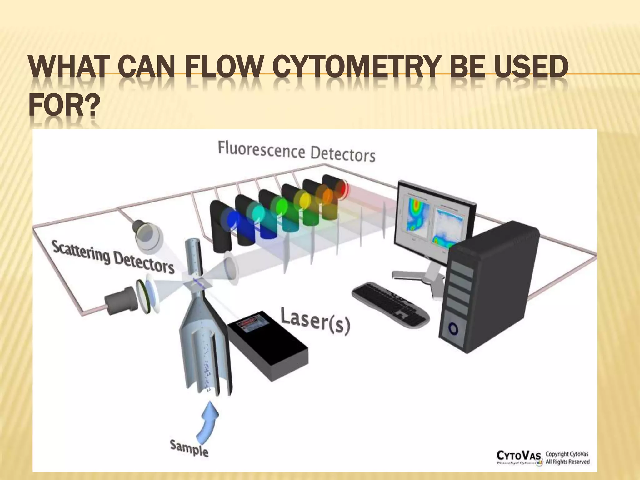
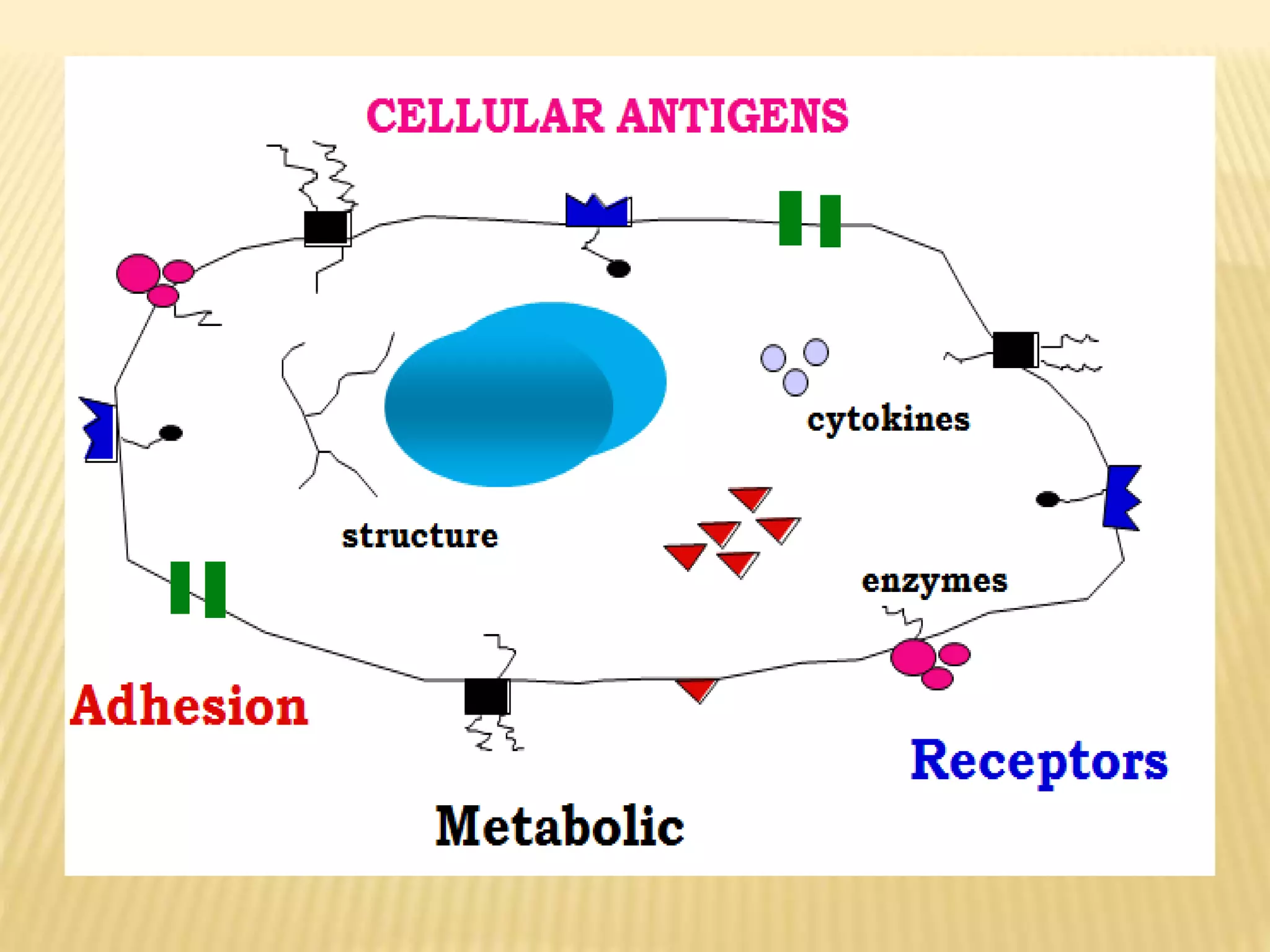
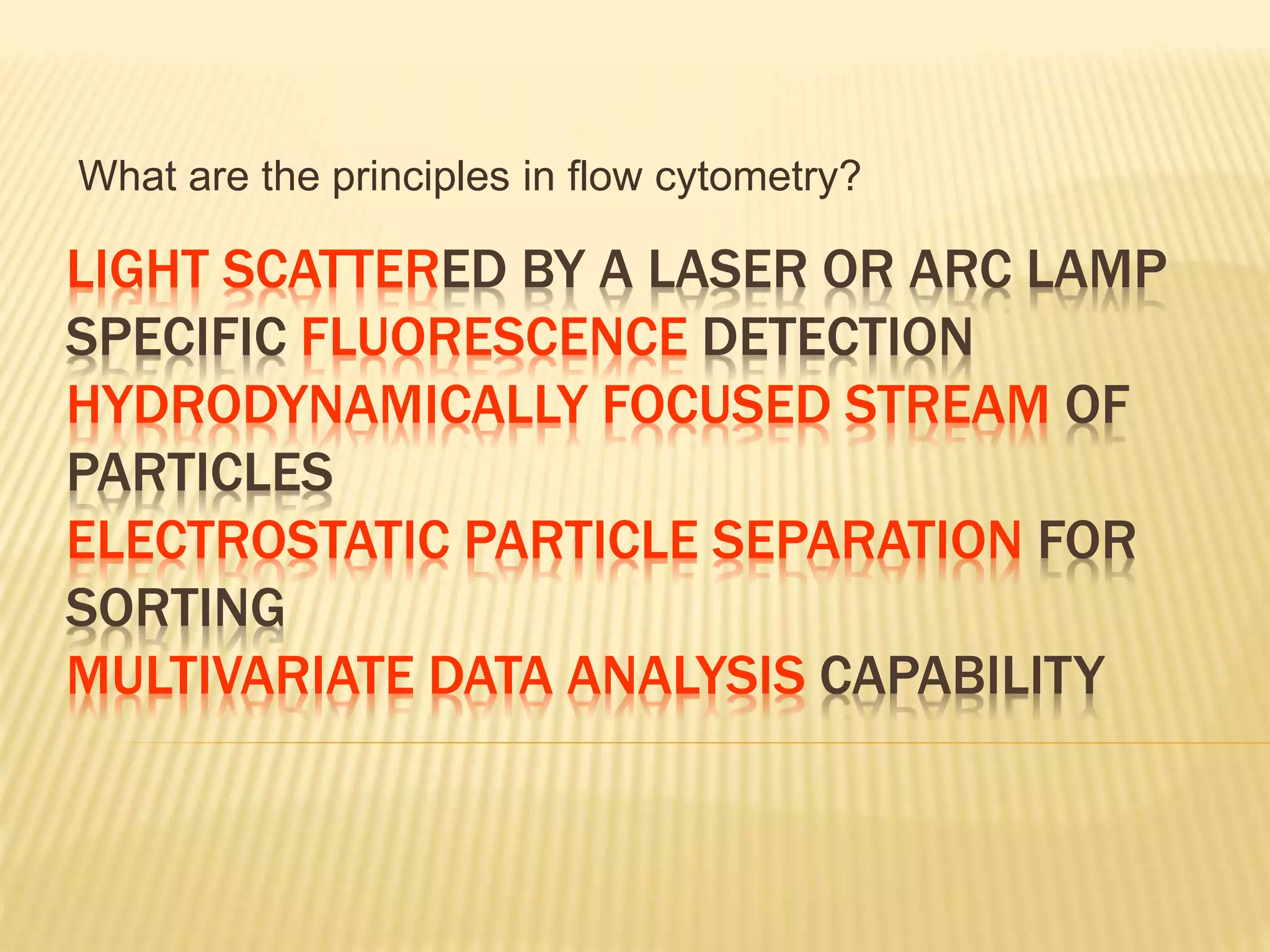
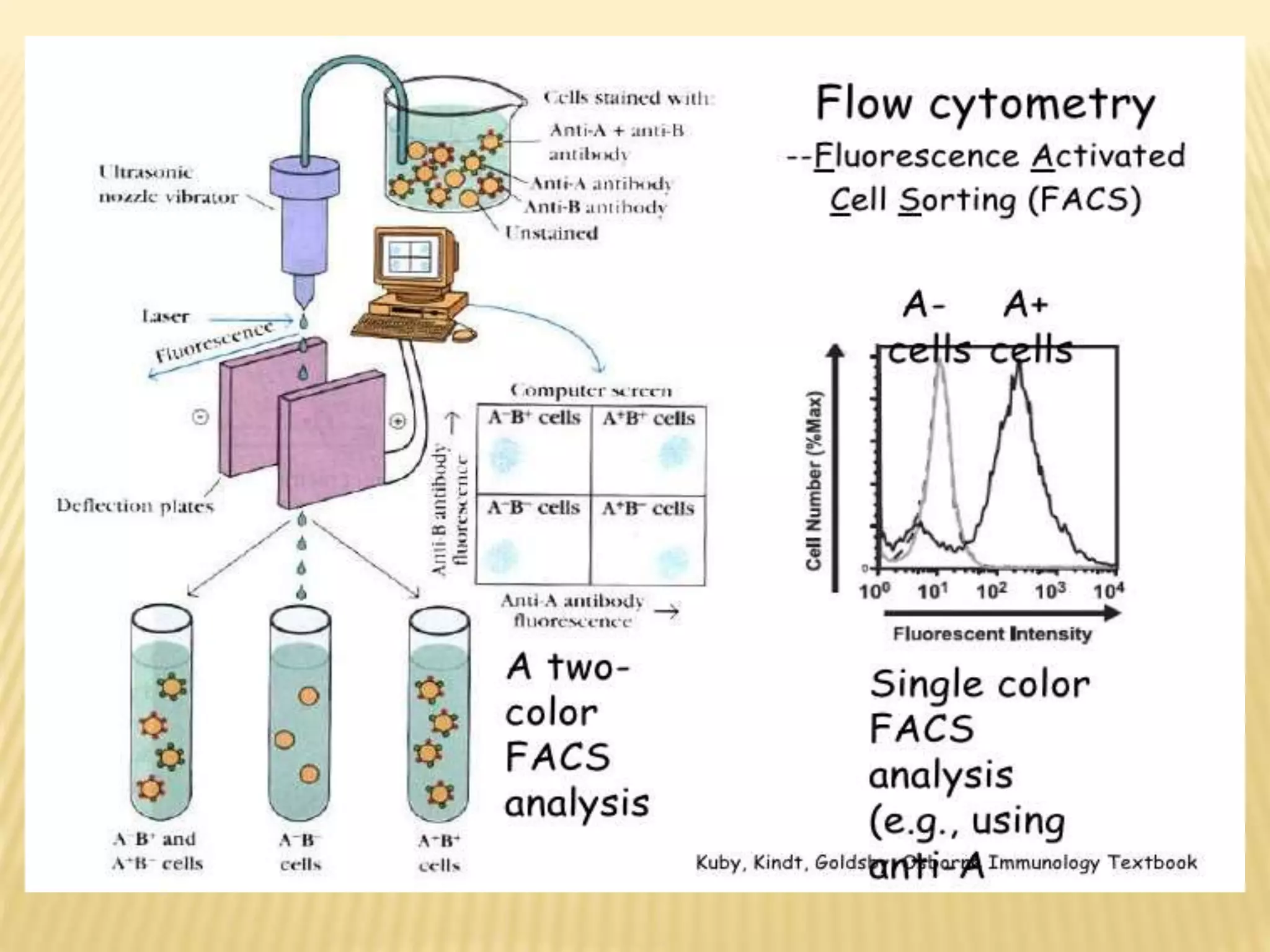
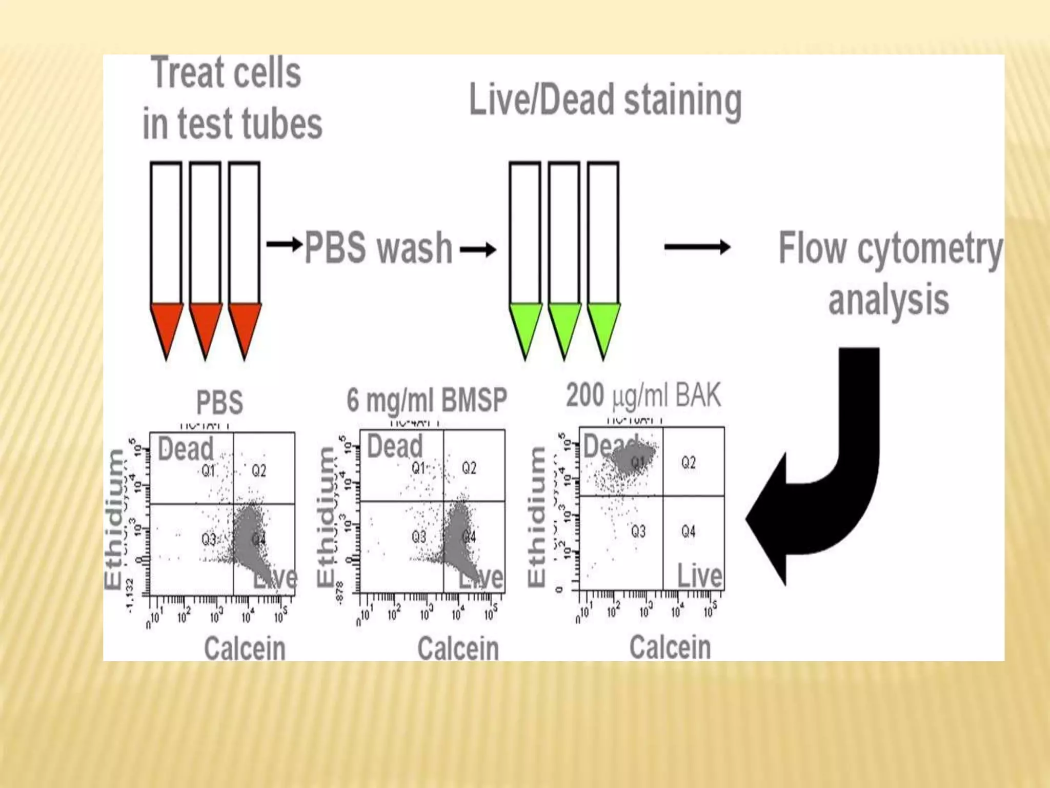
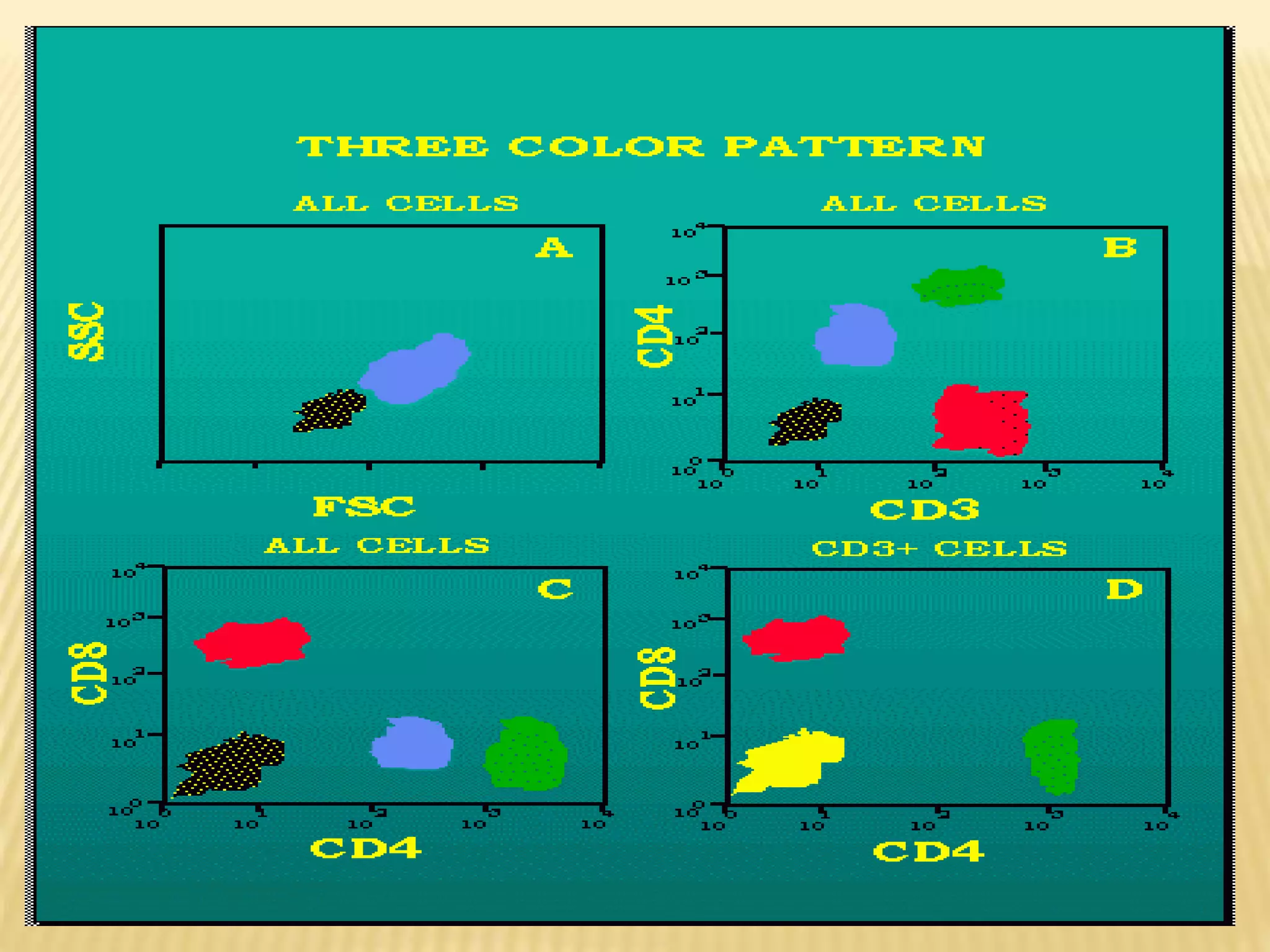
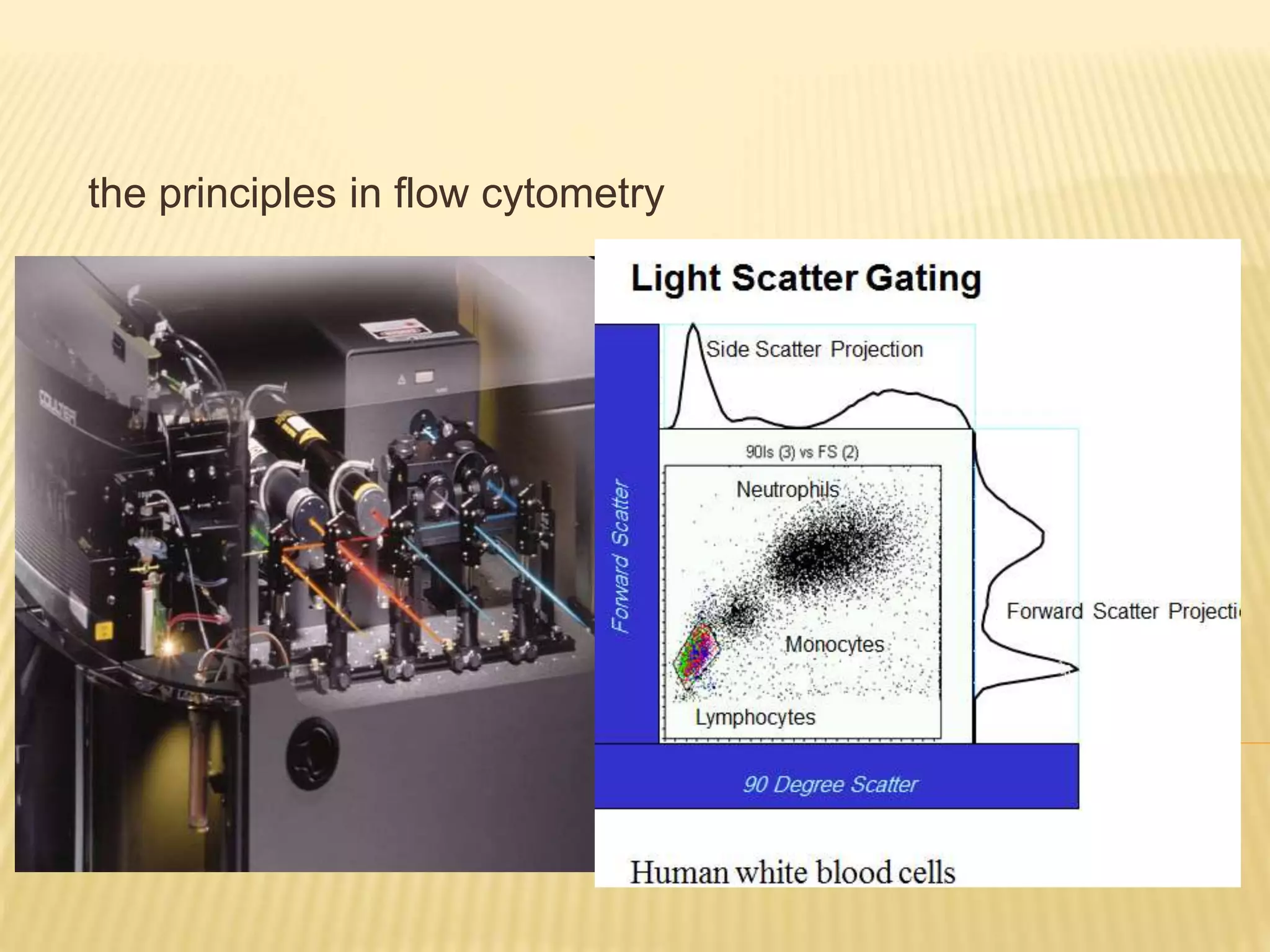
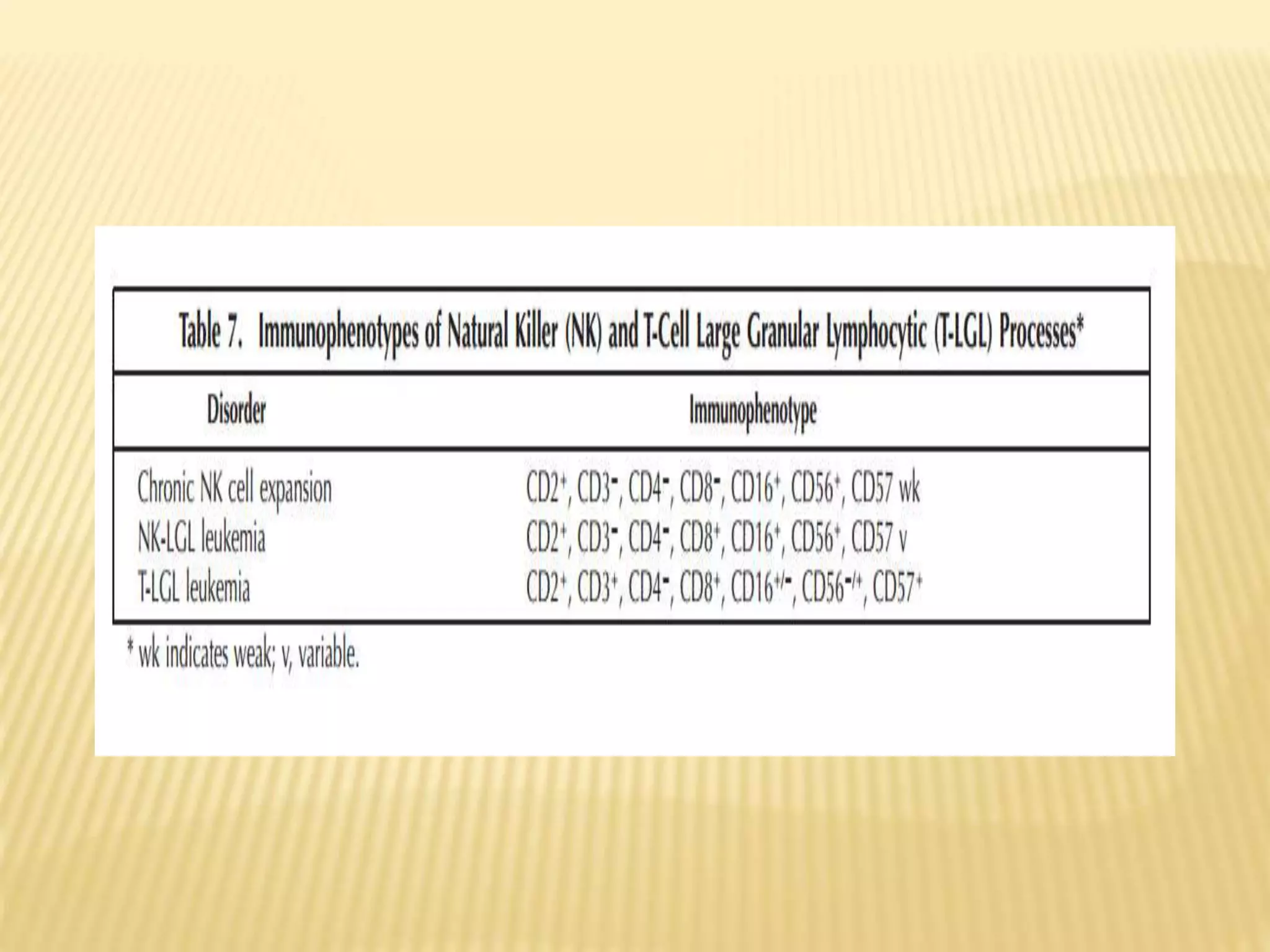
FLOWCYTOMET
RY
Name:Eman Abd El-Raouf Ahmed
WHAT CAN FLOW CYTOMETRY BE USED
FOR?
THE CONTENT
Definition
Advantages
Disadvantages
Some clinical
application
CLUSTER DESIGNATIONS
• These are based on the Immunology
• Each antigen that is defined on cells is
given a unique number
• Until a final number is agreed, antigens can
be designated CDw (w=workshop a
tentative designation)
LIGHT SCATTERED BY A LASER OR ARC LAMP
SPECIFIC FLUORESCENCE DETECTION
HYDRODYNAMICALLY FOCUSED STREAM OF
PARTICLES
ELECTROSTATIC PARTICLE SEPARATION FOR
SORTING
MULTIVARIATE DATA ANALYSIS CAPABILITY
What are the principles in flow cytometry?
the principles in flow cytometry
SOME CLINICAL APPLICATIONS
Analysis of leukemia and lymphoma
Immunophenotyping by flow cytometry is an important
tool in the diagnosis and staging of patients with a
hematological neoplasm. It is used in conjunction with
classical morphology.
In the bone marrow, normal blood cells develop from
stem cells in a progressive series of differentiations,
branching off to give different lineages of cells (for
example, myeloid or lymphoid, T cell or B cell), each of
which has a distinct maturation pathway.
STEM CELL ENUMERATIONHaemopoietic stem cells in the bone marrow can be
identified by their expression of CD34. Normally, the
number of such cells in bone marrow is low and is
negligible in peripheral blood. However, if mobilization
of CD34 positive cells from the bone marrow is
stimulated, stem cells can be harvested from the
peripheral blood as well as the marrow.
To assist in the identification of a small percentage of
CD34 positive cells, a double stain of CD34 and CD45 is
used. The method gives the percentage of CD34 +ve
cells. To obtain the absolute CD34+ concentration, the
total cell concentration is counted in a hematology
analyzer (two-platform method). The whole procedure
can be performed on a flow cytometer (single platform)
by spiking the sample with a known number of
fluorescent beads, which will enable the volume of blood
analyzed to be calculated
SOLID ORGAN TRANSPLANTATION
T CELL CROSS-MATCH
Flow cytometry can be used to cross match a recipient's
serum with donor lymphocytes to detect antibodies that
could interfere with engraftment (Bell et al., 1998). Prior to
organ transplantation, the organ donor's lymphocytes are
incubated with serum from the potential recipient of the
graft. After washing, bound immunoglobulin are detected
using an FITC-conjugated anti-human IgG antibody. The T
cells are identified using a PE-CD3 conjugate.
POSTOPERATIVE MONITORING
After the organ transplant, analysis of the peripheral blood
lymphocytes may help to indicate early rejection and bone
marrow toxicity during immunosuppressive therapies, and to
help in the differentiation of infections from transplant
rejection (Shanahan, 1997). A variety of cell surface markers
and activation antigens can be used depending on the clinical
condition and the organ transplanted.
Peripheral blood testing may also be used to monitor the
effectiveness of anti-rejection immunosuppressive therapy,
which may include, for example, antibodies designed to
destroy T cells. The number of circulating T-cells is usually
determined by measuring the cell surface marker CD3.
DETECTION OF AUTOANTIBODIES
Autoantibodies to leucocytes, platelets and erythrocytes may
be found in a variety of autoimmune conditions and can cause
anemia, leukopenia, or thrombocytopenia. They are detected
by immunofluorescence in either a direct or an indirect
assay. In the former, anti-human Ig antibodies are used to
detect Ig on the surface of the patient’s cells. In the indirect
assay, the reaction of antibodies in the patient’s serum with
cells from a normal person is observed. The procedures are
similar to those used for a T cell crossmatch
HIV INFECTION.
Determination of the numbers of CD4 +ve lymphocytes in the
peripheral blood is used to monitor patients with HIV infections
(Mandy et al., 2002). The percentage of CD4 +ve cells can be
obtained in a single tube by staining for CD45/CD3/CD4. A
cytogram of SS versus CD45 is used to identify the
lymphocytes and a cytogram of CD4 versus CD3 to
enumerate the CD4+ve T cells. An extended panel is used to
obtain a more complete picture of the peripheral blood
lymphocytes.
FOETO-MATERNAL HAEMORRHAGE
Foeto-maternal bleeding can sensitise a Rhesus blood
group D-ve mother to D+ve blood cells from the
foetus. In a subsequent pregnancy, haemolytic disease
of the new born child can be caused by the destruction
of Rhesus D +ve blood cells of the foetus by maternal
anti-D antibodies. Prophylactic anti-D given to the
Rhesus D -ve mother shortly after delivery of a Rhesus D
+ve child significantly reduces the incidence of anti-D
sensitisation in the mother and has led to the virtual
elimination of the disease from mothers so treated. Since
the dose of anti-D given is related to the size of the foeto-
maternal haemorrhage, quantitation of foetal-maternal
haemorrhage is therefore important.
IMMUNODEFICIENCY DISEASES
Diseases resulting from primary immunodeficiencies, which are usually found
in infants and young children, can be a result of defects in T-cells, B-cells,
granulocytes or monocytes. In many of these diseases surface or cytoplasmic
proteins are missing or have impaired function. They are characterized by
immunophenotyping, the selection of antibodies being based on the clinical
presentation.
Flow cytometry can also be used to detect functional abnormalities in
leucocytes. Possible functional tests include the analysis of oxidative burst
and phagocytic function in granulocytes and monocytes T and B cell mitogen
responses, NK cell tumour cell lytic function and platelet activation antigens.
Secondary immunodeficiencies are caused by another illness, condition,
medical treatment or intervention, the patient presenting with frequent and
often atypical infections. Some of the conditions and diseases which can
cause immunodeficiencies are severe malnutrition, biotin, B12 or zinc
deficiency, lymphoma, myasthenia gravis, myeloma, radiation or
chemotherapy, chronic alcoholism, drug abuse, cancer, splenectomy and
chronic viral illnesses. If a secondary immunodeficiency is suspected, the
immune status can be assessed by analysing the peripheral blood
lymphocytes. The panel of markers might include CD3, CD4 and CD8 (to
assess T cells), CD19 (for B cells), CD56 (NK cells) and, after incubation with a
mitogen such as PHA, either HLA-DR or CD69 (to assess lymphocyte
activation).
PAROXYSMAL NOCTURNAL
HAEMOGLOBINURIA
Paroxysmal nocturnal haemoglobinuria (PNH) is an acquired
disease characterised by the development of an abnormal
clone of precursor cells in the bone marrow.
The white cells and red cells produced are dysfunctional and
are susceptible to lysis. Analysis of this clonal abnormality by
flow cytometry, in general, can be accomplished by analysis of
CD55 and CD59 on red cells
SOME OTHER APPLICATIONS IN BLOOD
TRANSFUSION
Contaminating leucocytes
The presence of contaminating leukocytes in transfused
products of blood may cause a number of adverse effects.
These include:
• nonhaemolytic febrile reactions
• graft-versus-host disease
• cytomegalovirus transmission
• pulmonary oedema
• alloimmunization to HLA class I antigens
Flow cytometry offers a sensitive measure of the number of
remaining leucocytes in leucofiltered products, such as plasma
and red blood cells.
MEASUREMENTS ON RED BLOOD CELLS
(RBCS)
The measurements that can be made on red blood cells include:
• Detection and quantitation of RBC-bound proteins
• Quantitation of RBC-bound immunoglobulins
• Detection and quantitation of RBC antigens and antibodies
• Detection and quantitation of minor RBC populations,
including the detection and quantitation of transfused RBCs and
the detection and quantitation of fetal RBCs in maternal blood
PLATELET COUNTING AND FUNCTION
Flow cytometry can be used to count platelets and also to
measure their surface proteins (Harrison et al., 2001, 2005,
Michelson, 2006). The latter change during activation of the
platelets and can be used to measure their activation state.
Platelets are easily activated and blood has to be taken and
handled with care; for this reason, whole blood methods are
generally preferred.
Platelet analysis by flow cytometry can have application in
• Identification of inherited disorders
• Monitoring of anti-platelet therapy
• Monitoring clinical course of disease
• Monitoring platelet production in thrombocytopenia
• Identification of patients at risk of thrombosis
• Accurate platelet counting in thrombocytopenia
• Diagnosis of heparin induced thrombocytopenia
REFERENCES
http://flowbook.denovosoftware.com/Flow_Book/Chapter_7%3
A_Some_Clinical_Applications
http://www.bloodjournal.org/content/90/8/2863?sso-
checked=true
http://www.uth.tmc.edu/pathology/hematopathology/UsefulHem
eRefArticles/Flow-Immuno-In-Hematopathology.pdf
http://www.sysmex-partec.com/instrumentation.html
http://www.ncbi.nlm.nih.gov/pubmed/21204710

Flowcytometry

  • 2.
  • 3.
    WHAT CAN FLOWCYTOMETRY BE USED FOR?
  • 4.
  • 6.
    CLUSTER DESIGNATIONS • Theseare based on the Immunology • Each antigen that is defined on cells is given a unique number • Until a final number is agreed, antigens can be designated CDw (w=workshop a tentative designation)
  • 8.
    LIGHT SCATTERED BYA LASER OR ARC LAMP SPECIFIC FLUORESCENCE DETECTION HYDRODYNAMICALLY FOCUSED STREAM OF PARTICLES ELECTROSTATIC PARTICLE SEPARATION FOR SORTING MULTIVARIATE DATA ANALYSIS CAPABILITY What are the principles in flow cytometry?
  • 12.
    the principles inflow cytometry
  • 16.
    SOME CLINICAL APPLICATIONS Analysisof leukemia and lymphoma Immunophenotyping by flow cytometry is an important tool in the diagnosis and staging of patients with a hematological neoplasm. It is used in conjunction with classical morphology. In the bone marrow, normal blood cells develop from stem cells in a progressive series of differentiations, branching off to give different lineages of cells (for example, myeloid or lymphoid, T cell or B cell), each of which has a distinct maturation pathway.
  • 31.
    STEM CELL ENUMERATIONHaemopoieticstem cells in the bone marrow can be identified by their expression of CD34. Normally, the number of such cells in bone marrow is low and is negligible in peripheral blood. However, if mobilization of CD34 positive cells from the bone marrow is stimulated, stem cells can be harvested from the peripheral blood as well as the marrow. To assist in the identification of a small percentage of CD34 positive cells, a double stain of CD34 and CD45 is used. The method gives the percentage of CD34 +ve cells. To obtain the absolute CD34+ concentration, the total cell concentration is counted in a hematology analyzer (two-platform method). The whole procedure can be performed on a flow cytometer (single platform) by spiking the sample with a known number of fluorescent beads, which will enable the volume of blood analyzed to be calculated
  • 32.
    SOLID ORGAN TRANSPLANTATION TCELL CROSS-MATCH Flow cytometry can be used to cross match a recipient's serum with donor lymphocytes to detect antibodies that could interfere with engraftment (Bell et al., 1998). Prior to organ transplantation, the organ donor's lymphocytes are incubated with serum from the potential recipient of the graft. After washing, bound immunoglobulin are detected using an FITC-conjugated anti-human IgG antibody. The T cells are identified using a PE-CD3 conjugate.
  • 33.
    POSTOPERATIVE MONITORING After theorgan transplant, analysis of the peripheral blood lymphocytes may help to indicate early rejection and bone marrow toxicity during immunosuppressive therapies, and to help in the differentiation of infections from transplant rejection (Shanahan, 1997). A variety of cell surface markers and activation antigens can be used depending on the clinical condition and the organ transplanted. Peripheral blood testing may also be used to monitor the effectiveness of anti-rejection immunosuppressive therapy, which may include, for example, antibodies designed to destroy T cells. The number of circulating T-cells is usually determined by measuring the cell surface marker CD3.
  • 34.
    DETECTION OF AUTOANTIBODIES Autoantibodiesto leucocytes, platelets and erythrocytes may be found in a variety of autoimmune conditions and can cause anemia, leukopenia, or thrombocytopenia. They are detected by immunofluorescence in either a direct or an indirect assay. In the former, anti-human Ig antibodies are used to detect Ig on the surface of the patient’s cells. In the indirect assay, the reaction of antibodies in the patient’s serum with cells from a normal person is observed. The procedures are similar to those used for a T cell crossmatch
  • 35.
    HIV INFECTION. Determination ofthe numbers of CD4 +ve lymphocytes in the peripheral blood is used to monitor patients with HIV infections (Mandy et al., 2002). The percentage of CD4 +ve cells can be obtained in a single tube by staining for CD45/CD3/CD4. A cytogram of SS versus CD45 is used to identify the lymphocytes and a cytogram of CD4 versus CD3 to enumerate the CD4+ve T cells. An extended panel is used to obtain a more complete picture of the peripheral blood lymphocytes.
  • 36.
    FOETO-MATERNAL HAEMORRHAGE Foeto-maternal bleedingcan sensitise a Rhesus blood group D-ve mother to D+ve blood cells from the foetus. In a subsequent pregnancy, haemolytic disease of the new born child can be caused by the destruction of Rhesus D +ve blood cells of the foetus by maternal anti-D antibodies. Prophylactic anti-D given to the Rhesus D -ve mother shortly after delivery of a Rhesus D +ve child significantly reduces the incidence of anti-D sensitisation in the mother and has led to the virtual elimination of the disease from mothers so treated. Since the dose of anti-D given is related to the size of the foeto- maternal haemorrhage, quantitation of foetal-maternal haemorrhage is therefore important.
  • 37.
    IMMUNODEFICIENCY DISEASES Diseases resultingfrom primary immunodeficiencies, which are usually found in infants and young children, can be a result of defects in T-cells, B-cells, granulocytes or monocytes. In many of these diseases surface or cytoplasmic proteins are missing or have impaired function. They are characterized by immunophenotyping, the selection of antibodies being based on the clinical presentation. Flow cytometry can also be used to detect functional abnormalities in leucocytes. Possible functional tests include the analysis of oxidative burst and phagocytic function in granulocytes and monocytes T and B cell mitogen responses, NK cell tumour cell lytic function and platelet activation antigens. Secondary immunodeficiencies are caused by another illness, condition, medical treatment or intervention, the patient presenting with frequent and often atypical infections. Some of the conditions and diseases which can cause immunodeficiencies are severe malnutrition, biotin, B12 or zinc deficiency, lymphoma, myasthenia gravis, myeloma, radiation or chemotherapy, chronic alcoholism, drug abuse, cancer, splenectomy and chronic viral illnesses. If a secondary immunodeficiency is suspected, the immune status can be assessed by analysing the peripheral blood lymphocytes. The panel of markers might include CD3, CD4 and CD8 (to assess T cells), CD19 (for B cells), CD56 (NK cells) and, after incubation with a mitogen such as PHA, either HLA-DR or CD69 (to assess lymphocyte activation).
  • 38.
    PAROXYSMAL NOCTURNAL HAEMOGLOBINURIA Paroxysmal nocturnalhaemoglobinuria (PNH) is an acquired disease characterised by the development of an abnormal clone of precursor cells in the bone marrow. The white cells and red cells produced are dysfunctional and are susceptible to lysis. Analysis of this clonal abnormality by flow cytometry, in general, can be accomplished by analysis of CD55 and CD59 on red cells
  • 40.
    SOME OTHER APPLICATIONSIN BLOOD TRANSFUSION Contaminating leucocytes The presence of contaminating leukocytes in transfused products of blood may cause a number of adverse effects. These include: • nonhaemolytic febrile reactions • graft-versus-host disease • cytomegalovirus transmission • pulmonary oedema • alloimmunization to HLA class I antigens Flow cytometry offers a sensitive measure of the number of remaining leucocytes in leucofiltered products, such as plasma and red blood cells.
  • 41.
    MEASUREMENTS ON REDBLOOD CELLS (RBCS) The measurements that can be made on red blood cells include: • Detection and quantitation of RBC-bound proteins • Quantitation of RBC-bound immunoglobulins • Detection and quantitation of RBC antigens and antibodies • Detection and quantitation of minor RBC populations, including the detection and quantitation of transfused RBCs and the detection and quantitation of fetal RBCs in maternal blood
  • 42.
    PLATELET COUNTING ANDFUNCTION Flow cytometry can be used to count platelets and also to measure their surface proteins (Harrison et al., 2001, 2005, Michelson, 2006). The latter change during activation of the platelets and can be used to measure their activation state. Platelets are easily activated and blood has to be taken and handled with care; for this reason, whole blood methods are generally preferred. Platelet analysis by flow cytometry can have application in • Identification of inherited disorders • Monitoring of anti-platelet therapy • Monitoring clinical course of disease • Monitoring platelet production in thrombocytopenia • Identification of patients at risk of thrombosis • Accurate platelet counting in thrombocytopenia • Diagnosis of heparin induced thrombocytopenia
  • 44.