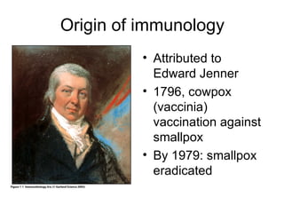
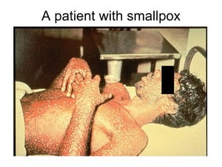
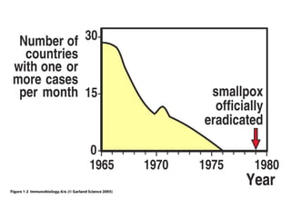
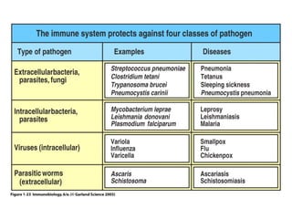
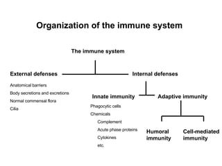
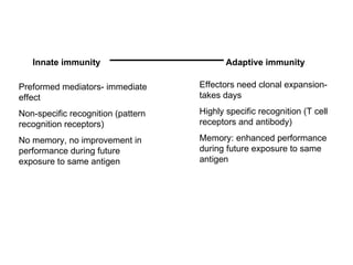
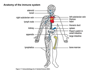
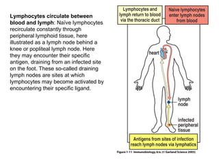
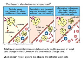
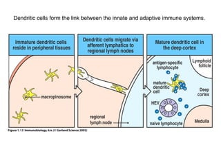
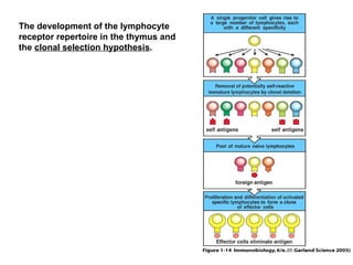
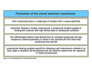
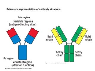
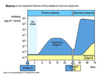
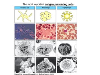
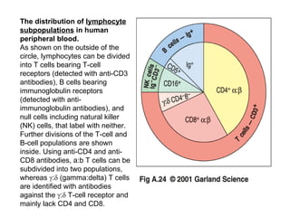
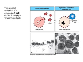
Haematology and Immunology Introduction to Immunology MB, ChB Phase II Prof Gerhard Walzl
Figure 1-1 Origin of immunology Attributed to Edward Jenner 1796, cowpox (vaccinia) vaccination against smallpox By 1979: smallpox eradicated
A patient with smallpox
Figure 1-2
History of immunology ‘ Infectious agents’: Robert Koch, late 19 th  century Louis Pasteur, 1880, vaccines against cholera in chicken, rabies in humans Emil von Behring and Shibasaburo Kitasano (1890’s): ‘antitoxic’ activity in serum of animals immune to diphtheria or tetanus
Functions of the immune system Protection against invaders Removal of dead or damaged tissues RBC Wounds Recognition and removal of abnormal or foreign cells or substances Malignancies Transplanted tissue Foreign bodies/substances
Figure 1-23
Some basic concepts in immunology
Organization of the immune system External defenses Internal defenses Anatomical barriers Body secretions and excretions Normal commensal flora Cilia Innate immunity Adaptive immunity Humoral immunity Cell-mediated immunity Phagocytic cells Chemicals Complement Acute phase proteins Cytokines etc. The immune system
… Internal defenses Innate immunity Adaptive immunity Humoral immunity B cells Plasma cells Antibody Cell-mediated immunity CD4+ T cells Th1 cells Th2 cells Treg cells Th17 cells CD8+ T cells Subtypes  NK T cells Phagocytic cells Neutrophils Macrophages Dendritic cells NK cells Chemicals Complement Acute phase proteins Cytokines
Innate immunity Adaptive immunity Preformed mediators- immediate effect Non-specific recognition (pattern recognition receptors) No memory, no improvement in performance during future exposure to same antigen Effectors need clonal expansion- takes days Highly specific recognition (T cell receptors and antibody) Memory: enhanced performance during future exposure to same antigen
Figure 1-7 Anatomy of the immune system
Figure 1-11 Lymphocytes circulate between blood and lymph : Naïve lymphocytes recirculate constantly through peripheral lymphoid tissue, here illustrated as a lymph node behind a knee or popliteal lymph node. Here they may encounter their specific antigen, draining from an infected site on the foot. These so-called draining lymph nodes are sites at which lymphocytes may become activated by encountering their specific ligand.
Figure 1-12 What happens when bacteria are phagocytosed? Cytokines = chemical messengers between cells, bind to receptors on target cells, change activation, behavior and differentiation of target cells. Chemokines = type of cytokine that  attracts  and activates target cells
Figure 1-13 Dendritic cells form the link between the innate and adaptive immune systems.
Figure 1-14 The development of the lymphocyte receptor repertoire in the thymus and the  clonal selection hypothesis .
Figure 1-15
Figure 1-16 Schematic representation of antibody structure. Fab region Fc region
Figure 1-20 Memory  is an important feature of the adaptive immune response.
Figure 1-22 The most important  antigen presenting cells .
The distribution of  lymphocyte subpopulations  in human peripheral blood. As shown on the outside of the circle, lymphocytes can be divided into T cells bearing T-cell receptors (detected with anti-CD3 antibodies), B cells bearing immunoglobulin receptors (detected with anti-immunoglobulin antibodies), and null cells including natural killer (NK) cells, that label with neither. Further divisions of the T-cell and B-cell populations are shown inside. Using anti-CD4 and anti-CD8 antibodies, a:b T cells can be subdivided into two populations, whereas   :   (gamma:delta) T cells are identified with antibodies against the   :   T-cell receptor and mainly lack CD4 and CD8.
The results of immune cell activation
Figure 1-25 The result of activation of a  cytotoxic T cell  (CD8+ T cell) by a virus infected cell:
Figure 1-26 The result of  activation of a T helper cell  by a macrophage infected by an intracellular pathogen like TB is that the T cell activates the macrophage, thereby enabling it to kill its invader.
Figure 1-27 Antigen presentation Antigen is recognized by T cells only if presented by antigen presenting cells on MHC molecules.
Figure 1-32 Response Deficient response

Immunology

  • 1.
    Haematology and ImmunologyIntroduction to Immunology MB, ChB Phase II Prof Gerhard Walzl
  • 2.
    Figure 1-1 Originof immunology Attributed to Edward Jenner 1796, cowpox (vaccinia) vaccination against smallpox By 1979: smallpox eradicated
  • 3.
  • 4.
  • 5.
    History of immunology‘ Infectious agents’: Robert Koch, late 19 th century Louis Pasteur, 1880, vaccines against cholera in chicken, rabies in humans Emil von Behring and Shibasaburo Kitasano (1890’s): ‘antitoxic’ activity in serum of animals immune to diphtheria or tetanus
  • 6.
    Functions of theimmune system Protection against invaders Removal of dead or damaged tissues RBC Wounds Recognition and removal of abnormal or foreign cells or substances Malignancies Transplanted tissue Foreign bodies/substances
  • 7.
  • 8.
    Some basic conceptsin immunology
  • 9.
    Organization of theimmune system External defenses Internal defenses Anatomical barriers Body secretions and excretions Normal commensal flora Cilia Innate immunity Adaptive immunity Humoral immunity Cell-mediated immunity Phagocytic cells Chemicals Complement Acute phase proteins Cytokines etc. The immune system
  • 10.
    … Internal defensesInnate immunity Adaptive immunity Humoral immunity B cells Plasma cells Antibody Cell-mediated immunity CD4+ T cells Th1 cells Th2 cells Treg cells Th17 cells CD8+ T cells Subtypes NK T cells Phagocytic cells Neutrophils Macrophages Dendritic cells NK cells Chemicals Complement Acute phase proteins Cytokines
  • 11.
    Innate immunity Adaptiveimmunity Preformed mediators- immediate effect Non-specific recognition (pattern recognition receptors) No memory, no improvement in performance during future exposure to same antigen Effectors need clonal expansion- takes days Highly specific recognition (T cell receptors and antibody) Memory: enhanced performance during future exposure to same antigen
  • 12.
    Figure 1-7 Anatomyof the immune system
  • 13.
    Figure 1-11 Lymphocytescirculate between blood and lymph : Naïve lymphocytes recirculate constantly through peripheral lymphoid tissue, here illustrated as a lymph node behind a knee or popliteal lymph node. Here they may encounter their specific antigen, draining from an infected site on the foot. These so-called draining lymph nodes are sites at which lymphocytes may become activated by encountering their specific ligand.
  • 14.
    Figure 1-12 Whathappens when bacteria are phagocytosed? Cytokines = chemical messengers between cells, bind to receptors on target cells, change activation, behavior and differentiation of target cells. Chemokines = type of cytokine that attracts and activates target cells
  • 15.
    Figure 1-13 Dendriticcells form the link between the innate and adaptive immune systems.
  • 16.
    Figure 1-14 Thedevelopment of the lymphocyte receptor repertoire in the thymus and the clonal selection hypothesis .
  • 17.
  • 18.
    Figure 1-16 Schematicrepresentation of antibody structure. Fab region Fc region
  • 19.
    Figure 1-20 Memory is an important feature of the adaptive immune response.
  • 20.
    Figure 1-22 Themost important antigen presenting cells .
  • 21.
    The distribution of lymphocyte subpopulations in human peripheral blood. As shown on the outside of the circle, lymphocytes can be divided into T cells bearing T-cell receptors (detected with anti-CD3 antibodies), B cells bearing immunoglobulin receptors (detected with anti-immunoglobulin antibodies), and null cells including natural killer (NK) cells, that label with neither. Further divisions of the T-cell and B-cell populations are shown inside. Using anti-CD4 and anti-CD8 antibodies, a:b T cells can be subdivided into two populations, whereas  :  (gamma:delta) T cells are identified with antibodies against the  :  T-cell receptor and mainly lack CD4 and CD8.
  • 22.
    The results ofimmune cell activation
  • 23.
    Figure 1-25 Theresult of activation of a cytotoxic T cell (CD8+ T cell) by a virus infected cell:
  • 24.
    Figure 1-26 Theresult of activation of a T helper cell by a macrophage infected by an intracellular pathogen like TB is that the T cell activates the macrophage, thereby enabling it to kill its invader.
  • 25.
    Figure 1-27 Antigenpresentation Antigen is recognized by T cells only if presented by antigen presenting cells on MHC molecules.
  • 26.
    Figure 1-32 ResponseDeficient response