Downloaded 721 times


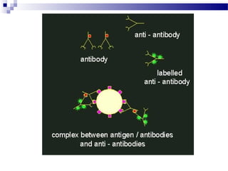


The document discusses immunofluorescence, a technique that uses fluorescent-labeled antibodies to detect specific antigens in tissues, viewed under a fluorescence microscope. It outlines the principles of direct and indirect immunofluorescence methods and their applications in clinical diagnoses, notably for skin diseases. Limitations include factors like antibody quality and specimen handling that can affect fluorescence signals.


