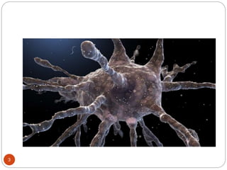
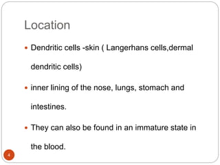
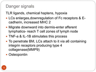
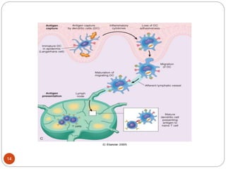
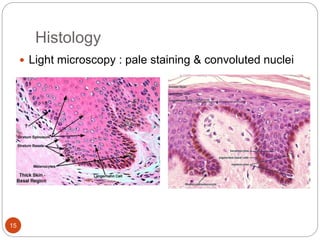
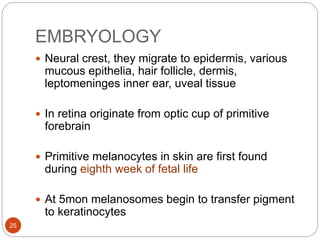
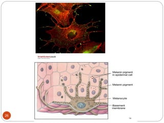
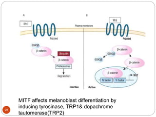
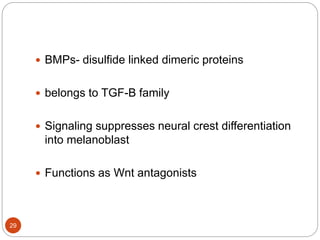
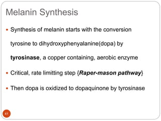
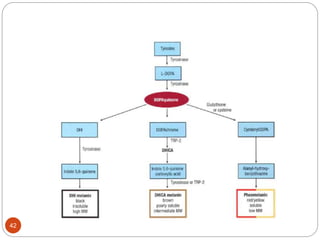
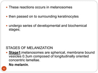
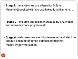
The document details the role and characteristics of dendritic cells, specifically their functions in the innate immune system, including antigen presentation, T cell activation, and immune memory maintenance. It discusses the different types of dendritic cells, their embryological development, histological identification, and their implications in various immune processes and diseases. Additionally, it covers melanocytes and Merkel cells, detailing their functions and significance within the skin's immune response and sensory mechanisms.


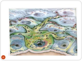
![Site specific melanocytes
1]Cutaneous Melanocytes
• Largest no. of melanocytes
• Skin & hair follicle
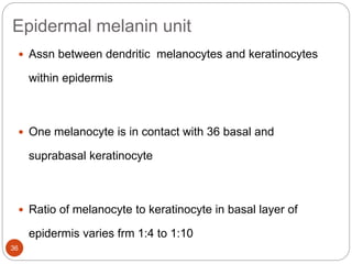
• Part of “epidermal melanin
unit”
2] Melanocyte stem cells
• present in hair follicle bulge
• Express TRP2 & nestin
• Other transcription factr-
SOX10 & Pax5
33](https://image.slidesharecdn.com/dendriticcells-151002185148-lva1-app6891/85/Dendritic-cells-33-320.jpg)
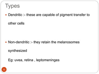
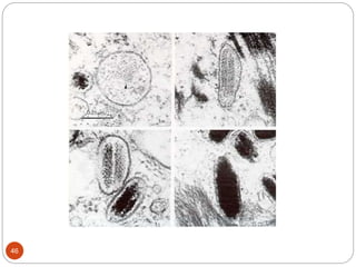
![3]Ocular melanocyte
uveal tract; they do not transfer their
melanosomes
required for proper routing of I/L & C/L neural
fibres in optic chiasm
Imp: visual abn in pts with albinism
34](https://image.slidesharecdn.com/dendriticcells-151002185148-lva1-app6891/85/Dendritic-cells-34-320.jpg)
![ 4]Otic Melannocyte
• reside in cochlea & imp for hearing
• Maintainence of endolymph through regulation of
K transport
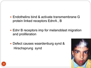
• Imp: Waardenburg synd
5]CEPHALIC Melanocyte
distributed through out the meninges & more
dense in leptomeninges above pons & medulla
oblongata.
35](https://image.slidesharecdn.com/dendriticcells-151002185148-lva1-app6891/85/Dendritic-cells-35-320.jpg)


![Deschambault[1]](https://cdn.slidesharecdn.com/ss_thumbnails/89882af6-e960-410c-be05-14e4d6adb648-160708012743-thumbnail.jpg?width=640&height=640&fit=bounds)