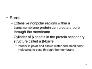
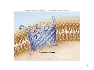
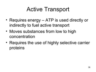
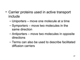
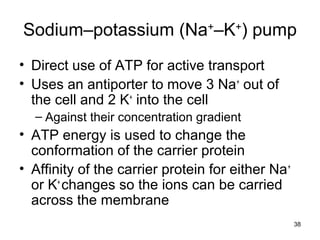
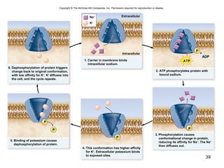
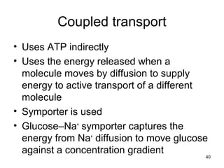
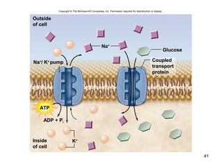
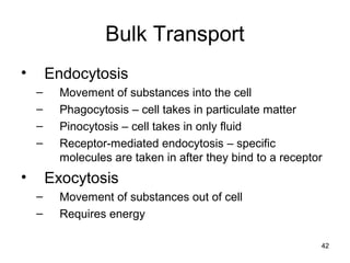
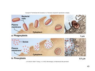
The document provides an outline and overview of key concepts about cell membranes:
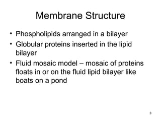
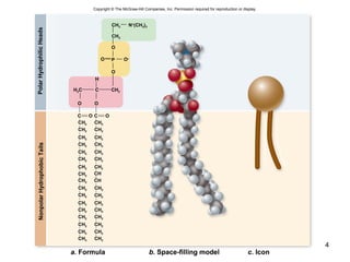
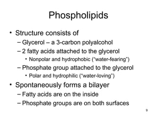
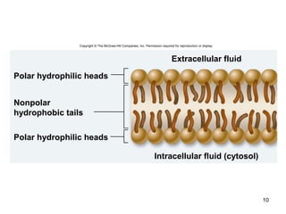
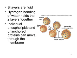
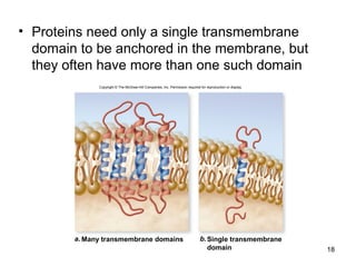
1. Membranes are composed of a phospholipid bilayer with embedded proteins. The fluid mosaic model describes membranes as a fluid bilayer with proteins floating within.
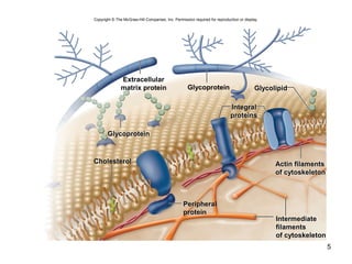
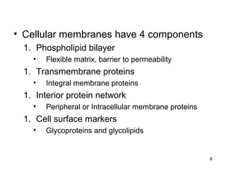
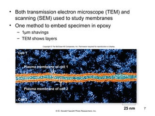
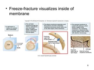
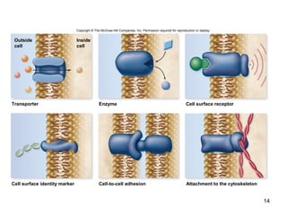
2. Membrane components include phospholipids, transmembrane proteins, peripheral proteins, and glycoproteins/glycolipids. Electron microscopy techniques like TEM and freeze-fracture are used to study membrane structure.
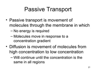
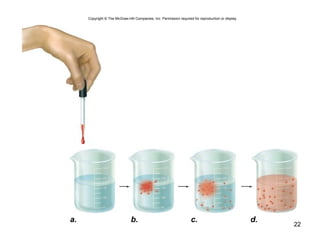
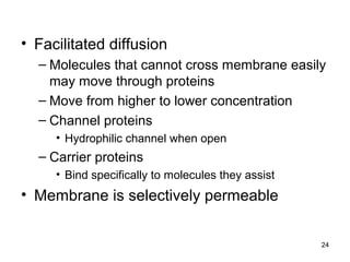
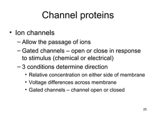
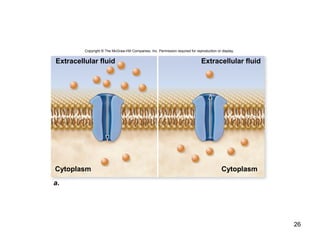
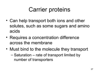
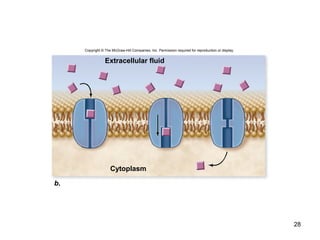
3. Passive transport across membranes, like diffusion and facilitated diffusion, moves molecules down concentration gradients through channel or carrier proteins without energy expenditure.


![04 [chapter 4 the tissue level of organization][11e]](https://cdn.slidesharecdn.com/ss_thumbnails/04chapter4thetissueleveloforganization11e-170828035609-thumbnail.jpg?width=640&height=640&fit=bounds)


![18 [chapter 18 the endocrine system]](https://cdn.slidesharecdn.com/ss_thumbnails/18chapter18theendocrinesystem-170828042016-thumbnail.jpg?width=640&height=640&fit=bounds)


