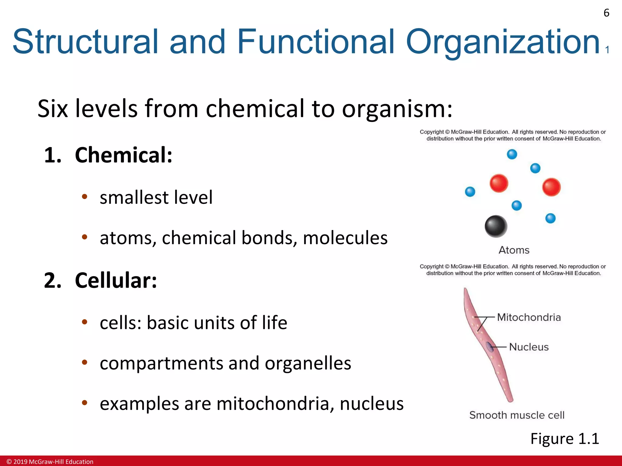
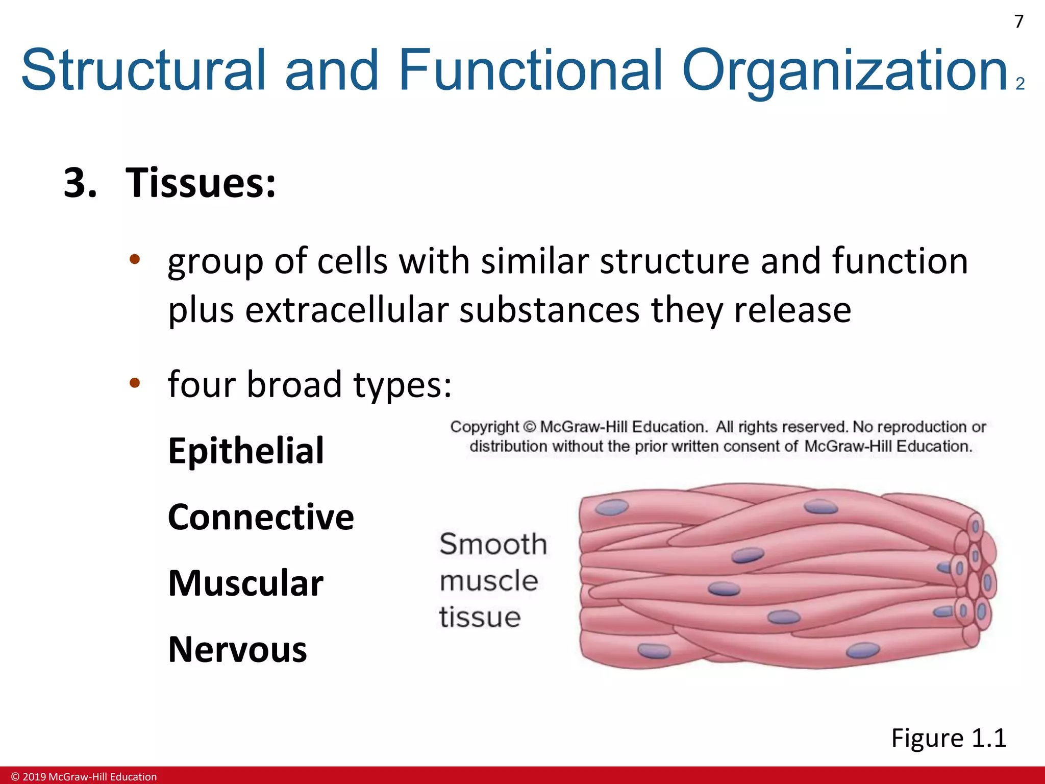
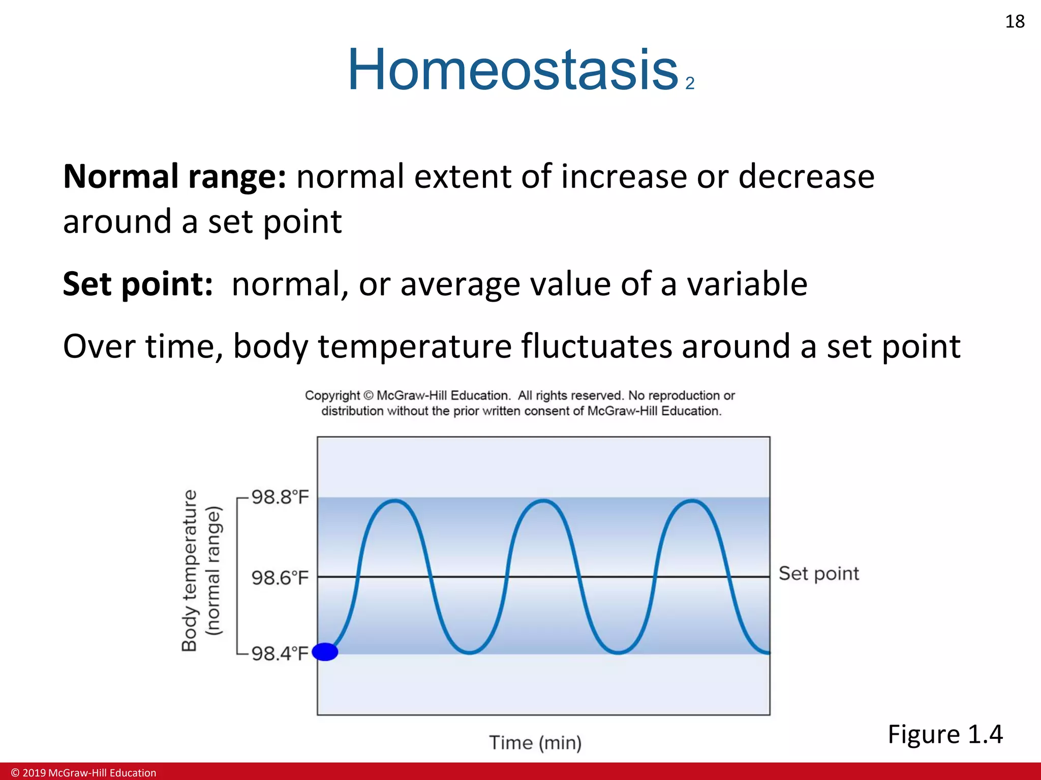
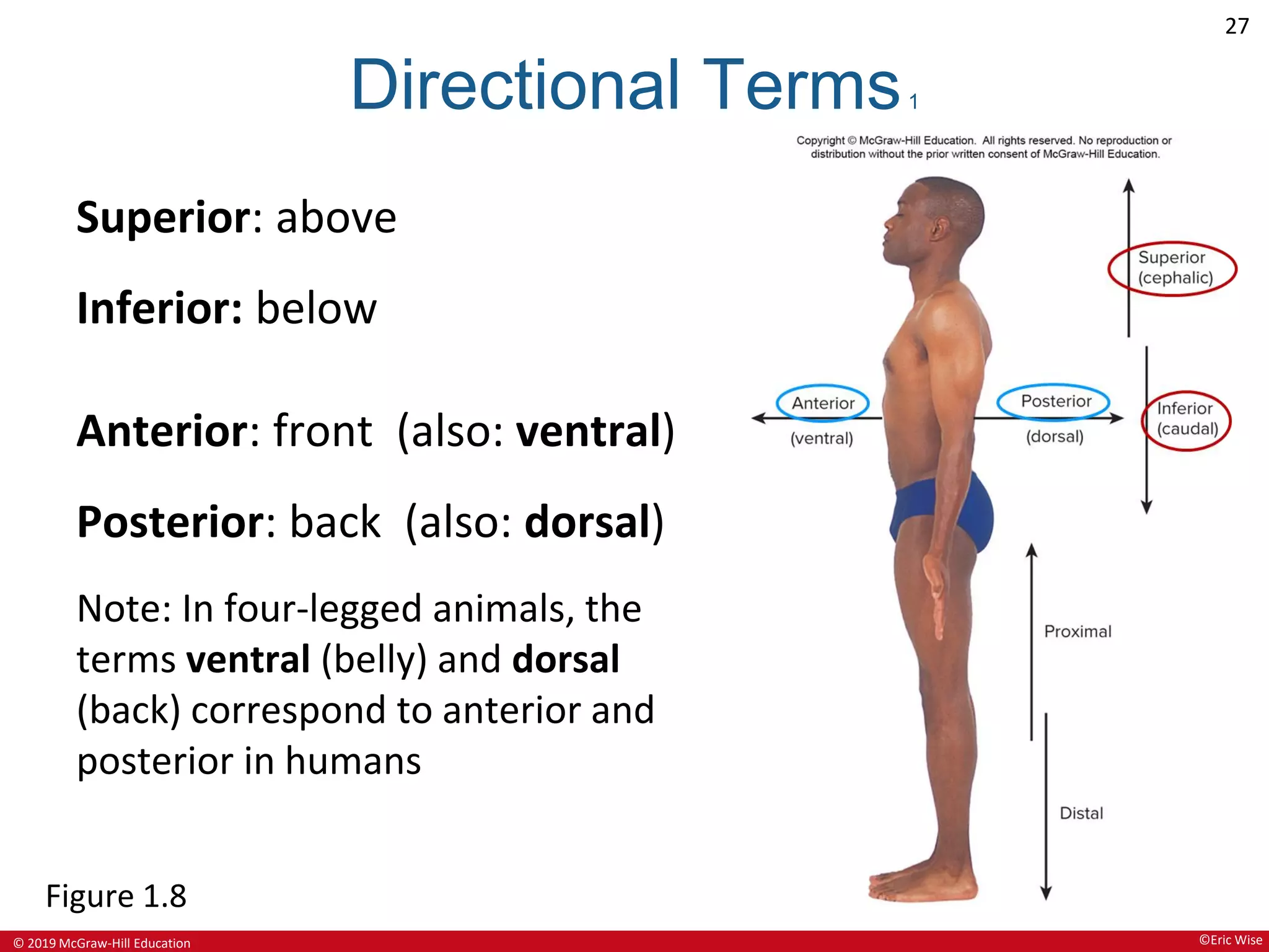
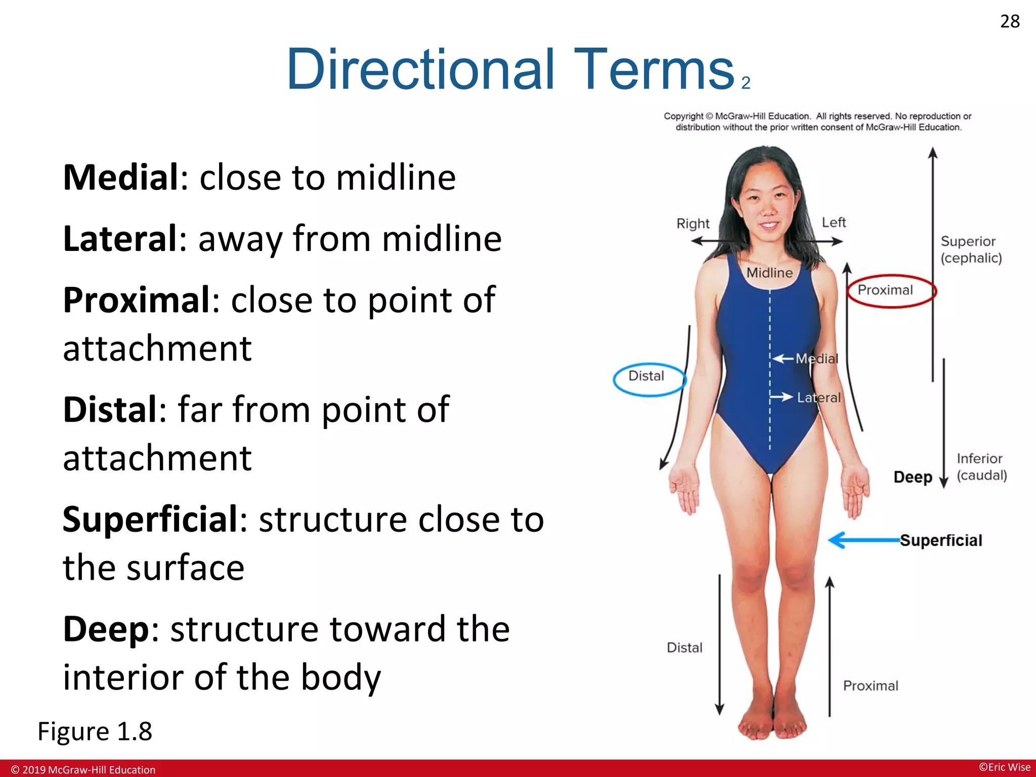
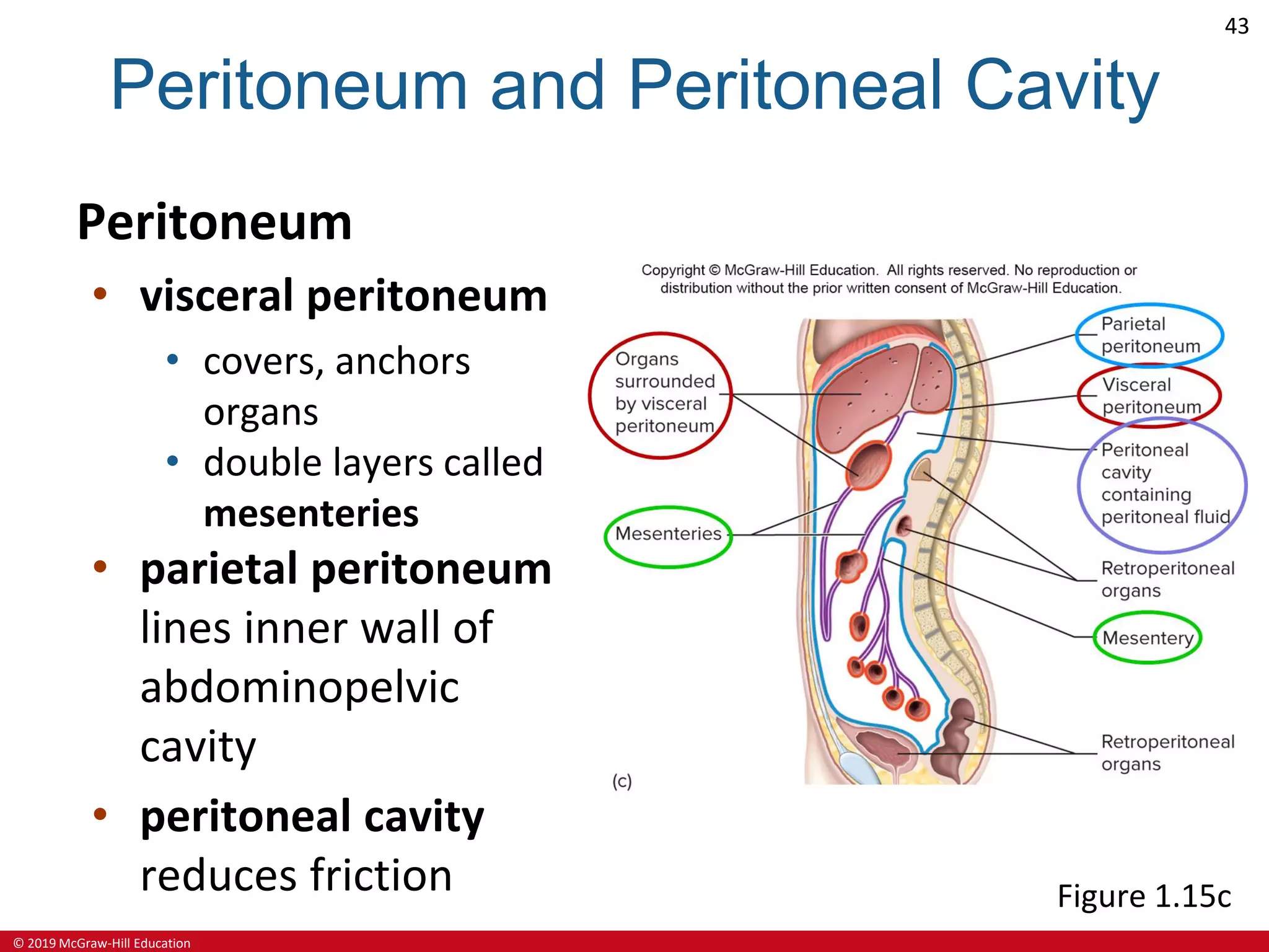
Seeley’s
ESSENTIALS OF
Anatomy &
Physiology
Tenth Edition
Cinnamon Vanputte
Jennifer Regan
Andrew Russo
See separate PowerPoint slides for all figures and tables
pre-inserted into PowerPoint without notes.
© 2019 McGraw-Hill Education. All rights reserved. Authorized only for instructor use in the classroom. No reproduction or further distribution permitted without the prior written consent of McGraw-Hill Education.
© 2019 McGraw-Hill Education
2
Chapter 1
The Human Organism
Lecture Outline
© 2019 McGraw-Hill Education
3
Anatomy and Physiology
Anatomy:
• investigates body structure
• the term means to dissect
Physiology:
• investigates processes and functions
• Human Physiology:
• studies the human organism
Systemic Physiology:
• studies body organ-systems
Cellular Physiology:
• studies body cells
© 2019 McGraw-Hill Education
4
Importance of Anatomy and Physiology
Understand how the body:
• responds to stimuli
• environmental changes
• environmental cues
• diseases
• injury
© 2019 McGraw-Hill Education
5
Types of Anatomy
Systemic:
• studies body organ-systems
Regional:
• studies body regions (medical schools)
Surface:
• studies external features, for example, bone
projections
Anatomical imaging:
• using technologies (x-rays, ultrasound, MRI)
© 2019 McGraw-Hill Education
6
Structural and Functional Organization1
Six levels from chemical to organism:
1. Chemical:
• smallest level
• atoms, chemical bonds, molecules
2. Cellular:
• cells: basic units of life
• compartments and organelles
• examples are mitochondria, nucleus
Figure 1.1
© 2019 McGraw-Hill Education
7
Structural and Functional Organization2
3. Tissues:
• group of cells with similar structure and function
plus extracellular substances they release
• four broad types:
Epithelial
Connective
Muscular
Nervous
Figure 1.1
© 2019 McGraw-Hill Education
8
Structural and Functional Organization3
4. Organs:
• two or more tissue types acting together to
perform function(s)
• Examples: stomach, heart, liver, ovary, bladder,
kidney
Figure 1.1
© 2019 McGraw-Hill Education
9
Structural and Functional Organization4
5. Organ-System:
• group of organs contributing to some function
• for example, digestive system, reproductive
system
Figure 1.1
© 2019 McGraw-Hill Education
10
Structural and Functional Organization5
6. Organism:
• all organ systems working together
• includes associated microorganisms such as
intestinal bacteria
Figure 1.1
©Bart Harris/Getty Images
© 2019 McGraw-Hill Education
11
Structural and Functional Organization6
Figure 1.1
1.1(6) ©Bart Harris/Getty Images
© 2019 McGraw-Hill Education
12
Major Organs of the Body
Figure 1.2
© 2019 McGraw-Hill Education
13
Organ Systems of the Body1
Figure 1.3
© 2019 McGraw-Hill Education
14
Organ Systems of the Body2
Figure 1.3
© 2019 McGraw-Hill Education
15
Characteristics of Life1
Organization:
• functional interrelationships between parts
Metabolism:
• sum of all chemical and physical changes sustaining an
organism
• ability to acquire and use energy in support of these
changes
Responsiveness:
• ability to sense and respond to environmental changes
• includes both internal and external environments
© 2019 McGraw-Hill Education
16
Characteristics of Life2
Growth:
• can increase in size
• size of cells, groups of cells, extracellular materials
Development:
• changes in form and size
• changes in cell structure and function from
generalized to specialized—differentiation
Reproduction:
• formation of new cells or new organisms
• generation of new individuals
• tissue repair
© 2019 McGraw-Hill Education
17
Homeostasis1
Homeostasis:
• maintenance of constant internal environment despite
fluctuations in the external or internal environment
Variables:
• measures of body properties that may change in value
Examples of variables:
body temperature
heart rate
blood pressure
© 2019 McGraw-Hill Education
18
Homeostasis2
Normal range: normal extent of increase or decrease
around a set point
Set point: normal, or average value of a variable
Over time, body temperature fluctuates around a set point
Figure 1.4
© 2019 McGraw-Hill Education
19
Homeostasis3
Set points for some variables can be temporarily
adjusted depending on body activities, as needed:
Examples Common cause of change
body temperature fever
heart rate, blood pressure
respiratory rate
exercise
© 2019 McGraw-Hill Education
20
Homeostasis4
Negative feedback is the main mechanism used
homeostatic regulation.
• A negative feedback response involves:
detection: of deviation away from set point
and
correction: reversal of deviation toward set point
and normal range
© 2019 McGraw-Hill Education
21
Homeostasis5
The components of feedback:
1. Receptor:
• detects changes in variable
2. Control center:
• receives receptor signal
• establishes set point
• sends signal to effector
3. Effector:
• directly causes change in variable
© 2019 McGraw-Hill Education
22
Homeostasis6
Figure 1.5
© 2019 McGraw-Hill Education
23
Negative Feedback Control of Body Temperature
Figure 1.6
© 2019 McGraw-Hill Education
24
Homeostasis7
Positive feedback mechanisms occur when the
initial stimulus further stimulates the response
• system response causes progressive deviation away from
• set point, outside of normal range
• not directly used for homeostasis
• some positive feedback occurs under normal conditions
Example: childbirth
• generally associated with injury, disease
• negative feedback mechanisms unable to maintain
homeostasis
© 2019 McGraw-Hill Education
25
Homeostasis8
Comparison of negative feedback and positive
feedback
Figure 1.7
© 2019 McGraw-Hill Education
26
Terminology and the Body Plan
Anatomical position:
• person standing erect with
face and palms forward
• all relational descriptions
based on the anatomical
position, regardless of
body orientation
Figure 1.8
©Eric Wise
© 2019 McGraw-Hill Education
27
Directional Terms1
Superior: above
Inferior: below
Anterior: front (also: ventral)
Posterior: back (also: dorsal)
Note: In four-legged animals, the
terms ventral (belly) and dorsal
(back) correspond to anterior and
posterior in humans
Figure 1.8
©Eric Wise
© 2019 McGraw-Hill Education
28
Directional Terms2
Medial: close to midline
Lateral: away from midline
Proximal: close to point of
attachment
Distal: far from point of
attachment
Superficial: structure close to
the surface
Deep: structure toward the
interior of the body
Figure 1.8
©Eric Wise
© 2019 McGraw-Hill Education
29
Directional Terms3
Figure 1.8
©Eric Wise
© 2019 McGraw-Hill Education
30
Body Planes1
Sagittal plane: separates the body
into right and left parts
Median plane: a sagittal plane
along the midline that divides body
into equal left and right halves
Transverse plane: a horizontal
plane that separates the body into
superior and inferior parts.
Frontal plane: a vertical plane that
separates the body into anterior
and posterior parts.
Figure 1.11
©Eric Wise
© 2019 McGraw-Hill Education
31
Body Planes2
Figure 1.11
(a) ©Eric Wise; (b,c,d) ©R. T. Hutchings
© 2019 McGraw-Hill Education
32
Planes of Section Through an Organ
Figure 1.12
© 2019 McGraw-Hill Education
33
Body Regions
Upper limbs:
• upper arm, forearm, wrist, hand
Lower limbs:
• thigh, lower leg, ankle, foot
Central region:
• head, neck, trunk
Figure 1.9
©Eric Wise
© 2019 McGraw-Hill Education
34
Body Parts and Regions1
Figure 1.9
©Eric Wise
© 2019 McGraw-Hill Education
35
Body Parts and Regions2
Figure 1.9
©Eric Wise
© 2019 McGraw-Hill Education
36
Subdivisions of the Abdomen
Figure 1.10
© 2019 McGraw-Hill Education
37
Body Cavities1
Thoracic cavity:
• space within chest wall
and diaphragm
• contains heart, lungs,
thymus gland,
esophagus, trachea
Mediastinum:
• space between lungs
• contains heart, thymus
gland, esophagus,
trachea Figure 1.13
© 2019 McGraw-Hill Education
38
Body Cavities2
Abdominal cavity:
• space between
diaphragm and pelvis
• contains stomach,
intestines, liver, spleen,
pancreas, kidneys
Pelvic cavity:
• space within pelvis
• contains urinary bladder,
reproductive organs,
part of large intestine Figure 1.13
© 2019 McGraw-Hill Education
39
Serous Membranes1
Line trunk cavities, cover organs
Structure:
• visceral serous membrane covers organs
• parietal serous membrane is the outer membrane
• cavity - a fluid-filled space between the membranes
Figure 1.14
© 2019 McGraw-Hill Education
40
Serous Membranes2
Three sets of serous membranes and cavities:
Membrane Cavity
Pericardium
around heart
Pericardial cavity
Pleura
around lungs
Pleural cavity
Peritoneum
around abdominopelvic
cavity and its organs
Peritoneal cavity
© 2019 McGraw-Hill Education
41
Pericardium and Pericardial Cavity
Pericardium
• visceral pericardium
covers heart
• parietal pericardium
thick, fibrous
• pericardial cavity
reduces friction
Figure 1.15a
© 2019 McGraw-Hill Education
42
Pleura and Pleural Cavity
Pleura
• visceral pleura
covers lungs
• parietal pleura
lines inner wall
of thorax
• pleural cavity
• reduces friction
• adheres lungs
to thoracic wall
Figure 1.15b
© 2019 McGraw-Hill Education
43
Peritoneum and Peritoneal Cavity
Peritoneum
• visceral peritoneum
• covers, anchors
organs
• double layers called
mesenteries
• parietal peritoneum
lines inner wall of
abdominopelvic
cavity
• peritoneal cavity
reduces friction Figure 1.15c

Chapter-1-The-Human-Organism-Lecture-Online.pdf

  • 1.
    Seeley’s ESSENTIALS OF Anatomy & Physiology TenthEdition Cinnamon Vanputte Jennifer Regan Andrew Russo See separate PowerPoint slides for all figures and tables pre-inserted into PowerPoint without notes. © 2019 McGraw-Hill Education. All rights reserved. Authorized only for instructor use in the classroom. No reproduction or further distribution permitted without the prior written consent of McGraw-Hill Education.
  • 2.
    © 2019 McGraw-HillEducation 2 Chapter 1 The Human Organism Lecture Outline
  • 3.
    © 2019 McGraw-HillEducation 3 Anatomy and Physiology Anatomy: • investigates body structure • the term means to dissect Physiology: • investigates processes and functions • Human Physiology: • studies the human organism Systemic Physiology: • studies body organ-systems Cellular Physiology: • studies body cells
  • 4.
    © 2019 McGraw-HillEducation 4 Importance of Anatomy and Physiology Understand how the body: • responds to stimuli • environmental changes • environmental cues • diseases • injury
  • 5.
    © 2019 McGraw-HillEducation 5 Types of Anatomy Systemic: • studies body organ-systems Regional: • studies body regions (medical schools) Surface: • studies external features, for example, bone projections Anatomical imaging: • using technologies (x-rays, ultrasound, MRI)
  • 6.
    © 2019 McGraw-HillEducation 6 Structural and Functional Organization1 Six levels from chemical to organism: 1. Chemical: • smallest level • atoms, chemical bonds, molecules 2. Cellular: • cells: basic units of life • compartments and organelles • examples are mitochondria, nucleus Figure 1.1
  • 7.
    © 2019 McGraw-HillEducation 7 Structural and Functional Organization2 3. Tissues: • group of cells with similar structure and function plus extracellular substances they release • four broad types: Epithelial Connective Muscular Nervous Figure 1.1
  • 8.
    © 2019 McGraw-HillEducation 8 Structural and Functional Organization3 4. Organs: • two or more tissue types acting together to perform function(s) • Examples: stomach, heart, liver, ovary, bladder, kidney Figure 1.1
  • 9.
    © 2019 McGraw-HillEducation 9 Structural and Functional Organization4 5. Organ-System: • group of organs contributing to some function • for example, digestive system, reproductive system Figure 1.1
  • 10.
    © 2019 McGraw-HillEducation 10 Structural and Functional Organization5 6. Organism: • all organ systems working together • includes associated microorganisms such as intestinal bacteria Figure 1.1 ©Bart Harris/Getty Images
  • 11.
    © 2019 McGraw-HillEducation 11 Structural and Functional Organization6 Figure 1.1 1.1(6) ©Bart Harris/Getty Images
  • 12.
    © 2019 McGraw-HillEducation 12 Major Organs of the Body Figure 1.2
  • 13.
    © 2019 McGraw-HillEducation 13 Organ Systems of the Body1 Figure 1.3
  • 14.
    © 2019 McGraw-HillEducation 14 Organ Systems of the Body2 Figure 1.3
  • 15.
    © 2019 McGraw-HillEducation 15 Characteristics of Life1 Organization: • functional interrelationships between parts Metabolism: • sum of all chemical and physical changes sustaining an organism • ability to acquire and use energy in support of these changes Responsiveness: • ability to sense and respond to environmental changes • includes both internal and external environments
  • 16.
    © 2019 McGraw-HillEducation 16 Characteristics of Life2 Growth: • can increase in size • size of cells, groups of cells, extracellular materials Development: • changes in form and size • changes in cell structure and function from generalized to specialized—differentiation Reproduction: • formation of new cells or new organisms • generation of new individuals • tissue repair
  • 17.
    © 2019 McGraw-HillEducation 17 Homeostasis1 Homeostasis: • maintenance of constant internal environment despite fluctuations in the external or internal environment Variables: • measures of body properties that may change in value Examples of variables: body temperature heart rate blood pressure
  • 18.
    © 2019 McGraw-HillEducation 18 Homeostasis2 Normal range: normal extent of increase or decrease around a set point Set point: normal, or average value of a variable Over time, body temperature fluctuates around a set point Figure 1.4
  • 19.
    © 2019 McGraw-HillEducation 19 Homeostasis3 Set points for some variables can be temporarily adjusted depending on body activities, as needed: Examples Common cause of change body temperature fever heart rate, blood pressure respiratory rate exercise
  • 20.
    © 2019 McGraw-HillEducation 20 Homeostasis4 Negative feedback is the main mechanism used homeostatic regulation. • A negative feedback response involves: detection: of deviation away from set point and correction: reversal of deviation toward set point and normal range
  • 21.
    © 2019 McGraw-HillEducation 21 Homeostasis5 The components of feedback: 1. Receptor: • detects changes in variable 2. Control center: • receives receptor signal • establishes set point • sends signal to effector 3. Effector: • directly causes change in variable
  • 22.
    © 2019 McGraw-HillEducation 22 Homeostasis6 Figure 1.5
  • 23.
    © 2019 McGraw-HillEducation 23 Negative Feedback Control of Body Temperature Figure 1.6
  • 24.
    © 2019 McGraw-HillEducation 24 Homeostasis7 Positive feedback mechanisms occur when the initial stimulus further stimulates the response • system response causes progressive deviation away from • set point, outside of normal range • not directly used for homeostasis • some positive feedback occurs under normal conditions Example: childbirth • generally associated with injury, disease • negative feedback mechanisms unable to maintain homeostasis
  • 25.
    © 2019 McGraw-HillEducation 25 Homeostasis8 Comparison of negative feedback and positive feedback Figure 1.7
  • 26.
    © 2019 McGraw-HillEducation 26 Terminology and the Body Plan Anatomical position: • person standing erect with face and palms forward • all relational descriptions based on the anatomical position, regardless of body orientation Figure 1.8 ©Eric Wise
  • 27.
    © 2019 McGraw-HillEducation 27 Directional Terms1 Superior: above Inferior: below Anterior: front (also: ventral) Posterior: back (also: dorsal) Note: In four-legged animals, the terms ventral (belly) and dorsal (back) correspond to anterior and posterior in humans Figure 1.8 ©Eric Wise
  • 28.
    © 2019 McGraw-HillEducation 28 Directional Terms2 Medial: close to midline Lateral: away from midline Proximal: close to point of attachment Distal: far from point of attachment Superficial: structure close to the surface Deep: structure toward the interior of the body Figure 1.8 ©Eric Wise
  • 29.
    © 2019 McGraw-HillEducation 29 Directional Terms3 Figure 1.8 ©Eric Wise
  • 30.
    © 2019 McGraw-HillEducation 30 Body Planes1 Sagittal plane: separates the body into right and left parts Median plane: a sagittal plane along the midline that divides body into equal left and right halves Transverse plane: a horizontal plane that separates the body into superior and inferior parts. Frontal plane: a vertical plane that separates the body into anterior and posterior parts. Figure 1.11 ©Eric Wise
  • 31.
    © 2019 McGraw-HillEducation 31 Body Planes2 Figure 1.11 (a) ©Eric Wise; (b,c,d) ©R. T. Hutchings
  • 32.
    © 2019 McGraw-HillEducation 32 Planes of Section Through an Organ Figure 1.12
  • 33.
    © 2019 McGraw-HillEducation 33 Body Regions Upper limbs: • upper arm, forearm, wrist, hand Lower limbs: • thigh, lower leg, ankle, foot Central region: • head, neck, trunk Figure 1.9 ©Eric Wise
  • 34.
    © 2019 McGraw-HillEducation 34 Body Parts and Regions1 Figure 1.9 ©Eric Wise
  • 35.
    © 2019 McGraw-HillEducation 35 Body Parts and Regions2 Figure 1.9 ©Eric Wise
  • 36.
    © 2019 McGraw-HillEducation 36 Subdivisions of the Abdomen Figure 1.10
  • 37.
    © 2019 McGraw-HillEducation 37 Body Cavities1 Thoracic cavity: • space within chest wall and diaphragm • contains heart, lungs, thymus gland, esophagus, trachea Mediastinum: • space between lungs • contains heart, thymus gland, esophagus, trachea Figure 1.13
  • 38.
    © 2019 McGraw-HillEducation 38 Body Cavities2 Abdominal cavity: • space between diaphragm and pelvis • contains stomach, intestines, liver, spleen, pancreas, kidneys Pelvic cavity: • space within pelvis • contains urinary bladder, reproductive organs, part of large intestine Figure 1.13
  • 39.
    © 2019 McGraw-HillEducation 39 Serous Membranes1 Line trunk cavities, cover organs Structure: • visceral serous membrane covers organs • parietal serous membrane is the outer membrane • cavity - a fluid-filled space between the membranes Figure 1.14
  • 40.
    © 2019 McGraw-HillEducation 40 Serous Membranes2 Three sets of serous membranes and cavities: Membrane Cavity Pericardium around heart Pericardial cavity Pleura around lungs Pleural cavity Peritoneum around abdominopelvic cavity and its organs Peritoneal cavity
  • 41.
    © 2019 McGraw-HillEducation 41 Pericardium and Pericardial Cavity Pericardium • visceral pericardium covers heart • parietal pericardium thick, fibrous • pericardial cavity reduces friction Figure 1.15a
  • 42.
    © 2019 McGraw-HillEducation 42 Pleura and Pleural Cavity Pleura • visceral pleura covers lungs • parietal pleura lines inner wall of thorax • pleural cavity • reduces friction • adheres lungs to thoracic wall Figure 1.15b
  • 43.
    © 2019 McGraw-HillEducation 43 Peritoneum and Peritoneal Cavity Peritoneum • visceral peritoneum • covers, anchors organs • double layers called mesenteries • parietal peritoneum lines inner wall of abdominopelvic cavity • peritoneal cavity reduces friction Figure 1.15c