Downloaded 341 times
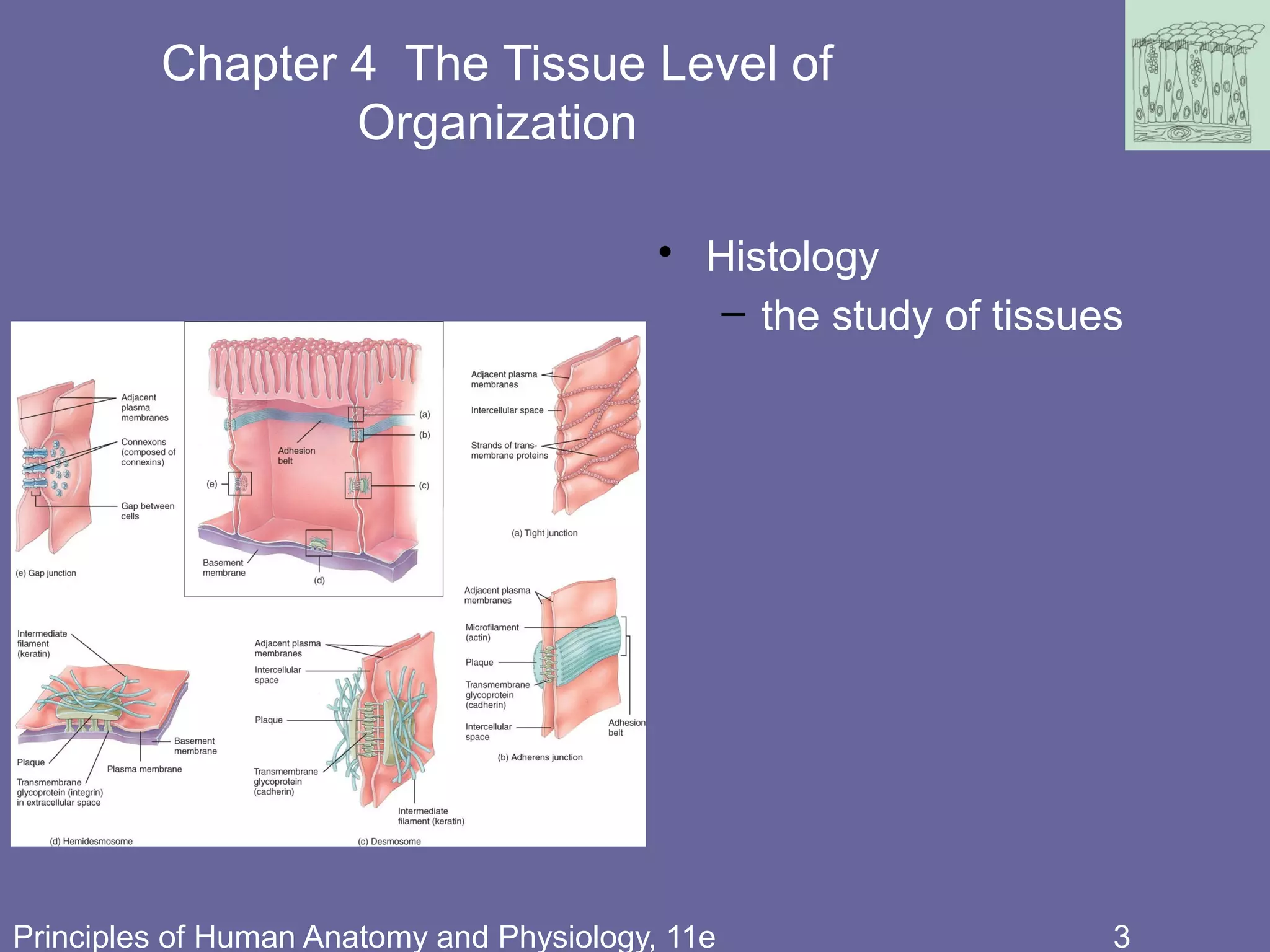


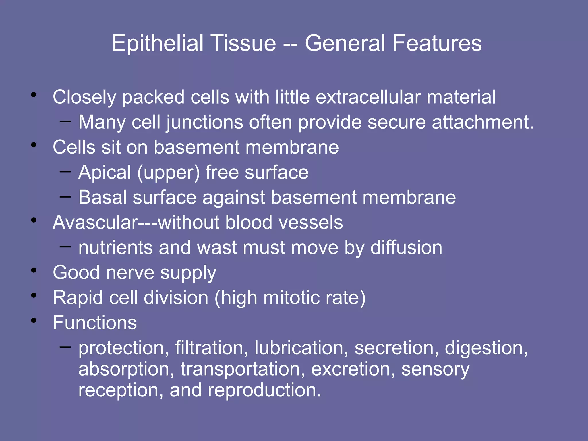
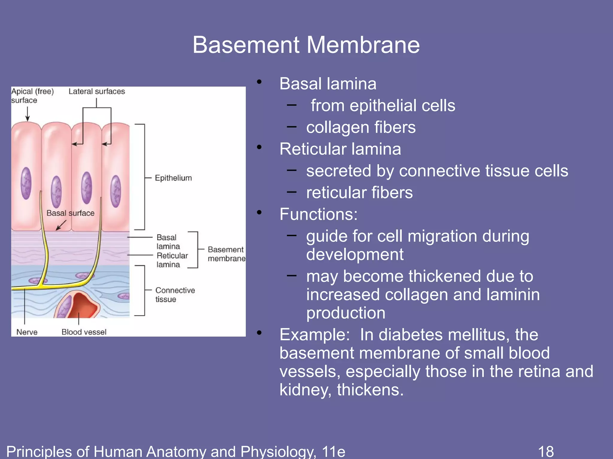


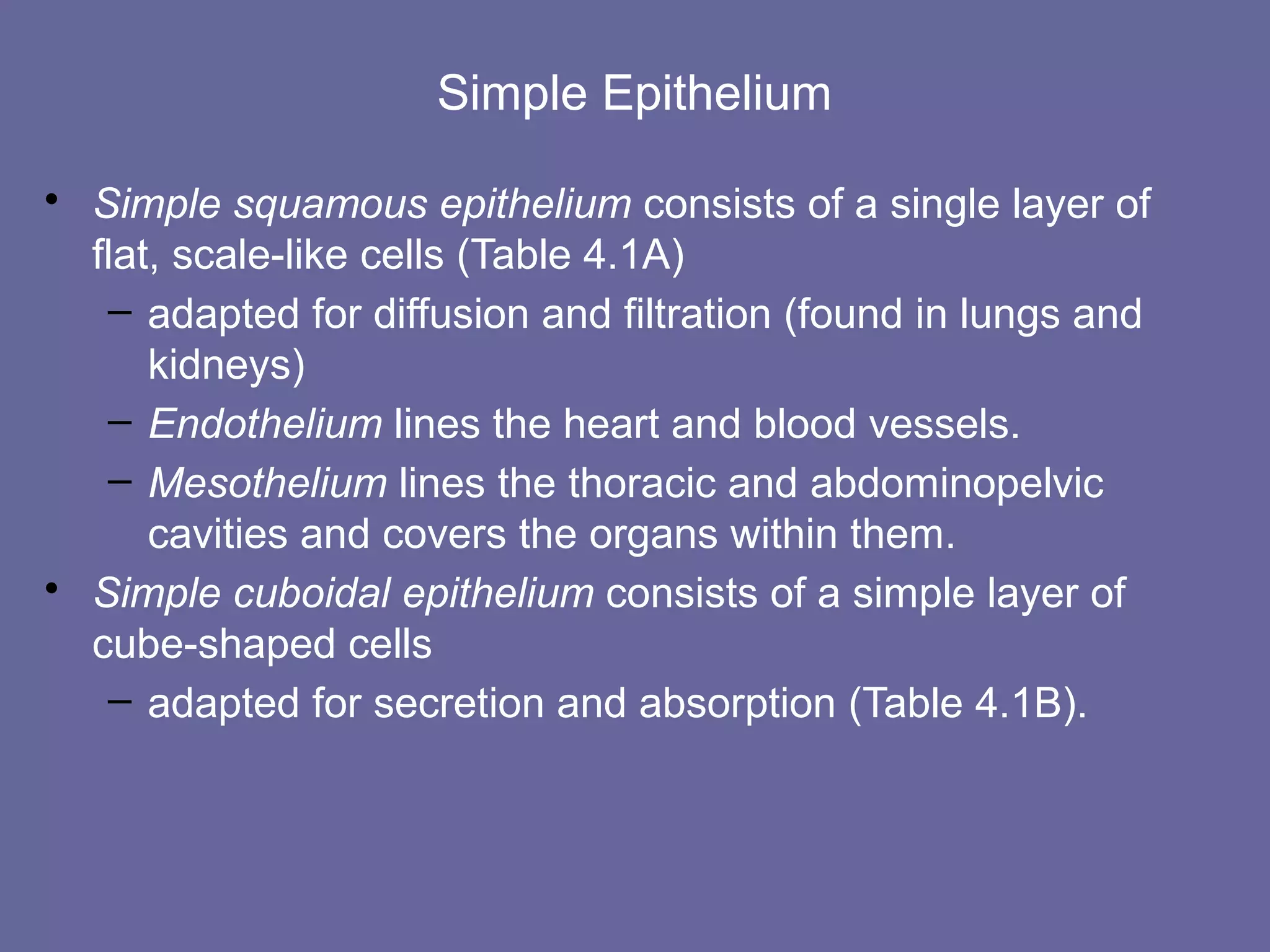

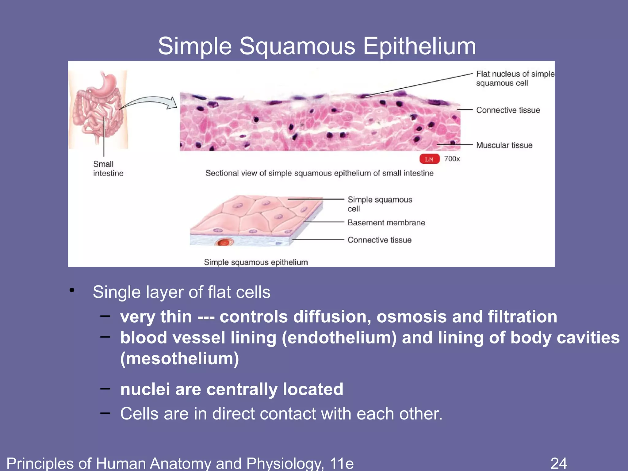


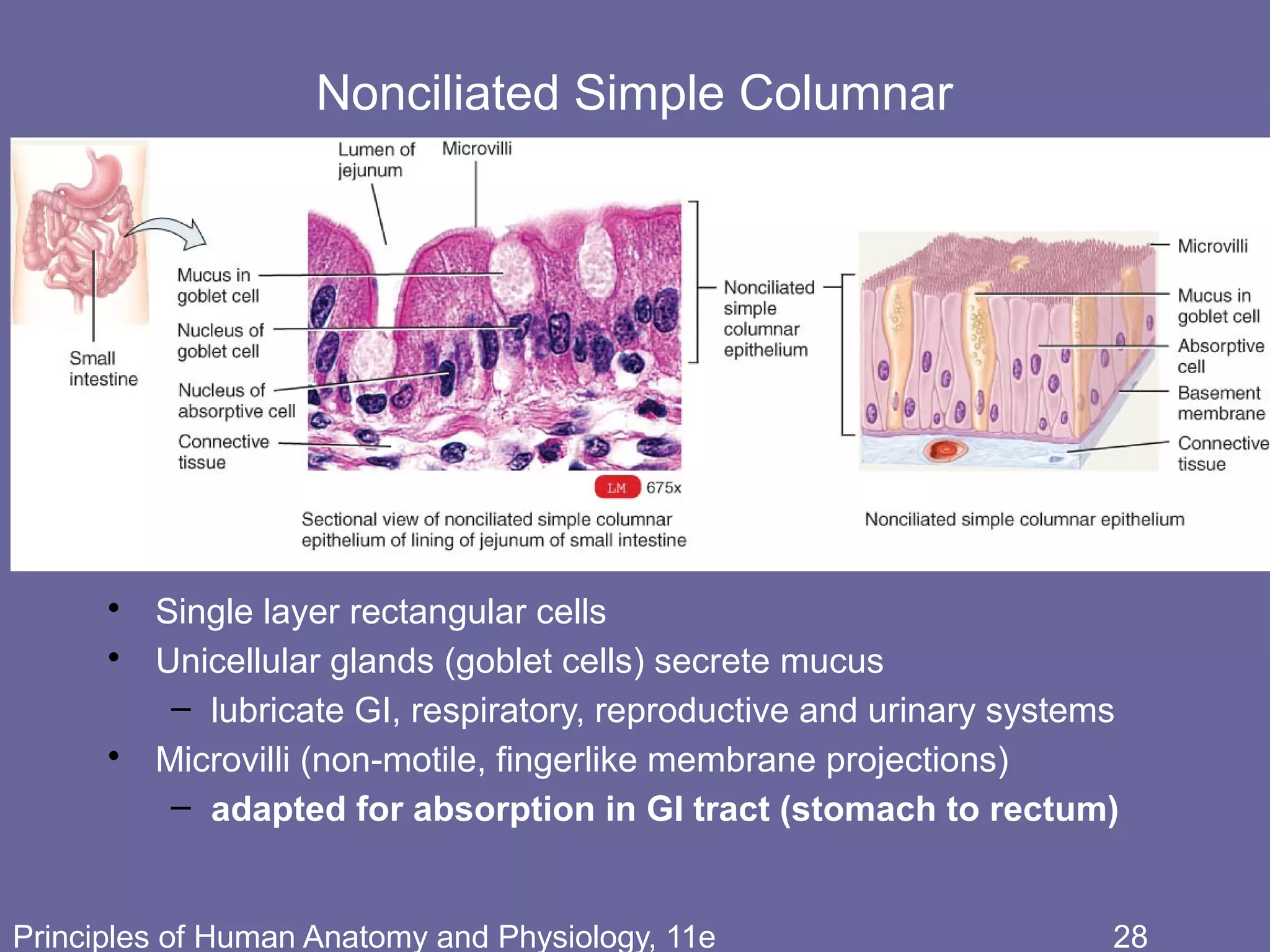


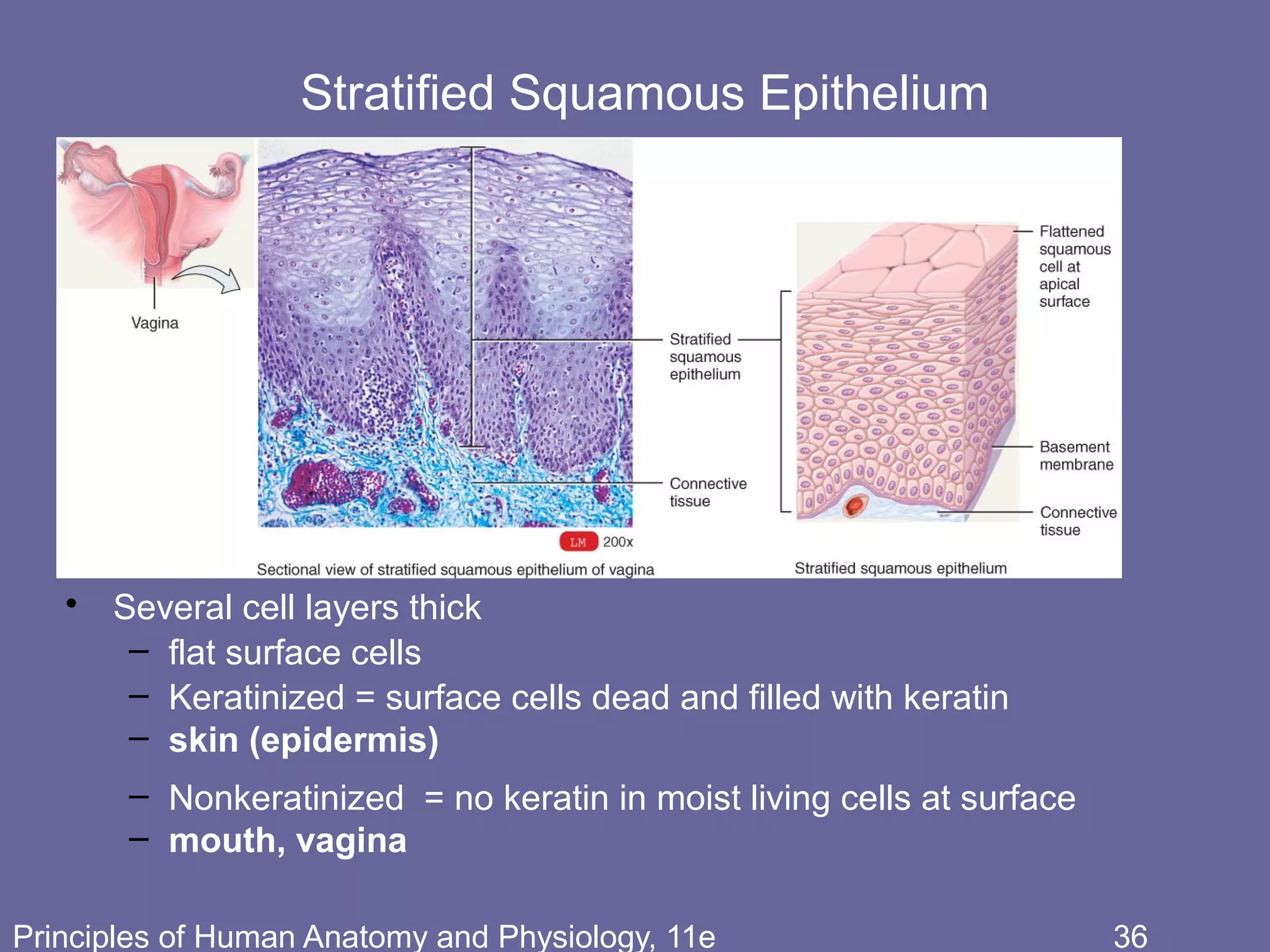


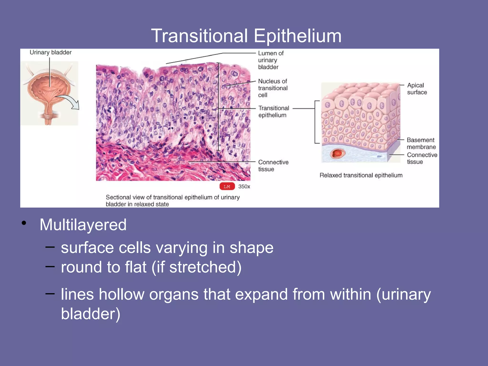


This document discusses the different types of tissues in the human body. It begins by defining what a tissue is and describing the four main types: epithelial, connective, muscle and nervous tissue. It then focuses on epithelial tissues, describing the different classifications of epithelium based on cell shape and layering. Simple epithelia are one cell layer thick, while stratified epithelia have two or more layers. Specific examples of each epithelial type are provided, along with diagrams to illustrate their structure.

![03 [chapter 3 the cellular level of organization]](https://cdn.slidesharecdn.com/ss_thumbnails/03chapter3thecellularleveloforganization-170828035521-thumbnail.jpg?width=640&height=640&fit=bounds)
![05 [chapter 5 the integumentary system]](https://cdn.slidesharecdn.com/ss_thumbnails/05chapter5theintegumentarysystem-170828035624-thumbnail.jpg?width=640&height=640&fit=bounds)
![10 [chapter 10 muscular tissue]](https://cdn.slidesharecdn.com/ss_thumbnails/10chapter10musculartissue-170828040153-thumbnail.jpg?width=640&height=640&fit=bounds)
![02 [chapter 2 the chemical level of organization]](https://cdn.slidesharecdn.com/ss_thumbnails/02chapter2thechemicalleveloforganization-170828035601-thumbnail.jpg?width=640&height=640&fit=bounds)
![01 [chapter 1 an introduction to the human body]](https://cdn.slidesharecdn.com/ss_thumbnails/01chapter1anintroductiontothehumanbody-170828035545-thumbnail.jpg?width=640&height=640&fit=bounds)
![06 [chapter 6 the skeletal system bone tissue]](https://cdn.slidesharecdn.com/ss_thumbnails/06chapter6theskeletalsystem-bonetissue-170828035633-thumbnail.jpg?width=640&height=640&fit=bounds)
![12 [chapter 12 nervous tissue]](https://cdn.slidesharecdn.com/ss_thumbnails/12chapter12nervoustissue-170828041102-thumbnail.jpg?width=640&height=640&fit=bounds)


![22 [chapter 22 the lymphatic system and immunity]](https://cdn.slidesharecdn.com/ss_thumbnails/22chapter22thelymphaticsystemandimmunity-170828153258-thumbnail.jpg?width=640&height=640&fit=bounds)

![21 [chapter 21 the cardiovascular system blood vessels and hemodynamics][11e]](https://cdn.slidesharecdn.com/ss_thumbnails/21chapter21thecardiovascularsystem-bloodvesselsandhemodynamics11e-170828043342-thumbnail.jpg?width=640&height=640&fit=bounds)


![19 [chapter 19 the cardiovascular system the blood]](https://cdn.slidesharecdn.com/ss_thumbnails/19chapter19thecardiovascularsystem-theblood-170828042033-thumbnail.jpg?width=640&height=640&fit=bounds)
![26 [chapter 26 the urinary system]](https://cdn.slidesharecdn.com/ss_thumbnails/26chapter26theurinarysystem-170828044011-thumbnail.jpg?width=640&height=640&fit=bounds)

![11 [chapter 11 the muscular system]](https://cdn.slidesharecdn.com/ss_thumbnails/11chapter11themuscularsystem-170828041038-thumbnail.jpg?width=640&height=640&fit=bounds)
![20 [chapter 20 the cardiovascular system the heart]](https://cdn.slidesharecdn.com/ss_thumbnails/20chapter20thecardiovascularsystem-theheart-170828133506-thumbnail.jpg?width=640&height=640&fit=bounds)
![07 [chapter 7 the skeletal system the axial skeleton]](https://cdn.slidesharecdn.com/ss_thumbnails/07chapter7theskeletalsystem-theaxialskeleton-170828035650-thumbnail.jpg?width=640&height=640&fit=bounds)

![14 [chapter 14 the brain and cranial nerves]](https://cdn.slidesharecdn.com/ss_thumbnails/14chapter14thebrainandcranialnerves-170828133437-thumbnail.jpg?width=640&height=640&fit=bounds)

![23 [chapter 23 the respiratory system]](https://cdn.slidesharecdn.com/ss_thumbnails/23chapter23therespiratorysystem-170828043650-thumbnail.jpg?width=640&height=640&fit=bounds)
![09 [chapter 9 joints]](https://cdn.slidesharecdn.com/ss_thumbnails/09chapter9joints-170828041032-thumbnail.jpg?width=640&height=640&fit=bounds)
![16 [chapter 16 sensory, motor, and integrative systems]](https://cdn.slidesharecdn.com/ss_thumbnails/16chapter16sensorymotorandintegrativesystems-170828041940-thumbnail.jpg?width=640&height=640&fit=bounds)


![24 [chapter 24 the digestive system][11e]](https://cdn.slidesharecdn.com/ss_thumbnails/24chapter24thedigestivesystem11e-170828043714-thumbnail.jpg?width=640&height=640&fit=bounds)
![11 [chapter 11 the muscular system][11e]](https://cdn.slidesharecdn.com/ss_thumbnails/11chapter11themuscularsystem11e-170828040427-thumbnail.jpg?width=640&height=640&fit=bounds)
![17 [chapter 17 the special senses]](https://cdn.slidesharecdn.com/ss_thumbnails/17chapter17thespecialsenses-170828041636-thumbnail.jpg?width=640&height=640&fit=bounds)
![25 [chapter 25 metabolism and nutrition]](https://cdn.slidesharecdn.com/ss_thumbnails/25chapter25metabolismandnutrition-170828145139-thumbnail.jpg?width=640&height=640&fit=bounds)
![08 [chapter 8 the skeletal system appendicular skeleton]](https://cdn.slidesharecdn.com/ss_thumbnails/08chapter8theskeletalsystem-appendicularskeleton-170828041008-thumbnail.jpg?width=640&height=640&fit=bounds)
![15 [chapter 15 the autonomic nervous system]](https://cdn.slidesharecdn.com/ss_thumbnails/15chapter15theautonomicnervoussystem-170828041929-thumbnail.jpg?width=640&height=640&fit=bounds)
![18 [chapter 18 the endocrine system]](https://cdn.slidesharecdn.com/ss_thumbnails/18chapter18theendocrinesystem-170828042016-thumbnail.jpg?width=640&height=640&fit=bounds)
![13 [chapter 13 the spinal cord and spinal nerves]](https://cdn.slidesharecdn.com/ss_thumbnails/13chapter13thespinalcordandspinalnerves-170828040950-thumbnail.jpg?width=640&height=640&fit=bounds)
![29 [chapter 29 development and inheritance]](https://cdn.slidesharecdn.com/ss_thumbnails/29chapter29developmentandinheritance-170828044352-thumbnail.jpg?width=640&height=640&fit=bounds)
![28 [chapter 28 the reproductive system]](https://cdn.slidesharecdn.com/ss_thumbnails/28chapter28thereproductivesystem-170828134050-thumbnail.jpg?width=640&height=640&fit=bounds)
![27 [chapter 27 fluid, electrolyte and acid base homeostasis]](https://cdn.slidesharecdn.com/ss_thumbnails/27chapter27fluidelectrolyteandacid-basehomeostasis-170828044023-thumbnail.jpg?width=640&height=640&fit=bounds)


