Downloaded 444 times
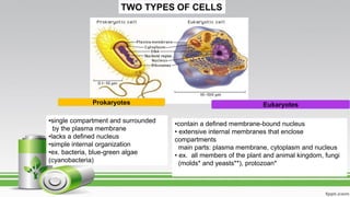
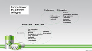
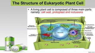
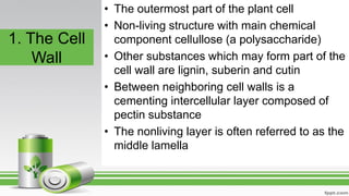
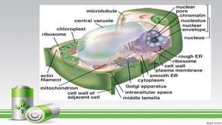
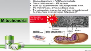


Plant cells come in two main types - eukaryotic and prokaryotic. Eukaryotic plant cells contain organelles like the cell wall, nucleus, chloroplasts, vacuoles, mitochondria and endoplasmic reticulum. The cell wall surrounds the plasma membrane and provides structure. Inside the plasma membrane is the cytoplasm and organelles. The nucleus contains the genetic material and directs cell activities. Chloroplasts perform photosynthesis using chlorophyll. Vacuoles store water and waste. Mitochondria generate energy. The endoplasmic reticulum modifies and transports proteins. Plant cells also contain inclusions like crystals and pigments.


