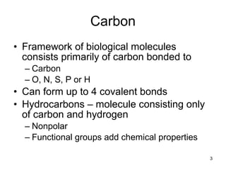
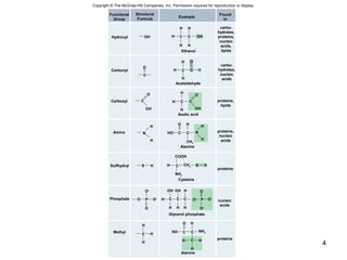
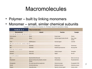
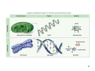
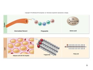
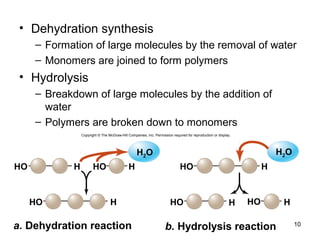
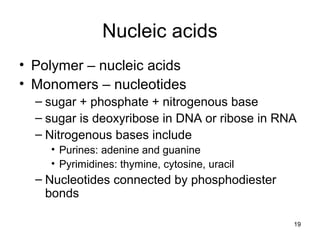
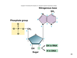
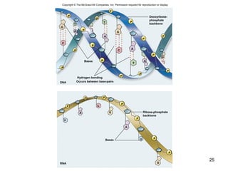
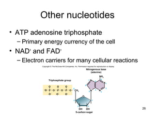
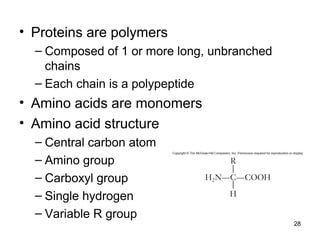
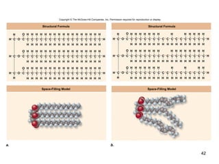
Chapter 03
Lecture and
Animation Outline
Copyright © The McGraw-Hill Companies, Inc. Permission required for reproduction or display.
To run the animations you must be in Slideshow View. Use
the buttons on the animation to play, pause, and turn
audio/text on or off.
Please Note: Once you have used any of the animation
functions (such as Play or Pause), you must first click on the
slide’s background before you can advance to the next slide.
1
The Chemical Building
Blocks of Life
Chapter 3
2
3
Carbon
• Framework of biological molecules
consists primarily of carbon bonded to
– Carbon
– O, N, S, P or H
• Can form up to 4 covalent bonds
• Hydrocarbons – molecule consisting only
of carbon and hydrogen
– Nonpolar
– Functional groups add chemical properties
4
Copyright © The McGraw-Hill Companies, Inc. Permission required for reproduction or display.
Hydroxyl
Carbonyl
Carboxyl
Amino
Sulfhydryl
Phosphate
Methyl
Example
OH
O
C
O
OH
C
H
H
N
S H
O
proteins
proteins
H
H
Glycerol phosphate
Alanine
Cysteine
Alanine
Acetic acid
Acetaldehyde
Ethanol
COOH
NH2
HH
H
HH
C OHC
O
H C C
H
H
H
H C
O
OHH
C
O H
N
H
H
CH3
H C HSCH2
NH2
OH OH H
P
O
P O–
O–
O
OCCCH
H H H
O H
HO C C
H C H
H
O–
O–
H
C
HO C C
H
Found
In
carbo-
hydrates,
proteins,
nucleic
acids,
lipids
carbo-
hydrates,
nucleic
acids
proteins,
lipids
proteins,
nucleic
acids
nucleic
acids
Functional
Group
Structural
Formula
5
Isomers
• Molecules with the same molecular or
empirical formula
– Structural isomers
– Stereoisomers – differ in how groups attached
• Enantiomers
– mirror image molecules
– chiral
– D-sugars and L-amino acids
6
Copyright © The McGraw-Hill Companies, Inc. Permission required for reproduction or display.
C
W
Y
W
Y
C
Z
X
Z
X
Mirror
7
Macromolecules
• Polymer – built by linking monomers
• Monomer – small, similar chemical subunits
8
Copyright © The McGraw-Hill Companies, Inc. Permission required for reproduction or display.
O
P
P
OH
HO
H
OH
H
OH
OH
H
H
H
O
Cellular Structure Polymer Monomer
Chromosome DNA strand Nucleotide
MonosaccharideStarchStarch grains in a chloroplast
5-carbon sugar
Phosphate
group
Nitrogenous base
CH2OH
CarbohydrateNucleicAcid
P
P
P
P
P
P
P
G
C
T
A
A
T
P
A
P
P
P
P
G
C
P
P
T
A
G
C
P
9
Ala
Ala
Val
Val
Ser O
N
H
C
H
OH
H
C
CH3
Intermediate filament Polypeptide Amino acid
Protein
Copyright © The McGraw-Hill Companies, Inc. Permission required for reproduction or display.
HO H H H H H H H H H H
HHO C C C C C C C C C C C C
H H H H H H H H H H H
Adipose cell with fat droplets
Triglyceride
Fatty acid
Lipid
10
• Dehydration synthesis
– Formation of large molecules by the removal of water
– Monomers are joined to form polymers
• Hydrolysis
– Breakdown of large molecules by the addition of
water
– Polymers are broken down to monomers
HO
HO H
HO HH HHO
HO H HHO
a. Dehydration reaction b. Hydrolysis reaction
H2O H2O
Copyright © The McGraw-Hill Companies, Inc. Permission required for reproduction or display.
11
Carbohydrates
• Molecules with a 1:2:1 ratio of carbon,
hydrogen, oxygen
• Empirical formula (CH2O)n
• C—H covalent bonds hold much energy
– Carbohydrates are good energy storage
molecules
– Examples: sugars, starch, glucose
12
Monosaccharides
• Simplest carbohydrate
• 6 carbon sugars play important roles
• Glucose C6H12O6
• Fructose is a structural isomer of glucose
• Galactose is a stereoisomer of glucose
• Enzymes that act on different sugars can
distinguish structural and stereoisomers of
this basic six-carbon skeleton
13
H
CH2OH
H
OH
OH H
H
H
C
CC
C
C
C
C
H
C H
HO
OHH
OHH C
OHH C
OHH C
H
OH
C HH
OH
H
OH
H
OH
H
OH
H
OH H
H
H
C
CC
C
C O
OH
OH H
H
H
C
CC
C
OC
OH
OH
O
H
23
5
14
6
5
4
3
2
1
23
5
6
14
23
5
14
α-glucose
or
β-glucose
O
O
CH2OH
Copyright © The McGraw-Hill Companies, Inc. Permission required for reproduction or display.
14
Fructose Glucose Galactose
H OH
O
C
C
C
C
C
C
H
H
HO H
H OH
H OH
H OH
H
OC
C
C
C
C
H
H
HO H
OH
H OH
H OH
OC
C
C
C
C
C
H
H
H
HO H
OH
H OH
H OH
CH OH HO H
Stereo-
isomer
Structural
isomer
Copyright © The McGraw-Hill Companies, Inc. Permission required for reproduction or display.
15
Disaccharides
• 2 monosaccharides linked together by
dehydration synthesis
• Used for sugar transport or energy storage
• Examples: sucrose, lactose, maltose
OH
SucroseFructoseα-glucose
HO
CH2OH
H
OH
H
OH
H
H
H
O
H
OH H
H
O
HO
H
OH
H
OH OH
H
H
O
H OH
OH H
HO
+
a. b.
Maltose
HO
H
OH
H
OH OH
H
H
O H
OH
H
OH
OH
H
H
H
O
OH
HO
H2O
CH2OH CH2OH CH2OH CH2OH CH2OH
CH2OH CH2OH
Copyright © The McGraw-Hill Companies, Inc. Permission required for reproduction or display.
16
Polysaccharides
• Long chains of monosaccharides
– Linked through dehydration synthesis
• Energy storage
– Plants use starch
– Animals use glycogen
• Structural support
– Plants use cellulose
– Arthropods and fungi use chitin
17
Glycogen
Amylose + Amylopectin
b.
a. c. 3.3 µm
Copyright © The McGraw-Hill Companies, Inc. Permission required for reproduction or display.
α-glucose
HO
CH2OH
H
OH
H
OH H
H
H
O
OH
4 1
α-1 4 linkages
CH2OH
OH
H
OH H
H
O
CH2OH
OH
H
OH H
H
O
CH2OH
OH
H
OH H
H
O
O O
H
α-1 4 linkage
a-1
CH2OH
O
H
OH
H
OH H
H
H
O
CH2
H
OH
H
OH H
H
H
O
O
CH2OH
H
OH
H
OH H
H
H
O
6 linkage
b: © Asa Th oresen/Photo Researchers, Inc.; c: © J. Carson/Custom Medical Stock Photo
7.5 µm
18
HO
CH2OH
H
OH
H
OH H
H
H
O
CH2OH
H H
OH
H
OH H
H
H
O
O
CH2OH
H
OH
H
OH H
H
H
O
OHH
H
OH H
H
CH2OH
O
β-1 4 linkages
OH
O O
O
4 1
Copyright © The McGraw-Hill Companies, Inc. Permission required for reproduction or display.
b: © Science VU/Visuals Unlimited
β-glucose
500 µmb.
a.
Nucleic acids
• Polymer – nucleic acids
• Monomers – nucleotides
– sugar + phosphate + nitrogenous base
– sugar is deoxyribose in DNA or ribose in RNA
– Nitrogenous bases include
• Purines: adenine and guanine
• Pyrimidines: thymine, cytosine, uracil
– Nucleotides connected by phosphodiester
bonds
19
20
5 ́
2
8
7 6
39
4
5
1
NH2
O
P
O-
–
O O CH2
Phosphate group
Sugar
Nitrogenous base
OH
NN
N
O
4 ́ 1 ́
2 ́3 ́
H in DNA
OH in RNA
Copyright © The McGraw-Hill Companies, Inc. Permission required for reproduction or display.
21
5′
3′
P
P
P
P
OH
Phosphate group
O
O
NH2CC
NN
N
C
H
N
C
CH
O
H
H
OC
NC
H
N
C
NH2
H
CH
a.
b.
O
O
O
C
NC
H
N
C
H3C
CH
H
O
O
C
NC
H
N
C
H
CH
CC
NN
N
C
H
N
C
CH
H
O
4′
5′
1′
3′ 2′
4′
5′
1′
3′ 2′
4′
5′
1′
3′ 2′
4′
5′
1′
3′
2′
Phosphodiester bonds
5-carbon sugar
Nitrogenous
base
NH2
GuanineAdenine
PurinesPyrimidines
Copyright © The McGraw-Hill Companies, Inc. Permission required for reproduction or display.
Cytosine
(both DNA and RNA)
Thymine
(DNA only)
Uracil
(RNA only)
Deoxyribonucleic acid (DNA)
• Encodes information for amino acid
sequence of proteins
– Sequence of bases
• Double helix – 2 polynucleotide strands
connected by hydrogen bonds
– Base-pairing rules
• A with T (or U in RNA)
• C with G
22
23
Copyright © The McGraw-Hill Companies, Inc. Permission required for reproduction or display.
5′ end
P
P
P
P
P
C
G
A
A
OH
T
T
Phosphodiester
bonds
3′ end
Sugar–phosphate
“backbone”
Hydrogenbonds
between
nitrogenous bases
24
Ribonucleic acid (RNA)
• RNA similar to DNA except
– Contains ribose instead of deoxyribose
– Contains uracil instead of thymine
• Single polynucleotide strand
• RNA uses information in DNA to specify
sequence of amino acids in proteins
25
Copyright © The McGraw-Hill Companies, Inc. Permission required for reproduction or display.
P
P
P
P
P
P
P
P
P
P
P
P
P
P
P
G
C
G
C
T
A
A
A
A
T
T
G
Bases
Hydrogen bonding
Occurs between base-pairs
P
P
PP
P
P
P
G
G
C
A
A
U
U
Bases
Deoxyribose-
phosphate
backbone
Ribose-phosphate
backbone
RNA
DNA
26
Other nucleotides
• ATP adenosine triphosphate
– Primary energy currency of the cell
• NAD+
and FAD+
– Electron carriers for many cellular reactions
4′
5
6
2
39
4
8
7
1′
3′ 2′
5′
1
CH2
OOO
O–
OH OH
NH2
N
N
N
N
O
–
O O O OP P P
Triphosphate group
5-carbon sugar
O
Nitrogenous base
(adenine)
O–
O–
Copyright © The McGraw-Hill Companies, Inc. Permission required for reproduction or display.
27
Proteins
Protein functions include:
1. Enzyme catalysis
2. Defense
3. Transport
4. Support
5. Motion
6. Regulation
7. Storage
28
• Proteins are polymers
– Composed of 1 or more long, unbranched
chains
– Each chain is a polypeptide
• Amino acids are monomers
• Amino acid structure
– Central carbon atom
– Amino group
– Carboxyl group
– Single hydrogen
– Variable R group
29
CH2
CH2
CH3
CH2
OH H O
S H
S
CH2
CH2
O
CH2
NH2
+
C C O–
H3N+
C C O–
H3N+
C C O–
H3N+
C C O–
H3N+
C C O–
H3N+
C C O–
H3N+
C C O–
H3N+
C C O–
H3N+
C C O–
H3N+
C C O–
H3N+
C C O–
H3N+
C C O–
H3N+
C C O–
H3N+
C C O–
H3N+
C C O–
H3N+
C C O–
H3N+ C C O–
H3N+
C C O–
H3N+
CH C O–
C C O–
NH3
+
CH3
OH
CH2
OH
OH
H
OH
OH
CH3CH3
CH
CH2
OH
CH3CH3
CH
CH2
OH
NH2O
C
CH2
OH
O–
O
C
CH2
CH2
CH2 NH3
+
OH
CH2
CH2
CH2
OH
O–
O
C
CH2
CH2
OH
NH2O
C
H C CH3
CH2
CH3
OH
H C OH
CH3
OH
CH2
CH
C N
HC NH+
H O
H
CH2
NH
C
H O
CH2
H O
OH
CH2
H O
CH2
CH2
NH
NH2
C
OH
CH2
NH3
+
Nonpolar Polar uncharged Charged
Proline
(Pro)
Methionine
(Met)
Cysteine
(Cys)
Tryptophan
(Trp)
Tyrosine
(Tyr)
Phenylalanine
(Phe)
Glycine
(Gly)
Leucine
(Leu)
Glutamine
(Gln)
Isoleucine
(Ile)
Asparagine
(Asn)
Asparticacid
(Asp)
Histidine
(His)
Lysine
(Lys)
Glutamic acid
(Glu)
Threonine
(Thr) Arginine
(Arg)
(Ser)
Serine
(Ala)
Alanine
NonaromaticAromaticSpecialfunction
Copyright © The McGraw-Hill Companies, Inc. Permission required for reproduction or display.
Valine
(Val)
30
• Amino acids joined by dehydration
synthesis
– Peptide bond
RH
H
R
H O
R
OH
RH
OH
H2O
H
O
H
OH OH
OHH
CC C C
CC CC
N
N N
NH H
Amino acid
Dipeptide
Amino acid
31
4 Levels of structure
• The shape of a protein determines its
function
1. Primary structure – sequence of amino
acids
2. Secondary structure – interaction of groups
in the peptide backbone
α helix
β sheet
4 Levels of structure
3. Tertiary structure – final folded shape of a
globular protein
– Stabilized by a number of forces
– Final level of structure for proteins consisting
of only a single polypeptide chain
4. Quaternary structure – arrangement of
individual chains (subunits) in a protein with
2 or more polypeptide chains
32
33
•
•
•
•
•
•
•
•
•
•
••
•
•
•
•
•
•
•
•
•
•
•
•
•
•
•
•
•
•
•
•
•
•
•
•
•
•
•
•
•
•
•
•
•
•
•
•
•
••
•
•
•
•
•
•
•
•
•
•
•
•
•
•
•
•
•
•
•
•
•
•
•
•
•
•
•
Primary Structure
Secondary Structure Secondary Structure
Tertiary Structure Quaternary Structure
N
H
H H
C C C N C C NC
O
O H
OH H
R
R
R
β-pleated sheet α-helix
C
H
R
Copyright © The McGraw-Hill Companies, Inc. Permission required for reproduction or display.
The primary structure can fold
Into a pleated sheet, or turn into a helix
Additional structural characteristics
• Motifs
– Common elements of secondary structure
seen in many polypeptides
– Useful in determining the function of unknown
proteins
• Domains
– Functional units within a larger structure
– Most proteins made of multiple domains that
perform different parts of the protein’s function
34
35
Domain 3 Domain 2
Domain 1
DomainsMotifs
Copyright © The McGraw-Hill Companies, Inc. Permission required for reproduction or display.
β-α-β
motif
Helix-turn-Helix
motif
36
• Once thought newly made proteins folded
spontaneously
• Chaperone proteins help protein fold
correctly
• Deficiencies in chaperone proteins
implicated in certain diseases
– Cystic fibrosis is a hereditary disorder
• In some individuals, protein appears to have correct
amino acid sequence but fails to fold
Chaperones
37
Chance for protein to refold
ADP + P
Cap
ATP
Copyright © The McGraw-Hill Companies, Inc. Permission required for reproduction or display.
Correctly
folded
protein
Misfolded
protein
Chaperone
protein
Denaturation
• Protein loses
structure and function
• Due to environmental
conditions
– pH
– Temperature
– Ionic concentration of
solution
38
Properly folded protein
Denaturation
Denatured
protein
Copyright © The McGraw-Hill Companies, Inc. Permission required for reproduction or display.
39
Please note that due to differing
operating systems, some animations
will not appear until the presentation is
viewed in Presentation Mode (Slide
Show view). You may see blank slides
in the “Normal” or “Slide Sorter” views.
All animations will appear after viewing
in Presentation Mode and playing each
animation. Most animations will require
the latest version of the Flash Player,
which is available at
http://get.adobe.com/flashplayer.
40
Lipids
• Loosely defined group of molecules with
one main chemical characteristic
– They are insoluble in water
• High proportion of nonpolar C—H bonds
causes the molecule to be hydrophobic
• Fats, oils, waxes, and even some vitamins
41
Fats
• Triglycerides
– Composed of 1 glycerol and 3 fatty acids
• Fatty acids
– Need not be identical
– Chain length varies
– Saturated – no double bonds between carbon
atoms
• Higher melting point, animal origin
– Unsaturated – 1 or more double bonds
• Low melting point, plant origin
– Trans fats produced industrially
42
C
C
H
H
C
H
H
C
H
H
O
C
H
CH C
H
H
C
H
H
C
H
H
C
H
H
C
H
H
C
H
H
C
H
C
H H
C
H
C C
HH H H
C
H
C C
H
O
H
HH H
C
H
H
C
H
H
O
C
H
CH C
H
H
C
H
H
C
H
H
C
H
H
C
H
H
C
H
H
C
H
C
H
C
H
C
H
C
HH H
C
H
C
H
C
H H
O
H
H H
C
H
H
C
H
H
O
C
H
CH C
H
H
C
H
H
C
H
H
C
H
H
C
H
H
C
H
H
C
H
C
H H
C
H H
C
H
C
H
H
C
H H
C
HH
C
H H
CO
O
C
H
C
H
H
C
H
H
C
H
H
CH C
H
H
C
H
H
C
H
H
C
H
H
C
H
H
C
H
H
C
H
H
C
H
H
C
H
H
C
H
H
C
H
H
C
H
O
O
C
H
C
H
H
C
H
H
C
H
H
CH C
H
H
C
H
H
C
H
H
C
H
H
C
H
H
C
H
H
C
H
H
C
H
H
C
H
H
C
H
H
C
H
H
C
H
O
O
C
H
C
H
H
C
H
H
C
H
H
CH C
H
H
C
H
H
C
H
H
C
H
H
C
H
H
C
H
H
C
H
H
C
H
H
C
H
H
C
H
H
C
H
H
C
H
H
C
H
H
C
H
H
H
C
H
H
C
H
H
H
C
H
H
C
H
HO
H
H
Structural Formula Structural Formula
Space-Filling Model Space-Filling Model
a. b.
Copyright © The McGraw-Hill Companies, Inc. Permission required for reproduction or display.
43
Phospholipids
• Composed of
– Glycerol
– 2 fatty acids – nonpolar “tails”
– A phosphate group – polar “head”
• Form all biological membranes
44
NonpolarHydrophobicTailsPolarHydrophilicHeads
Choline
Phosphate
Glycerol
a. b. c. d.
CH2
CH2
CH2
H2C
O
O
O P
C
O O
OO
C
H
C
O-
N+
(CH3)3
Copyright © The McGraw-Hill Companies, Inc. Permission required for reproduction or display.
F
a
t
t
y
a
c
i
d
F
a
t
t
y
a
c
i
d
CH
CH2
CH2
CH2
CH2
CH2
CH2
CH2
CH2
CH2
CH2
CH2
CH2
CH2
CH2
CH2
CH3
CH
CH2
CH2
CH2
CH2
CH2
CH2
CH
CH
CH2
CH2
CH2
CH2
CH2
CH2
CH2
CH3
45
• Micelles – lipid molecules orient with polar
(hydrophilic) head toward water and
nonpolar (hydrophobic) tails away from
water
a.
Water
Lipid head
(hydrophilic)
Lipid tail
(hydrophobic)
Copyright © The McGraw-Hill Companies, Inc. Permission required for reproduction or display.
• Phospholipid bilayer – more complicated
structure where 2 layers form
– Hydrophilic heads point outward
– Hydrophobic tails point inward toward each
other
46
b.
Water
Water
Copyright © The McGraw-Hill Companies, Inc. Permission required for reproduction or display.

Ch 3 PowerPoint Biology 201

  • 1.
    Chapter 03 Lecture and AnimationOutline Copyright © The McGraw-Hill Companies, Inc. Permission required for reproduction or display. To run the animations you must be in Slideshow View. Use the buttons on the animation to play, pause, and turn audio/text on or off. Please Note: Once you have used any of the animation functions (such as Play or Pause), you must first click on the slide’s background before you can advance to the next slide. 1
  • 2.
    The Chemical Building Blocksof Life Chapter 3 2
  • 3.
    3 Carbon • Framework ofbiological molecules consists primarily of carbon bonded to – Carbon – O, N, S, P or H • Can form up to 4 covalent bonds • Hydrocarbons – molecule consisting only of carbon and hydrogen – Nonpolar – Functional groups add chemical properties
  • 4.
    4 Copyright © TheMcGraw-Hill Companies, Inc. Permission required for reproduction or display. Hydroxyl Carbonyl Carboxyl Amino Sulfhydryl Phosphate Methyl Example OH O C O OH C H H N S H O proteins proteins H H Glycerol phosphate Alanine Cysteine Alanine Acetic acid Acetaldehyde Ethanol COOH NH2 HH H HH C OHC O H C C H H H H C O OHH C O H N H H CH3 H C HSCH2 NH2 OH OH H P O P O– O– O OCCCH H H H O H HO C C H C H H O– O– H C HO C C H Found In carbo- hydrates, proteins, nucleic acids, lipids carbo- hydrates, nucleic acids proteins, lipids proteins, nucleic acids nucleic acids Functional Group Structural Formula
  • 5.
    5 Isomers • Molecules withthe same molecular or empirical formula – Structural isomers – Stereoisomers – differ in how groups attached • Enantiomers – mirror image molecules – chiral – D-sugars and L-amino acids
  • 6.
    6 Copyright © TheMcGraw-Hill Companies, Inc. Permission required for reproduction or display. C W Y W Y C Z X Z X Mirror
  • 7.
    7 Macromolecules • Polymer –built by linking monomers • Monomer – small, similar chemical subunits
  • 8.
    8 Copyright © TheMcGraw-Hill Companies, Inc. Permission required for reproduction or display. O P P OH HO H OH H OH OH H H H O Cellular Structure Polymer Monomer Chromosome DNA strand Nucleotide MonosaccharideStarchStarch grains in a chloroplast 5-carbon sugar Phosphate group Nitrogenous base CH2OH CarbohydrateNucleicAcid P P P P P P P G C T A A T P A P P P P G C P P T A G C P
  • 9.
    9 Ala Ala Val Val Ser O N H C H OH H C CH3 Intermediate filamentPolypeptide Amino acid Protein Copyright © The McGraw-Hill Companies, Inc. Permission required for reproduction or display. HO H H H H H H H H H H HHO C C C C C C C C C C C C H H H H H H H H H H H Adipose cell with fat droplets Triglyceride Fatty acid Lipid
  • 10.
    10 • Dehydration synthesis –Formation of large molecules by the removal of water – Monomers are joined to form polymers • Hydrolysis – Breakdown of large molecules by the addition of water – Polymers are broken down to monomers HO HO H HO HH HHO HO H HHO a. Dehydration reaction b. Hydrolysis reaction H2O H2O Copyright © The McGraw-Hill Companies, Inc. Permission required for reproduction or display.
  • 11.
    11 Carbohydrates • Molecules witha 1:2:1 ratio of carbon, hydrogen, oxygen • Empirical formula (CH2O)n • C—H covalent bonds hold much energy – Carbohydrates are good energy storage molecules – Examples: sugars, starch, glucose
  • 12.
    12 Monosaccharides • Simplest carbohydrate •6 carbon sugars play important roles • Glucose C6H12O6 • Fructose is a structural isomer of glucose • Galactose is a stereoisomer of glucose • Enzymes that act on different sugars can distinguish structural and stereoisomers of this basic six-carbon skeleton
  • 13.
    13 H CH2OH H OH OH H H H C CC C C C C H C H HO OHH OHHC OHH C OHH C H OH C HH OH H OH H OH H OH H OH H H H C CC C C O OH OH H H H C CC C OC OH OH O H 23 5 14 6 5 4 3 2 1 23 5 6 14 23 5 14 α-glucose or β-glucose O O CH2OH Copyright © The McGraw-Hill Companies, Inc. Permission required for reproduction or display.
  • 14.
    14 Fructose Glucose Galactose HOH O C C C C C C H H HO H H OH H OH H OH H OC C C C C H H HO H OH H OH H OH OC C C C C C H H H HO H OH H OH H OH CH OH HO H Stereo- isomer Structural isomer Copyright © The McGraw-Hill Companies, Inc. Permission required for reproduction or display.
  • 15.
    15 Disaccharides • 2 monosaccharideslinked together by dehydration synthesis • Used for sugar transport or energy storage • Examples: sucrose, lactose, maltose OH SucroseFructoseα-glucose HO CH2OH H OH H OH H H H O H OH H H O HO H OH H OH OH H H O H OH OH H HO + a. b. Maltose HO H OH H OH OH H H O H OH H OH OH H H H O OH HO H2O CH2OH CH2OH CH2OH CH2OH CH2OH CH2OH CH2OH Copyright © The McGraw-Hill Companies, Inc. Permission required for reproduction or display.
  • 16.
    16 Polysaccharides • Long chainsof monosaccharides – Linked through dehydration synthesis • Energy storage – Plants use starch – Animals use glycogen • Structural support – Plants use cellulose – Arthropods and fungi use chitin
  • 17.
    17 Glycogen Amylose + Amylopectin b. a.c. 3.3 µm Copyright © The McGraw-Hill Companies, Inc. Permission required for reproduction or display. α-glucose HO CH2OH H OH H OH H H H O OH 4 1 α-1 4 linkages CH2OH OH H OH H H O CH2OH OH H OH H H O CH2OH OH H OH H H O O O H α-1 4 linkage a-1 CH2OH O H OH H OH H H H O CH2 H OH H OH H H H O O CH2OH H OH H OH H H H O 6 linkage b: © Asa Th oresen/Photo Researchers, Inc.; c: © J. Carson/Custom Medical Stock Photo 7.5 µm
  • 18.
    18 HO CH2OH H OH H OH H H H O CH2OH H H OH H OHH H H O O CH2OH H OH H OH H H H O OHH H OH H H CH2OH O β-1 4 linkages OH O O O 4 1 Copyright © The McGraw-Hill Companies, Inc. Permission required for reproduction or display. b: © Science VU/Visuals Unlimited β-glucose 500 µmb. a.
  • 19.
    Nucleic acids • Polymer– nucleic acids • Monomers – nucleotides – sugar + phosphate + nitrogenous base – sugar is deoxyribose in DNA or ribose in RNA – Nitrogenous bases include • Purines: adenine and guanine • Pyrimidines: thymine, cytosine, uracil – Nucleotides connected by phosphodiester bonds 19
  • 20.
    20 5 ́ 2 8 7 6 39 4 5 1 NH2 O P O- – OO CH2 Phosphate group Sugar Nitrogenous base OH NN N O 4 ́ 1 ́ 2 ́3 ́ H in DNA OH in RNA Copyright © The McGraw-Hill Companies, Inc. Permission required for reproduction or display.
  • 21.
    21 5′ 3′ P P P P OH Phosphate group O O NH2CC NN N C H N C CH O H H OC NC H N C NH2 H CH a. b. O O O C NC H N C H3C CH H O O C NC H N C H CH CC NN N C H N C CH H O 4′ 5′ 1′ 3′ 2′ 4′ 5′ 1′ 3′2′ 4′ 5′ 1′ 3′ 2′ 4′ 5′ 1′ 3′ 2′ Phosphodiester bonds 5-carbon sugar Nitrogenous base NH2 GuanineAdenine PurinesPyrimidines Copyright © The McGraw-Hill Companies, Inc. Permission required for reproduction or display. Cytosine (both DNA and RNA) Thymine (DNA only) Uracil (RNA only)
  • 22.
    Deoxyribonucleic acid (DNA) •Encodes information for amino acid sequence of proteins – Sequence of bases • Double helix – 2 polynucleotide strands connected by hydrogen bonds – Base-pairing rules • A with T (or U in RNA) • C with G 22
  • 23.
    23 Copyright © TheMcGraw-Hill Companies, Inc. Permission required for reproduction or display. 5′ end P P P P P C G A A OH T T Phosphodiester bonds 3′ end Sugar–phosphate “backbone” Hydrogenbonds between nitrogenous bases
  • 24.
    24 Ribonucleic acid (RNA) •RNA similar to DNA except – Contains ribose instead of deoxyribose – Contains uracil instead of thymine • Single polynucleotide strand • RNA uses information in DNA to specify sequence of amino acids in proteins
  • 25.
    25 Copyright © TheMcGraw-Hill Companies, Inc. Permission required for reproduction or display. P P P P P P P P P P P P P P P G C G C T A A A A T T G Bases Hydrogen bonding Occurs between base-pairs P P PP P P P G G C A A U U Bases Deoxyribose- phosphate backbone Ribose-phosphate backbone RNA DNA
  • 26.
    26 Other nucleotides • ATPadenosine triphosphate – Primary energy currency of the cell • NAD+ and FAD+ – Electron carriers for many cellular reactions 4′ 5 6 2 39 4 8 7 1′ 3′ 2′ 5′ 1 CH2 OOO O– OH OH NH2 N N N N O – O O O OP P P Triphosphate group 5-carbon sugar O Nitrogenous base (adenine) O– O– Copyright © The McGraw-Hill Companies, Inc. Permission required for reproduction or display.
  • 27.
    27 Proteins Protein functions include: 1.Enzyme catalysis 2. Defense 3. Transport 4. Support 5. Motion 6. Regulation 7. Storage
  • 28.
    28 • Proteins arepolymers – Composed of 1 or more long, unbranched chains – Each chain is a polypeptide • Amino acids are monomers • Amino acid structure – Central carbon atom – Amino group – Carboxyl group – Single hydrogen – Variable R group
  • 29.
    29 CH2 CH2 CH3 CH2 OH H O SH S CH2 CH2 O CH2 NH2 + C C O– H3N+ C C O– H3N+ C C O– H3N+ C C O– H3N+ C C O– H3N+ C C O– H3N+ C C O– H3N+ C C O– H3N+ C C O– H3N+ C C O– H3N+ C C O– H3N+ C C O– H3N+ C C O– H3N+ C C O– H3N+ C C O– H3N+ C C O– H3N+ C C O– H3N+ C C O– H3N+ CH C O– C C O– NH3 + CH3 OH CH2 OH OH H OH OH CH3CH3 CH CH2 OH CH3CH3 CH CH2 OH NH2O C CH2 OH O– O C CH2 CH2 CH2 NH3 + OH CH2 CH2 CH2 OH O– O C CH2 CH2 OH NH2O C H C CH3 CH2 CH3 OH H C OH CH3 OH CH2 CH C N HC NH+ H O H CH2 NH C H O CH2 H O OH CH2 H O CH2 CH2 NH NH2 C OH CH2 NH3 + Nonpolar Polar uncharged Charged Proline (Pro) Methionine (Met) Cysteine (Cys) Tryptophan (Trp) Tyrosine (Tyr) Phenylalanine (Phe) Glycine (Gly) Leucine (Leu) Glutamine (Gln) Isoleucine (Ile) Asparagine (Asn) Asparticacid (Asp) Histidine (His) Lysine (Lys) Glutamic acid (Glu) Threonine (Thr) Arginine (Arg) (Ser) Serine (Ala) Alanine NonaromaticAromaticSpecialfunction Copyright © The McGraw-Hill Companies, Inc. Permission required for reproduction or display. Valine (Val)
  • 30.
    30 • Amino acidsjoined by dehydration synthesis – Peptide bond RH H R H O R OH RH OH H2O H O H OH OH OHH CC C C CC CC N N N NH H Amino acid Dipeptide Amino acid
  • 31.
    31 4 Levels ofstructure • The shape of a protein determines its function 1. Primary structure – sequence of amino acids 2. Secondary structure – interaction of groups in the peptide backbone α helix β sheet
  • 32.
    4 Levels ofstructure 3. Tertiary structure – final folded shape of a globular protein – Stabilized by a number of forces – Final level of structure for proteins consisting of only a single polypeptide chain 4. Quaternary structure – arrangement of individual chains (subunits) in a protein with 2 or more polypeptide chains 32
  • 33.
    33 • • • • • • • • • • •• • • • • • • • • • • • • • • • • • • • • • • • • • • • • • • • • • • • • • •• • • • • • • • • • • • • • • • • • • • • • • • • • • • Primary Structure Secondary StructureSecondary Structure Tertiary Structure Quaternary Structure N H H H C C C N C C NC O O H OH H R R R β-pleated sheet α-helix C H R Copyright © The McGraw-Hill Companies, Inc. Permission required for reproduction or display. The primary structure can fold Into a pleated sheet, or turn into a helix
  • 34.
    Additional structural characteristics •Motifs – Common elements of secondary structure seen in many polypeptides – Useful in determining the function of unknown proteins • Domains – Functional units within a larger structure – Most proteins made of multiple domains that perform different parts of the protein’s function 34
  • 35.
    35 Domain 3 Domain2 Domain 1 DomainsMotifs Copyright © The McGraw-Hill Companies, Inc. Permission required for reproduction or display. β-α-β motif Helix-turn-Helix motif
  • 36.
    36 • Once thoughtnewly made proteins folded spontaneously • Chaperone proteins help protein fold correctly • Deficiencies in chaperone proteins implicated in certain diseases – Cystic fibrosis is a hereditary disorder • In some individuals, protein appears to have correct amino acid sequence but fails to fold Chaperones
  • 37.
    37 Chance for proteinto refold ADP + P Cap ATP Copyright © The McGraw-Hill Companies, Inc. Permission required for reproduction or display. Correctly folded protein Misfolded protein Chaperone protein
  • 38.
    Denaturation • Protein loses structureand function • Due to environmental conditions – pH – Temperature – Ionic concentration of solution 38 Properly folded protein Denaturation Denatured protein Copyright © The McGraw-Hill Companies, Inc. Permission required for reproduction or display.
  • 39.
    39 Please note thatdue to differing operating systems, some animations will not appear until the presentation is viewed in Presentation Mode (Slide Show view). You may see blank slides in the “Normal” or “Slide Sorter” views. All animations will appear after viewing in Presentation Mode and playing each animation. Most animations will require the latest version of the Flash Player, which is available at http://get.adobe.com/flashplayer.
  • 40.
    40 Lipids • Loosely definedgroup of molecules with one main chemical characteristic – They are insoluble in water • High proportion of nonpolar C—H bonds causes the molecule to be hydrophobic • Fats, oils, waxes, and even some vitamins
  • 41.
    41 Fats • Triglycerides – Composedof 1 glycerol and 3 fatty acids • Fatty acids – Need not be identical – Chain length varies – Saturated – no double bonds between carbon atoms • Higher melting point, animal origin – Unsaturated – 1 or more double bonds • Low melting point, plant origin – Trans fats produced industrially
  • 42.
    42 C C H H C H H C H H O C H CH C H H C H H C H H C H H C H H C H H C H C H H C H CC HH H H C H C C H O H HH H C H H C H H O C H CH C H H C H H C H H C H H C H H C H H C H C H C H C H C HH H C H C H C H H O H H H C H H C H H O C H CH C H H C H H C H H C H H C H H C H H C H C H H C H H C H C H H C H H C HH C H H CO O C H C H H C H H C H H CH C H H C H H C H H C H H C H H C H H C H H C H H C H H C H H C H H C H O O C H C H H C H H C H H CH C H H C H H C H H C H H C H H C H H C H H C H H C H H C H H C H H C H O O C H C H H C H H C H H CH C H H C H H C H H C H H C H H C H H C H H C H H C H H C H H C H H C H H C H H C H H H C H H C H H H C H H C H HO H H Structural Formula Structural Formula Space-Filling Model Space-Filling Model a. b. Copyright © The McGraw-Hill Companies, Inc. Permission required for reproduction or display.
  • 43.
    43 Phospholipids • Composed of –Glycerol – 2 fatty acids – nonpolar “tails” – A phosphate group – polar “head” • Form all biological membranes
  • 44.
    44 NonpolarHydrophobicTailsPolarHydrophilicHeads Choline Phosphate Glycerol a. b. c.d. CH2 CH2 CH2 H2C O O O P C O O OO C H C O- N+ (CH3)3 Copyright © The McGraw-Hill Companies, Inc. Permission required for reproduction or display. F a t t y a c i d F a t t y a c i d CH CH2 CH2 CH2 CH2 CH2 CH2 CH2 CH2 CH2 CH2 CH2 CH2 CH2 CH2 CH2 CH3 CH CH2 CH2 CH2 CH2 CH2 CH2 CH CH CH2 CH2 CH2 CH2 CH2 CH2 CH2 CH3
  • 45.
    45 • Micelles –lipid molecules orient with polar (hydrophilic) head toward water and nonpolar (hydrophobic) tails away from water a. Water Lipid head (hydrophilic) Lipid tail (hydrophobic) Copyright © The McGraw-Hill Companies, Inc. Permission required for reproduction or display.
  • 46.
    • Phospholipid bilayer– more complicated structure where 2 layers form – Hydrophilic heads point outward – Hydrophobic tails point inward toward each other 46 b. Water Water Copyright © The McGraw-Hill Companies, Inc. Permission required for reproduction or display.