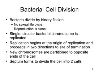
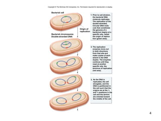
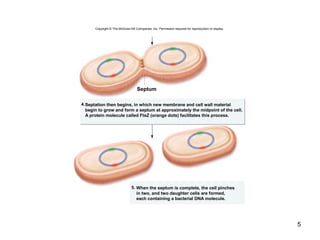
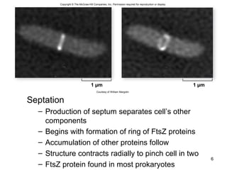
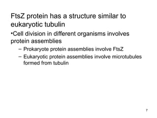
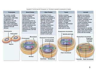
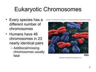
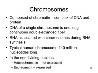
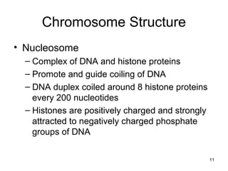
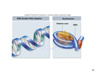
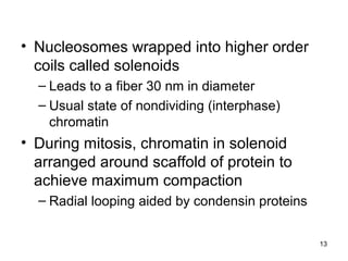
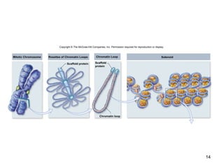
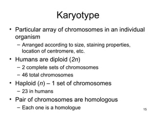
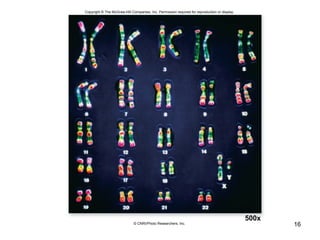
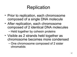
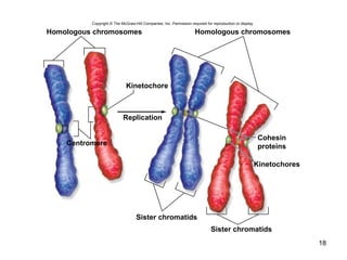
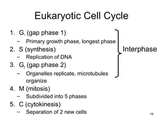
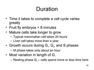
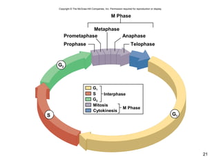
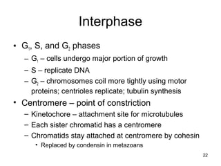
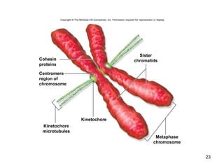
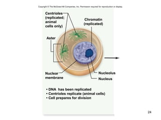
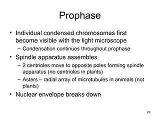
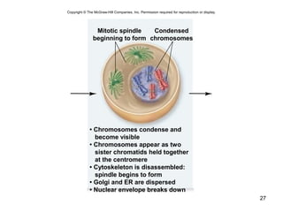
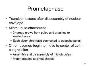
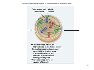
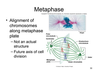
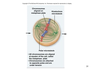
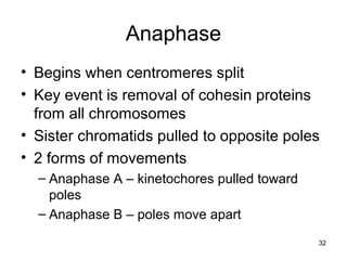
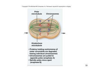
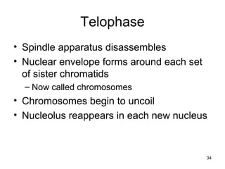
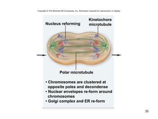
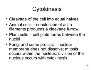
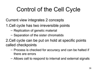
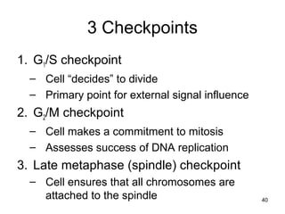
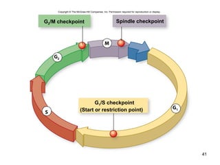
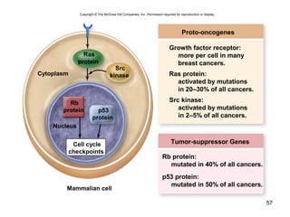
SlideShare a Scribd company logo
Chapter 10
Lecture and
Animation Outline
Copyright © The McGraw-Hill Companies, Inc. Permission required for reproduction or display.
See separate PowerPoint slides for all figures and
tables pre-inserted into PowerPoint without notes and
animations.
To run the animations you must be in Slideshow View. Use
the buttons on the animation to play, pause, and turn
audio/text on or off.
Please Note: Once you have used any of the animation
functions (such as Play or Pause), you must first click on the
slide’s background before you can advance to the next slide.
How Cells Divide
Chapter 10
3
Bacterial Cell Division
• Bacteria divide by binary fission
– No sexual life cycle
– Reproduction is clonal
• Single, circular bacterial chromosome is
replicated
• Replication begins at the origin of replication and
proceeds in two directions to site of termination
• New chromosomes are partitioned to opposite
ends of the cell
• Septum forms to divide the cell into 2 cells
4
Prior to cell division,
the bacterial DNA
molecule replicates.
The replication of the
double-stranded,
Circular DNA mole-
cule that constitutes
the genome of a
bacterium begins at a
specific site, called
the origin of replica-
tion (green area).
The replication
enzymes move out
in both directions
from that site and
make copies of each
strand in the DNA
duplex. The enzymes
continue until they
meet at another
specific site, the
terminus of replication
(red area).
As the DNA is
replicated, the cell
elongates, and the
DNA is partitioned in
the cell such that the
origins are at the ¼
and ¾ positions in the
cell and the termini
are oriented toward
the middle of the cell.
1.
2.
3.
Bacterial cell
Bacterial chromosome:
Double-stranded DNA
Origin of
replication
Copyright © The McGraw-Hill Companies, Inc. Permission required for reproduction or display.
5
5.
Septation then begins, in which new membrane and cell wall material
begin to grow and form a septum at approximately the midpoint of the cell.
A protein molecule called FtsZ (orange dots) facilitates this process.
When the septum is complete, the cell pinches
in two, and two daughter cells are formed,
each containing a bacterial DNA molecule.
4.
Septum
Copyright © The McGraw-Hill Companies, Inc. Permission required for reproduction or display.
Septation
– Production of septum separates cell’s other
components
– Begins with formation of ring of FtsZ proteins
– Accumulation of other proteins follow
– Structure contracts radially to pinch cell in two
– FtsZ protein found in most prokaryotes
6
1 µm 1 µm
Copyright © The McGraw-Hill Companies, Inc. Permission required for reproduction or display.
Courtesy of William Margolin
7
FtsZ protein has a structure similar to
eukaryotic tubulin
•Cell division in different organisms involves
protein assemblies
– Prokaryote protein assemblies involve FtsZ
– Eukaryotic protein assemblies involve microtubules
formed from tubulin
8
Copyright © The McGraw-Hill Companies, Inc. Permission required for reproduction or display.
Septum
FtsZ protein
Chromosome
Chromosome
Microtubule
Nucleus
Kinetochore microtubule
Centrioles Kinetochore
Polar microtubule
Spindle pole body
Kinetochore microtubule
Centriole
Prokaryotes Some Protists Other Protists Animals
Kinetochore microtubule
Polar microtubule
No nucleus, usually
have single circular
chromosome. After DNA
is replicated, it is
partitioned in the cell.
After cell elongation,
FtsZ protein assembles
into a ring and facilitates
septation and cell
division.
Nucleus present and
nuclear envelope
remains intact during
cell division.
Chromosomes line up.
Microtubule fibers pass
through tunnels in the
nuclear membrane and
set up an axis for
separation of replicated
chromosomes, and cell
division.
A spindle of micro-
tubules forms between
two pairs of centrioles at
opposite ends of the
cell. The spindle passes
through one tunnel in
the intact nuclear
envelope. Kinetochore
microtubules form
between kinetochores
on the chromosomes
and the spindle poles
and pull the chromo-
somes to each pole.
Nuclear envelope
remains intact; spindle
microtubules form
inside the nucleus
between spindle pole
bodies. A single
kinetochore microtubule
attaches to each
chromosome and
pulls each to a pole.
Spindle microtubules
begin to form between
centrioles outside of
nucleus. Centrioles move
to the poles and the
nuclear envelope breaks
down. Kinetochore
microtubules attach
kinetochores of
chromosomes to spindle
poles. Polar microtubules
extend toward the center
of the cell and overlap.
Yeasts
Central spindle
of microtubules
Fragments
of nuclear
envelope
Eukaryotic Chromosomes
• Every species has a
different number of
chromosomes
• Humans have 46
chromosomes in 23
nearly identical pairs
– Additional/missing
chromosomes usually
fatal
9
950x
Copyright © The McGraw-Hill Companies, Inc. Permission required for reproduction or display.
© Biophoto Associates/Photo Researchers, Inc.
Chromosomes
• Composed of chromatin – complex of DNA and
protein
• DNA of a single chromosome is one long
continuous double-stranded fiber
• RNA associated with chromosomes during RNA
synthesis
• Typical human chromosome 140 million
nucleotides long
• In the nondividing nucleus
– Heterochromatin – not expressed
– Euchromatin – expressed 10
Chromosome Structure
• Nucleosome
– Complex of DNA and histone proteins
– Promote and guide coiling of DNA
– DNA duplex coiled around 8 histone proteins
every 200 nucleotides
– Histones are positively charged and strongly
attracted to negatively charged phosphate
groups of DNA
11
12
DNA Double Helix (duplex) Nucleosome
Histone core
DNA
Copyright © The McGraw-Hill Companies, Inc. Permission required for reproduction or display.
• Nucleosomes wrapped into higher order
coils called solenoids
– Leads to a fiber 30 nm in diameter
– Usual state of nondividing (interphase)
chromatin
• During mitosis, chromatin in solenoid
arranged around scaffold of protein to
achieve maximum compaction
– Radial looping aided by condensin proteins
13
14
Copyright © The McGraw-Hill Companies, Inc. Permission required for reproduction or display.
Mitotic Chromosome
Chromatin loop
Solenoid
Scaffold
protein
Scaffold protein
Chromatin LoopRosettes of Chromatin Loops
Karyotype
• Particular array of chromosomes in an individual
organism
– Arranged according to size, staining properties,
location of centromere, etc.
• Humans are diploid (2n)
– 2 complete sets of chromosomes
– 46 total chromosomes
• Haploid (n) – 1 set of chromosomes
– 23 in humans
• Pair of chromosomes are homologous
– Each one is a homologue 15
16
500x
Copyright © The McGraw-Hill Companies, Inc. Permission required for reproduction or display.
© CNRI/Photo Researchers, Inc.
Replication
• Prior to replication, each chromosome
composed of a single DNA molecule
• After replication, each chromosome
composed of 2 identical DNA molecules
– Held together by cohesin proteins
• Visible as 2 strands held together as
chromosome becomes more condensed
– One chromosome composed of 2 sister
chromatids
17
18
Copyright © The McGraw-Hill Companies, Inc. Permission required for reproduction or display.
Homologous chromosomes Homologous chromosomes
Cohesin
proteins
Kinetochores
Sister chromatids
Sister chromatids
Kinetochore
Centromere
Replication
Eukaryotic Cell Cycle
1. G1 (gap phase 1)
– Primary growth phase, longest phase
2. S (synthesis)
– Replication of DNA
3. G2 (gap phase 2)
– Organelles replicate, microtubules
organize
4. M (mitosis)
– Subdivided into 5 phases
5. C (cytokinesis)
– Separation of 2 new cells 19
Interphase
20
Duration
• Time it takes to complete a cell cycle varies
greatly
• Fruit fly embryos = 8 minutes
• Mature cells take longer to grow
– Typical mammalian cell takes 24 hours
– Liver cell takes more than a year
• Growth occurs during G1, G2, and S phases
– M phase takes only about an hour
• Most variation in length of G1
– Resting phase G0 – cells spend more or less time here
21
Copyright © The McGraw-Hill Companies, Inc. Permission required for reproduction or display.
M Phase
Metaphase
Anaphase
Telophase
Prometaphase
Prophase
S
G2
G1
Interphase
M Phase
G1
Cytokinesis
Mitosis
S
G2
22
Interphase
• G1, S, and G2 phases
– G1 – cells undergo major portion of growth
– S – replicate DNA
– G2 – chromosomes coil more tightly using motor
proteins; centrioles replicate; tubulin synthesis
• Centromere – point of constriction
– Kinetochore – attachment site for microtubules
– Each sister chromatid has a centromere
– Chromatids stay attached at centromere by cohesin
• Replaced by condensin in metazoans
23
Copyright © The McGraw-Hill Companies, Inc. Permission required for reproduction or display.
Cohesin
proteins
Centromere
region of
chromosome
Kinetochore
microtubules
Kinetochore
Metaphase
chromosome
Sister
chromatids
24
Nucleus
Nucleolus
Aster
Centrioles
(replicated;
animal
cells only)
Nuclear
membrane
• DNA has been replicated
• Centrioles replicate (animal cells)
• Cell prepares for division
Chromatin
(replicated)
Copyright © The McGraw-Hill Companies, Inc. Permission required for reproduction or display.
25
M phase
Mitosis is divided into 5 phases:
1. Prophase
2. Prometaphase
3. Metaphase
4. Anaphase
5. Telophase
26
Prophase
• Individual condensed chromosomes first
become visible with the light microscope
– Condensation continues throughout prophase
• Spindle apparatus assembles
– 2 centrioles move to opposite poles forming spindle
apparatus (no centrioles in plants)
– Asters – radial array of microtubules in animals (not
plants)
• Nuclear envelope breaks down
27
• Chromosomes condense and
become visible
• Chromosomes appear as two
sister chromatids held together
at the centromere
• Cytoskeleton is disassembled:
spindle begins to form
• Golgi and ER are dispersed
• Nuclear envelope breaks down
Mitotic spindle
beginning to form
Condensed
chromosomes
Copyright © The McGraw-Hill Companies, Inc. Permission required for reproduction or display.
28
Prometaphase
• Transition occurs after disassembly of nuclear
envelope
• Microtubule attachment
– 2nd
group grows from poles and attaches to
kinetochores
– Each sister chromatid connected to opposite poles
• Chromosomes begin to move to center of cell –
congression
– Assembly and disassembly of microtubules
– Motor proteins at kinetochores
29
• Chromosomes attach to
microtubules at the kinetochores
• Each chromosome is oriented
such that the kinetochores
of sister chromatids are
attached to microtubules
from opposite poles.
• Chromosomes move to
equator of the cell
Centromere and
kinetochore
Mitotic
spindle
Copyright © The McGraw-Hill Companies, Inc. Permission required for reproduction or display.
Metaphase
• Alignment of
chromosomes
along metaphase
plate
– Not an actual
structure
– Future axis of cell
division
30
Polar
microtubule
Centrioles
Metaphase
plate
Aster
Kinetochore
microtubule
Sister chromatids
57µm
Copyright © The McGraw-Hill Companies, Inc. Permission required for reproduction or display.
© Andrew S. Bajer, University of Oregon
31
• All chromosomes are aligned
at equator of the cell, called
the metaphase plate
• Chromosomes are attached
to opposite poles and are
under tension
Polar microtubule
Chromosomes
aligned on
metaphase plate
Kinetochore
microtubule
Copyright © The McGraw-Hill Companies, Inc. Permission required for reproduction or display.
32
Anaphase
• Begins when centromeres split
• Key event is removal of cohesin proteins
from all chromosomes
• Sister chromatids pulled to opposite poles
• 2 forms of movements
– Anaphase A – kinetochores pulled toward
poles
– Anaphase B – poles move apart
33
Chromosomes
• Proteins holding centromeres of
sister chromatids are degraded,
freeing individual chromosomes
• Chromosomes are pulled to
opposite poles (anaphase A)
• Spindle poles move apart
(anaphase B)
Kinetochore
microtubule
Polar
microtubule
Copyright © The McGraw-Hill Companies, Inc. Permission required for reproduction or display.
34
Telophase
• Spindle apparatus disassembles
• Nuclear envelope forms around each set
of sister chromatids
– Now called chromosomes
• Chromosomes begin to uncoil
• Nucleolus reappears in each new nucleus
35
Polar microtubule
Nucleus reforming
• Chromosomes are clustered at
opposite poles and decondense
• Nuclear envelopes re-form around
chromosomes
• Golgi complex and ER re-form
Kinetochore
microtubule
Copyright © The McGraw-Hill Companies, Inc. Permission required for reproduction or display.
36
Cytokinesis
• Cleavage of the cell into equal halves
• Animal cells – constriction of actin
filaments produces a cleavage furrow
• Plant cells – cell plate forms between the
nuclei
• Fungi and some protists – nuclear
membrane does not dissolve; mitosis
occurs within the nucleus; division of the
nucleus occurs with cytokinesis
37
Copyright © The McGraw-Hill Companies, Inc. Permission required for reproduction or display.
25 µmb.325 µma.
a: © David M. Phillips/Visuals Unlimited; b: © Guenter Albrecht-Buehler, Northwestern University, Chicago
38
Copyright © The McGraw-Hill Companies, Inc. Permission required for reproduction or display.
Vesicles containing
membrane components
fusing to form cell plate
Cell wall
19,000×
(top): © E.H. Newcomb & W.P. Wergin/Biological Photo Service
39
Control of the Cell Cycle
Current view integrates 2 concepts
1.Cell cycle has two irreversible points
– Replication of genetic material
– Separation of the sister chromatids
2.Cell cycle can be put on hold at specific points
called checkpoints
– Process is checked for accuracy and can be halted if
there are errors
– Allows cell to respond to internal and external signals
3 Checkpoints
1. G1/S checkpoint
– Cell “decides” to divide
– Primary point for external signal influence
2. G2/M checkpoint
– Cell makes a commitment to mitosis
– Assesses success of DNA replication
3. Late metaphase (spindle) checkpoint
– Cell ensures that all chromosomes are
attached to the spindle 40
41
Copyright © The McGraw-Hill Companies, Inc. Permission required for reproduction or display.
G2
M
S
G2/M checkpoint Spindle checkpoint
G1/S checkpoint
(Start or restriction point) G1
42
Cyclin-dependent kinases (Cdks)
• Enzymes that phosphorylate proteins
• Primary mechanism of cell cycle control
• Cdks partner with different cyclins at different
points in the cell cycle
• For many years, a common view was that
cyclins drove the cell cycle – that is, the periodic
synthesis and destruction of cyclins acted as a
clock
• Now clear that Cdk itself is also controlled by
phosphorylation
43
Copyright © The McGraw-Hill Companies, Inc. Permission required for reproduction or display.
G2/M Checkpoint
Cdc2/Mitotic Cyclin
• DNA integrity
• Replication
completed
Spindle Checkpoint
APC
• Chromosomes
attached at
metaphase plate
M
G2
G1
S
G1/S Checkpoint
• Size of cell
• Nutritional state
of cell
• Growth factors
Cdc2/G1 Cyclin
• Cdk – cyclin complex
– Also called mitosis-promoting factor (MPF)
• Activity of Cdk is also controlled by the pattern of
phosphorylation
– Phosphorylation at one site (red) inactivates Cdk
– Phosphorylation at another site (green) activates Cdk
44
Cyclin-dependent kinase
(Cdk)
Cyclin
P
P
Copyright © The McGraw-Hill Companies, Inc. Permission required for reproduction or display.
45
MPF
• Once thought that MPF was controlled solely by
the level of the M phase-specific cyclins
• Although M phase cyclin is necessary for MPF
function, activity is controlled by inhibitory
phosphorylation of the kinase component, Cdc2
• Damage to DNA acts through a complex
pathway to tip the balance toward the inhibitory
phosphorylation of MPF
46
Anaphase-promoting complex (APC)
• Also called cyclosome (APC/C)
• At the spindle checkpoint, presence of all
chromosomes at the metaphase plate and
the tension on the microtubules between
opposite poles are both important
• Function of the APC/C is to trigger
anaphase itself
• Marks securin for destruction; no inhibition
of separase; separase destroys cohesin
47
Control in multicellular eukaryotes
• Multiple Cdks control the cycle as
opposed to the single Cdk in yeasts
• Animal cells respond to a greater variety
of external signals than do yeasts, which
primarily respond to signals necessary for
mating
• More complex controls allow the
integration of more input into control of the
cycle
48
Copyright © The McGraw-Hill Companies, Inc. Permission required for reproduction or display.
G2/M Checkpoint
Cdk1/Cyclin B
• DNA integrity
• Replication
completed
Spindle Checkpoint
APC
• Chromosomes
attached at
metaphase plate
M
G2
G1
S
G1/S Checkpoint
• Size of cell
• Nutritional state
of cell
• Growth factors
Cdk2/Cyclin E
Growth factors
• Act by triggering intracellular signaling
systems
• Platelet-derived growth factor (PDGF) one
of the first growth factors to be identified
• PDGF receptor is a receptor tyrosine
kinase (RTK) that initiates a MAP kinase
cascade to stimulate cell division
• Growth factors can override cellular
controls that otherwise inhibit cell division
49
50
GTP
5. MAP kinase (ERK)
activates proteins to
produce cellular
responses, including
transcription factors
that alter gene expression
4. MEK activates
MAP kinases (ERK)
1. Proteins bound to
receptor activate
Ras by exchanging
GDP for GTP.
2. Ras activates
the first
kinase (Raf)
3. Raf activates
the second
Kinase (MEK)
Growth factor
RAS
Cyclins/
proteins
for Sphase
ChromosomeP
Rb
Nucleus
E2F
Rb
ERK
MEK
P
P P
P
RAF
MEK
RAF
ERK
P
P
E2F
P
P
P
P
RAS
Copyright © The McGraw-Hill Companies, Inc. Permission required for reproduction or display.
GDP
51
Cancer
Unrestrained, uncontrolled growth of cells
•Failure of cell cycle control
•Two kinds of genes can disturb the cell
cycle when they are mutated
1. Tumor-suppressor genes
2. Proto-oncogenes
52
Tumor-suppressor genes
• p53 plays a key role in G1 checkpoint
• p53 protein monitors integrity of DNA
– If DNA damaged, cell division halted and repair
enzymes stimulated
– If DNA damage is irreparable, p53 directs cell to kill
itself
• Prevent the development of mutated cells
containing mutations
• p53 is absent or damaged in many cancerous
cells
53
Copyright © The McGraw-Hill Companies, Inc. Permission required for reproduction or display.
1. DNA damage is caused by
heat, radiation, or chemicals.
2. Cell division stops, and p53 triggers
enzymes to repair damaged region.
3. p53 triggers the destruction of
cells damaged beyond repair.
p53 allows cells with
repaired DNA to divide.
1. DNA damage is caused by
heat, radiation, or chemicals.
2. The p53 protein fails to stop cell
division and repair DNA. Cell divides
without repair to damaged DNA.
3. Damaged cells continue to divide.
If other damage accumulates, the
cell can turn cancerous.
DNA repair enzyme
Cancer cell
p53
protein
Normalp53Abnormalp53
Abnormal
p53 protein
54
Proto-oncogenes
• Normal cellular genes that become oncogenes
when mutated
– Oncogenes can cause cancer
• Some encode receptors for growth factors
– If receptor is mutated in “on,” cell no longer depends
on growth factors
• Some encode signal transduction proteins
• Only one copy of a proto-oncogene needs to
undergo this mutation for uncontrolled division to
take place
Tumor-suppressor genes
• p53 gene and many others
• Both copies of a tumor-suppressor gene
must lose function for the cancerous
phenotype to develop
• First tumor-suppressor identified was the
retinoblastoma susceptibility gene (Rb)
– Predisposes individuals for a rare form of
cancer that affects the retina of the eye
55
• Inheriting a single mutant copy of Rb means the
individual has only one “good” copy left
– During the hundreds of thousands of divisions that
occur to produce the retina, any error that damages
the remaining good copy leads to a cancerous cell
– Single cancerous cell in the retina then leads to the
formation of a retinoblastoma tumor
• Rb protein integrates signals from growth factors
– Role to bind important regulatory proteins and prevent
stimulation of cyclin or Cdk production
56
57
Copyright © The McGraw-Hill Companies, Inc. Permission required for reproduction or display.
Proto-oncogenes
Growth factor receptor:
more per cell in many
breast cancers.
Ras protein:
activated by mutations
in 20–30% of all cancers.
Src kinase:
activated by mutations
in 2–5% of all cancers.
Tumor-suppressor Genes
Rb protein:
mutated in 40% of all cancers.
p53 protein:
mutated in 50% of all cancers.
Cell cycle
checkpoints
Ras
protein
Rb
protein p53
protein
Src
kinase
Mammalian cell
Nucleus
Cytoplasm
58
Please note that due to differing
operating systems, some animations
will not appear until the presentation is
viewed in Presentation Mode (Slide
Show view). You may see blank slides
in the “Normal” or “Slide Sorter” views.
All animations will appear after viewing
in Presentation Mode and playing each
animation. Most animations will require
the latest version of the Flash Player,
which is available at
http://get.adobe.com/flashplayer.

More Related Content

What's hot

Cell theory By KK Sahu Sir
Cell theory By KK Sahu SirCell theory By KK Sahu Sir
Cell theory By KK Sahu Sir
KAUSHAL SAHU
 
Is Human Cloning Ethical?
Is Human Cloning Ethical?Is Human Cloning Ethical?
Is Human Cloning Ethical?
Karim Frenn
 
Substrates for animal cellculture
Substrates for animal cellcultureSubstrates for animal cellculture
Substrates for animal cellculture
Vipin Shukla
 
IB Biology 1.4 Slides: Cell Transport
IB Biology 1.4 Slides: Cell TransportIB Biology 1.4 Slides: Cell Transport
IB Biology 1.4 Slides: Cell Transport
Jacob Cedarbaum
 
1.6. Cell division
1.6. Cell division1.6. Cell division
1.6. Cell division
Miltiadis Kitsos
 
Cell-Cell Interaction (Part 1: Cell Junctions)
Cell-Cell Interaction (Part 1: Cell Junctions)Cell-Cell Interaction (Part 1: Cell Junctions)
Cell-Cell Interaction (Part 1: Cell Junctions)
Varij Nayan
 
Chapter 1 cell biology intro cell
Chapter 1 cell biology intro cellChapter 1 cell biology intro cell
Chapter 1 cell biology intro cellElle Phant
 
1.5 origin of the cells
1.5 origin of the cells 1.5 origin of the cells
1.5 origin of the cells
Bob Smullen
 
Microtubules & filaments.pptx 2
Microtubules & filaments.pptx 2Microtubules & filaments.pptx 2
Microtubules & filaments.pptx 2
Arv080
 
Cloning
CloningCloning
Cloning
Dinesh Kumar
 
Ultra structure of plant cell (2)
Ultra structure of plant cell (2)Ultra structure of plant cell (2)
Ultra structure of plant cell (2)
PILLAI ASWATHY VISWANATH
 
1.1 introduction to the cell
1.1 introduction to the cell 1.1 introduction to the cell
1.1 introduction to the cell
Bob Smullen
 
2.7 dna replication, transcription, and translation
2.7 dna replication, transcription, and translation2.7 dna replication, transcription, and translation
2.7 dna replication, transcription, and translation
Bob Smullen
 
Nucleus
NucleusNucleus
Nucleus
Aftab Badshah
 
Lymphoid organs ppt
Lymphoid organs  pptLymphoid organs  ppt
Lymphoid organs ppt
Mercy Biotech
 
4.1 Species, communities and ecosystems
 4.1 Species, communities and ecosystems 4.1 Species, communities and ecosystems
4.1 Species, communities and ecosystems
Miltiadis Kitsos
 

What's hot (20)

Cell theory By KK Sahu Sir
Cell theory By KK Sahu SirCell theory By KK Sahu Sir
Cell theory By KK Sahu Sir
 
Golgi apparatus ppt
Golgi apparatus pptGolgi apparatus ppt
Golgi apparatus ppt
 
Is Human Cloning Ethical?
Is Human Cloning Ethical?Is Human Cloning Ethical?
Is Human Cloning Ethical?
 
Substrates for animal cellculture
Substrates for animal cellcultureSubstrates for animal cellculture
Substrates for animal cellculture
 
IB Biology 1.4 Slides: Cell Transport
IB Biology 1.4 Slides: Cell TransportIB Biology 1.4 Slides: Cell Transport
IB Biology 1.4 Slides: Cell Transport
 
1.6. Cell division
1.6. Cell division1.6. Cell division
1.6. Cell division
 
Cell-Cell Interaction (Part 1: Cell Junctions)
Cell-Cell Interaction (Part 1: Cell Junctions)Cell-Cell Interaction (Part 1: Cell Junctions)
Cell-Cell Interaction (Part 1: Cell Junctions)
 
Cell Differentiation
Cell  DifferentiationCell  Differentiation
Cell Differentiation
 
Chapter 1 cell biology intro cell
Chapter 1 cell biology intro cellChapter 1 cell biology intro cell
Chapter 1 cell biology intro cell
 
1.5 origin of the cells
1.5 origin of the cells 1.5 origin of the cells
1.5 origin of the cells
 
Microtubules & filaments.pptx 2
Microtubules & filaments.pptx 2Microtubules & filaments.pptx 2
Microtubules & filaments.pptx 2
 
Cloning
CloningCloning
Cloning
 
Lecture 7
Lecture 7Lecture 7
Lecture 7
 
Ultra structure of plant cell (2)
Ultra structure of plant cell (2)Ultra structure of plant cell (2)
Ultra structure of plant cell (2)
 
1.1 introduction to the cell
1.1 introduction to the cell 1.1 introduction to the cell
1.1 introduction to the cell
 
2.7 dna replication, transcription, and translation
2.7 dna replication, transcription, and translation2.7 dna replication, transcription, and translation
2.7 dna replication, transcription, and translation
 
Nucleus
NucleusNucleus
Nucleus
 
Lymphoid organs ppt
Lymphoid organs  pptLymphoid organs  ppt
Lymphoid organs ppt
 
4.1 Species, communities and ecosystems
 4.1 Species, communities and ecosystems 4.1 Species, communities and ecosystems
4.1 Species, communities and ecosystems
 
Cloning
CloningCloning
Cloning
 

Viewers also liked

Biol 201 006_w_007w_fa16_hudon
Biol 201 006_w_007w_fa16_hudonBiol 201 006_w_007w_fa16_hudon
Biol 201 006_w_007w_fa16_hudon
stephaniehudon
 
photosynthesis basics study for class 10th
photosynthesis basics study for class 10thphotosynthesis basics study for class 10th
photosynthesis basics study for class 10th
Chinmaya Pattnaik
 
Animal phys chapter 5 part 2
Animal phys chapter 5 part 2Animal phys chapter 5 part 2
Animal phys chapter 5 part 2
stephaniehudon
 
Animal phys chapter 5
Animal phys chapter 5Animal phys chapter 5
Animal phys chapter 5
stephaniehudon
 
Biology 201 Chapter 11
Biology 201 Chapter 11Biology 201 Chapter 11
Biology 201 Chapter 11
stephaniehudon
 
Biology 201 Chapter 12 PowerPoint
Biology 201 Chapter 12 PowerPointBiology 201 Chapter 12 PowerPoint
Biology 201 Chapter 12 PowerPoint
stephaniehudon
 
Animal Physiology chapter 1
Animal Physiology chapter 1Animal Physiology chapter 1
Animal Physiology chapter 1
stephaniehudon
 
Biology 201 Chapter 15
Biology 201 Chapter 15Biology 201 Chapter 15
Biology 201 Chapter 15
stephaniehudon
 
Biology 201 Chapter 8
Biology 201 Chapter 8Biology 201 Chapter 8
Biology 201 Chapter 8
stephaniehudon
 
Biology 201 Chapter 7
Biology 201 Chapter 7Biology 201 Chapter 7
Biology 201 Chapter 7
stephaniehudon
 
PSY 150 401 Chapter 10 SLIDES
PSY 150 401 Chapter 10 SLIDESPSY 150 401 Chapter 10 SLIDES
PSY 150 401 Chapter 10 SLIDESkimappel
 
Statistics Math project class 10th
Statistics Math project class 10thStatistics Math project class 10th
Statistics Math project class 10th
Riya Singh
 
DNA Replication PowerPoint
DNA Replication PowerPointDNA Replication PowerPoint
DNA Replication PowerPointBiologyIB
 

Viewers also liked (14)

Biol 201 006_w_007w_fa16_hudon
Biol 201 006_w_007w_fa16_hudonBiol 201 006_w_007w_fa16_hudon
Biol 201 006_w_007w_fa16_hudon
 
photosynthesis basics study for class 10th
photosynthesis basics study for class 10thphotosynthesis basics study for class 10th
photosynthesis basics study for class 10th
 
Animal phys chapter 5 part 2
Animal phys chapter 5 part 2Animal phys chapter 5 part 2
Animal phys chapter 5 part 2
 
Animal phys chapter 5
Animal phys chapter 5Animal phys chapter 5
Animal phys chapter 5
 
Biology 201 Chapter 11
Biology 201 Chapter 11Biology 201 Chapter 11
Biology 201 Chapter 11
 
Animal Physiology
Animal PhysiologyAnimal Physiology
Animal Physiology
 
Biology 201 Chapter 12 PowerPoint
Biology 201 Chapter 12 PowerPointBiology 201 Chapter 12 PowerPoint
Biology 201 Chapter 12 PowerPoint
 
Animal Physiology chapter 1
Animal Physiology chapter 1Animal Physiology chapter 1
Animal Physiology chapter 1
 
Biology 201 Chapter 15
Biology 201 Chapter 15Biology 201 Chapter 15
Biology 201 Chapter 15
 
Biology 201 Chapter 8
Biology 201 Chapter 8Biology 201 Chapter 8
Biology 201 Chapter 8
 
Biology 201 Chapter 7
Biology 201 Chapter 7Biology 201 Chapter 7
Biology 201 Chapter 7
 
PSY 150 401 Chapter 10 SLIDES
PSY 150 401 Chapter 10 SLIDESPSY 150 401 Chapter 10 SLIDES
PSY 150 401 Chapter 10 SLIDES
 
Statistics Math project class 10th
Statistics Math project class 10thStatistics Math project class 10th
Statistics Math project class 10th
 
DNA Replication PowerPoint
DNA Replication PowerPointDNA Replication PowerPoint
DNA Replication PowerPoint
 

Similar to Biology 201 Chapter 10

Cell division
Cell divisionCell division
Cell division
Kesar Shroff
 
Cell_Division_Mitosis_Meiosis_ppt.pptx
Cell_Division_Mitosis_Meiosis_ppt.pptxCell_Division_Mitosis_Meiosis_ppt.pptx
Cell_Division_Mitosis_Meiosis_ppt.pptx
CharlesIbus
 
Cell_Division_Mitosis_Meiosis_ppt_{SIS7DDA7B647659}.pptx
Cell_Division_Mitosis_Meiosis_ppt_{SIS7DDA7B647659}.pptxCell_Division_Mitosis_Meiosis_ppt_{SIS7DDA7B647659}.pptx
Cell_Division_Mitosis_Meiosis_ppt_{SIS7DDA7B647659}.pptx
AnnalieLobiano1
 
Bio 201 chapter 4 powerpoint
Bio 201 chapter 4 powerpointBio 201 chapter 4 powerpoint
Bio 201 chapter 4 powerpoint
stephaniehudon
 
Bio201chapter4powerpoint 150914185916-lva1-app6892
Bio201chapter4powerpoint 150914185916-lva1-app6892Bio201chapter4powerpoint 150914185916-lva1-app6892
Bio201chapter4powerpoint 150914185916-lva1-app6892
Cleophas Rwemera
 
The Cell Cycle Mitosis 2015
The Cell Cycle   Mitosis 2015The Cell Cycle   Mitosis 2015
The Cell Cycle Mitosis 2015
Dobbs Ferry High School
 
Celldivision
CelldivisionCelldivision
Cell division in eukaryotic and prokaryotics I.pptx
Cell division in eukaryotic and prokaryotics I.pptxCell division in eukaryotic and prokaryotics I.pptx
Cell division in eukaryotic and prokaryotics I.pptx
MuhammadMusharaf17
 
Bio1308 mitosis
Bio1308 mitosisBio1308 mitosis
Bio1308 mitosis
Kevinn Lloyd Sepada
 
Mitossis and meosis -cell cycle -
Mitossis and meosis -cell cycle - Mitossis and meosis -cell cycle -
Mitossis and meosis -cell cycle -
malaika20
 
cell division.pptx
cell division.pptxcell division.pptx
cell division.pptx
JanhaviBurade
 
Mitosis ppt
Mitosis pptMitosis ppt
Mitosis ppt
Allison Miller
 
12_Lecture_Presentation.ppt
12_Lecture_Presentation.ppt12_Lecture_Presentation.ppt
12_Lecture_Presentation.ppt
AnandHarsh3
 
the nucleus.pptx
the nucleus.pptxthe nucleus.pptx
the nucleus.pptx
abdiasis omar mohamed
 
Chapter 10 notes - Cell Growth and Division
Chapter 10 notes - Cell Growth and DivisionChapter 10 notes - Cell Growth and Division
Chapter 10 notes - Cell Growth and Divisionpetersbiology
 
08 lecture presentation
08 lecture presentation08 lecture presentation
08 lecture presentationUconn Stamford
 
Chapt11lectureanim 151027004158-lva1-app6892
Chapt11lectureanim 151027004158-lva1-app6892Chapt11lectureanim 151027004158-lva1-app6892
Chapt11lectureanim 151027004158-lva1-app6892
Cleophas Rwemera
 
Ch4cellreproduction
Ch4cellreproductionCh4cellreproduction
Ch4cellreproduction
CDA-PamelaOrtiz
 
Dna replication, mitosis and the cell cycle
Dna replication, mitosis and the cell cycleDna replication, mitosis and the cell cycle
Dna replication, mitosis and the cell cycle
lumenalexis
 
DNA Replication, Mitosis, meiosis, and the Cell Cycle
DNA Replication, Mitosis, meiosis, and the Cell CycleDNA Replication, Mitosis, meiosis, and the Cell Cycle
DNA Replication, Mitosis, meiosis, and the Cell Cycle
Lumen Learning
 

Similar to Biology 201 Chapter 10 (20)

Cell division
Cell divisionCell division
Cell division
 
Cell_Division_Mitosis_Meiosis_ppt.pptx
Cell_Division_Mitosis_Meiosis_ppt.pptxCell_Division_Mitosis_Meiosis_ppt.pptx
Cell_Division_Mitosis_Meiosis_ppt.pptx
 
Cell_Division_Mitosis_Meiosis_ppt_{SIS7DDA7B647659}.pptx
Cell_Division_Mitosis_Meiosis_ppt_{SIS7DDA7B647659}.pptxCell_Division_Mitosis_Meiosis_ppt_{SIS7DDA7B647659}.pptx
Cell_Division_Mitosis_Meiosis_ppt_{SIS7DDA7B647659}.pptx
 
Bio 201 chapter 4 powerpoint
Bio 201 chapter 4 powerpointBio 201 chapter 4 powerpoint
Bio 201 chapter 4 powerpoint
 
Bio201chapter4powerpoint 150914185916-lva1-app6892
Bio201chapter4powerpoint 150914185916-lva1-app6892Bio201chapter4powerpoint 150914185916-lva1-app6892
Bio201chapter4powerpoint 150914185916-lva1-app6892
 
The Cell Cycle Mitosis 2015
The Cell Cycle   Mitosis 2015The Cell Cycle   Mitosis 2015
The Cell Cycle Mitosis 2015
 
Celldivision
CelldivisionCelldivision
Celldivision
 
Cell division in eukaryotic and prokaryotics I.pptx
Cell division in eukaryotic and prokaryotics I.pptxCell division in eukaryotic and prokaryotics I.pptx
Cell division in eukaryotic and prokaryotics I.pptx
 
Bio1308 mitosis
Bio1308 mitosisBio1308 mitosis
Bio1308 mitosis
 
Mitossis and meosis -cell cycle -
Mitossis and meosis -cell cycle - Mitossis and meosis -cell cycle -
Mitossis and meosis -cell cycle -
 
cell division.pptx
cell division.pptxcell division.pptx
cell division.pptx
 
Mitosis ppt
Mitosis pptMitosis ppt
Mitosis ppt
 
12_Lecture_Presentation.ppt
12_Lecture_Presentation.ppt12_Lecture_Presentation.ppt
12_Lecture_Presentation.ppt
 
the nucleus.pptx
the nucleus.pptxthe nucleus.pptx
the nucleus.pptx
 
Chapter 10 notes - Cell Growth and Division
Chapter 10 notes - Cell Growth and DivisionChapter 10 notes - Cell Growth and Division
Chapter 10 notes - Cell Growth and Division
 
08 lecture presentation
08 lecture presentation08 lecture presentation
08 lecture presentation
 
Chapt11lectureanim 151027004158-lva1-app6892
Chapt11lectureanim 151027004158-lva1-app6892Chapt11lectureanim 151027004158-lva1-app6892
Chapt11lectureanim 151027004158-lva1-app6892
 
Ch4cellreproduction
Ch4cellreproductionCh4cellreproduction
Ch4cellreproduction
 
Dna replication, mitosis and the cell cycle
Dna replication, mitosis and the cell cycleDna replication, mitosis and the cell cycle
Dna replication, mitosis and the cell cycle
 
DNA Replication, Mitosis, meiosis, and the Cell Cycle
DNA Replication, Mitosis, meiosis, and the Cell CycleDNA Replication, Mitosis, meiosis, and the Cell Cycle
DNA Replication, Mitosis, meiosis, and the Cell Cycle
 

More from stephaniehudon

Syllabus Biology 201 Fall 2016
Syllabus Biology 201 Fall 2016Syllabus Biology 201 Fall 2016
Syllabus Biology 201 Fall 2016
stephaniehudon
 
Su16 syl b227 002 stephanie hudon
Su16 syl b227  002 stephanie hudonSu16 syl b227  002 stephanie hudon
Su16 syl b227 002 stephanie hudon
stephaniehudon
 
Su16 Syllabus B227-002 Stephanie Hudon
Su16 Syllabus B227-002 Stephanie HudonSu16 Syllabus B227-002 Stephanie Hudon
Su16 Syllabus B227-002 Stephanie Hudon
stephaniehudon
 
Biol 201 003W and 005W syllabus
Biol 201 003W and 005W syllabusBiol 201 003W and 005W syllabus
Biol 201 003W and 005W syllabus
stephaniehudon
 
Biology 201 Chapter 14
Biology 201 Chapter 14Biology 201 Chapter 14
Biology 201 Chapter 14
stephaniehudon
 
Chapter 13 Biology 201
Chapter 13 Biology 201Chapter 13 Biology 201
Chapter 13 Biology 201
stephaniehudon
 
Chapter 9 Medical Terminology
Chapter 9 Medical TerminologyChapter 9 Medical Terminology
Chapter 9 Medical Terminology
stephaniehudon
 
Chapter 8 Medical Terminology
Chapter 8 Medical TerminologyChapter 8 Medical Terminology
Chapter 8 Medical Terminology
stephaniehudon
 
Medical Terminology Chapter 7
Medical Terminology Chapter 7Medical Terminology Chapter 7
Medical Terminology Chapter 7
stephaniehudon
 
Biology 201 Ch 6 PowerPoint
Biology 201 Ch 6 PowerPointBiology 201 Ch 6 PowerPoint
Biology 201 Ch 6 PowerPoint
stephaniehudon
 
Medical Terminology Chapter 6
Medical Terminology Chapter 6Medical Terminology Chapter 6
Medical Terminology Chapter 6
stephaniehudon
 
Medical Terminology Chapter 5
Medical Terminology Chapter 5Medical Terminology Chapter 5
Medical Terminology Chapter 5
stephaniehudon
 
Ch 5 PowerPoint Biology 201
Ch 5 PowerPoint Biology 201Ch 5 PowerPoint Biology 201
Ch 5 PowerPoint Biology 201
stephaniehudon
 
Medical Terminology Chapter 4
Medical Terminology Chapter 4Medical Terminology Chapter 4
Medical Terminology Chapter 4
stephaniehudon
 
Animal physiology chapter 2 2nd half
Animal physiology chapter 2   2nd halfAnimal physiology chapter 2   2nd half
Animal physiology chapter 2 2nd half
stephaniehudon
 
Medical Terminology Chapter 3
Medical Terminology Chapter 3Medical Terminology Chapter 3
Medical Terminology Chapter 3
stephaniehudon
 
Ch 3 PowerPoint Biology 201
Ch 3 PowerPoint Biology 201Ch 3 PowerPoint Biology 201
Ch 3 PowerPoint Biology 201
stephaniehudon
 
Animal physiology chapter 2 1st half
Animal physiology chapter 2   1st halfAnimal physiology chapter 2   1st half
Animal physiology chapter 2 1st half
stephaniehudon
 
Animal Physiology Intro
Animal Physiology IntroAnimal Physiology Intro
Animal Physiology Intro
stephaniehudon
 
Medical Terminology Chapter 2
Medical Terminology Chapter 2Medical Terminology Chapter 2
Medical Terminology Chapter 2
stephaniehudon
 

More from stephaniehudon (20)

Syllabus Biology 201 Fall 2016
Syllabus Biology 201 Fall 2016Syllabus Biology 201 Fall 2016
Syllabus Biology 201 Fall 2016
 
Su16 syl b227 002 stephanie hudon
Su16 syl b227  002 stephanie hudonSu16 syl b227  002 stephanie hudon
Su16 syl b227 002 stephanie hudon
 
Su16 Syllabus B227-002 Stephanie Hudon
Su16 Syllabus B227-002 Stephanie HudonSu16 Syllabus B227-002 Stephanie Hudon
Su16 Syllabus B227-002 Stephanie Hudon
 
Biol 201 003W and 005W syllabus
Biol 201 003W and 005W syllabusBiol 201 003W and 005W syllabus
Biol 201 003W and 005W syllabus
 
Biology 201 Chapter 14
Biology 201 Chapter 14Biology 201 Chapter 14
Biology 201 Chapter 14
 
Chapter 13 Biology 201
Chapter 13 Biology 201Chapter 13 Biology 201
Chapter 13 Biology 201
 
Chapter 9 Medical Terminology
Chapter 9 Medical TerminologyChapter 9 Medical Terminology
Chapter 9 Medical Terminology
 
Chapter 8 Medical Terminology
Chapter 8 Medical TerminologyChapter 8 Medical Terminology
Chapter 8 Medical Terminology
 
Medical Terminology Chapter 7
Medical Terminology Chapter 7Medical Terminology Chapter 7
Medical Terminology Chapter 7
 
Biology 201 Ch 6 PowerPoint
Biology 201 Ch 6 PowerPointBiology 201 Ch 6 PowerPoint
Biology 201 Ch 6 PowerPoint
 
Medical Terminology Chapter 6
Medical Terminology Chapter 6Medical Terminology Chapter 6
Medical Terminology Chapter 6
 
Medical Terminology Chapter 5
Medical Terminology Chapter 5Medical Terminology Chapter 5
Medical Terminology Chapter 5
 
Ch 5 PowerPoint Biology 201
Ch 5 PowerPoint Biology 201Ch 5 PowerPoint Biology 201
Ch 5 PowerPoint Biology 201
 
Medical Terminology Chapter 4
Medical Terminology Chapter 4Medical Terminology Chapter 4
Medical Terminology Chapter 4
 
Animal physiology chapter 2 2nd half
Animal physiology chapter 2   2nd halfAnimal physiology chapter 2   2nd half
Animal physiology chapter 2 2nd half
 
Medical Terminology Chapter 3
Medical Terminology Chapter 3Medical Terminology Chapter 3
Medical Terminology Chapter 3
 
Ch 3 PowerPoint Biology 201
Ch 3 PowerPoint Biology 201Ch 3 PowerPoint Biology 201
Ch 3 PowerPoint Biology 201
 
Animal physiology chapter 2 1st half
Animal physiology chapter 2   1st halfAnimal physiology chapter 2   1st half
Animal physiology chapter 2 1st half
 
Animal Physiology Intro
Animal Physiology IntroAnimal Physiology Intro
Animal Physiology Intro
 
Medical Terminology Chapter 2
Medical Terminology Chapter 2Medical Terminology Chapter 2
Medical Terminology Chapter 2
 

Recently uploaded

A Strategic Approach: GenAI in Education
A Strategic Approach: GenAI in EducationA Strategic Approach: GenAI in Education
A Strategic Approach: GenAI in Education
Peter Windle
 
Unit 2- Research Aptitude (UGC NET Paper I).pdf
Unit 2- Research Aptitude (UGC NET Paper I).pdfUnit 2- Research Aptitude (UGC NET Paper I).pdf
Unit 2- Research Aptitude (UGC NET Paper I).pdf
Thiyagu K
 
aaaaaaaaaaaaaaaaaaaaaaaaaaaaaaaaaaaaaaaaa
aaaaaaaaaaaaaaaaaaaaaaaaaaaaaaaaaaaaaaaaaaaaaaaaaaaaaaaaaaaaaaaaaaaaaaaaaaaaaaaaaa
aaaaaaaaaaaaaaaaaaaaaaaaaaaaaaaaaaaaaaaaa
siemaillard
 
How libraries can support authors with open access requirements for UKRI fund...
How libraries can support authors with open access requirements for UKRI fund...How libraries can support authors with open access requirements for UKRI fund...
How libraries can support authors with open access requirements for UKRI fund...
Jisc
 
STRAND 3 HYGIENIC PRACTICES.pptx GRADE 7 CBC
STRAND 3 HYGIENIC PRACTICES.pptx GRADE 7 CBCSTRAND 3 HYGIENIC PRACTICES.pptx GRADE 7 CBC
STRAND 3 HYGIENIC PRACTICES.pptx GRADE 7 CBC
kimdan468
 
1.4 modern child centered education - mahatma gandhi-2.pptx
1.4 modern child centered education - mahatma gandhi-2.pptx1.4 modern child centered education - mahatma gandhi-2.pptx
1.4 modern child centered education - mahatma gandhi-2.pptx
JosvitaDsouza2
 
BÀI TẬP BỔ TRỢ TIẾNG ANH GLOBAL SUCCESS LỚP 3 - CẢ NĂM (CÓ FILE NGHE VÀ ĐÁP Á...
BÀI TẬP BỔ TRỢ TIẾNG ANH GLOBAL SUCCESS LỚP 3 - CẢ NĂM (CÓ FILE NGHE VÀ ĐÁP Á...BÀI TẬP BỔ TRỢ TIẾNG ANH GLOBAL SUCCESS LỚP 3 - CẢ NĂM (CÓ FILE NGHE VÀ ĐÁP Á...
BÀI TẬP BỔ TRỢ TIẾNG ANH GLOBAL SUCCESS LỚP 3 - CẢ NĂM (CÓ FILE NGHE VÀ ĐÁP Á...
Nguyen Thanh Tu Collection
 
Honest Reviews of Tim Han LMA Course Program.pptx
Honest Reviews of Tim Han LMA Course Program.pptxHonest Reviews of Tim Han LMA Course Program.pptx
Honest Reviews of Tim Han LMA Course Program.pptx
timhan337
 
Multithreading_in_C++ - std::thread, race condition
Multithreading_in_C++ - std::thread, race conditionMultithreading_in_C++ - std::thread, race condition
Multithreading_in_C++ - std::thread, race condition
Mohammed Sikander
 
Thesis Statement for students diagnonsed withADHD.ppt
Thesis Statement for students diagnonsed withADHD.pptThesis Statement for students diagnonsed withADHD.ppt
Thesis Statement for students diagnonsed withADHD.ppt
EverAndrsGuerraGuerr
 
special B.ed 2nd year old paper_20240531.pdf
special B.ed 2nd year old paper_20240531.pdfspecial B.ed 2nd year old paper_20240531.pdf
special B.ed 2nd year old paper_20240531.pdf
Special education needs
 
Synthetic Fiber Construction in lab .pptx
Synthetic Fiber Construction in lab .pptxSynthetic Fiber Construction in lab .pptx
Synthetic Fiber Construction in lab .pptx
Pavel ( NSTU)
 
Chapter 3 - Islamic Banking Products and Services.pptx
Chapter 3 - Islamic Banking Products and Services.pptxChapter 3 - Islamic Banking Products and Services.pptx
Chapter 3 - Islamic Banking Products and Services.pptx
Mohd Adib Abd Muin, Senior Lecturer at Universiti Utara Malaysia
 
2024.06.01 Introducing a competency framework for languag learning materials ...
2024.06.01 Introducing a competency framework for languag learning materials ...2024.06.01 Introducing a competency framework for languag learning materials ...
2024.06.01 Introducing a competency framework for languag learning materials ...
Sandy Millin
 
S1-Introduction-Biopesticides in ICM.pptx
S1-Introduction-Biopesticides in ICM.pptxS1-Introduction-Biopesticides in ICM.pptx
S1-Introduction-Biopesticides in ICM.pptx
tarandeep35
 
Digital Tools and AI for Teaching Learning and Research
Digital Tools and AI for Teaching Learning and ResearchDigital Tools and AI for Teaching Learning and Research
Digital Tools and AI for Teaching Learning and Research
Vikramjit Singh
 
TESDA TM1 REVIEWER FOR NATIONAL ASSESSMENT WRITTEN AND ORAL QUESTIONS WITH A...
TESDA TM1 REVIEWER  FOR NATIONAL ASSESSMENT WRITTEN AND ORAL QUESTIONS WITH A...TESDA TM1 REVIEWER  FOR NATIONAL ASSESSMENT WRITTEN AND ORAL QUESTIONS WITH A...
TESDA TM1 REVIEWER FOR NATIONAL ASSESSMENT WRITTEN AND ORAL QUESTIONS WITH A...
EugeneSaldivar
 
Acetabularia Information For Class 9 .docx
Acetabularia Information For Class 9  .docxAcetabularia Information For Class 9  .docx
Acetabularia Information For Class 9 .docx
vaibhavrinwa19
 
Embracing GenAI - A Strategic Imperative
Embracing GenAI - A Strategic ImperativeEmbracing GenAI - A Strategic Imperative
Embracing GenAI - A Strategic Imperative
Peter Windle
 
Francesca Gottschalk - How can education support child empowerment.pptx
Francesca Gottschalk - How can education support child empowerment.pptxFrancesca Gottschalk - How can education support child empowerment.pptx
Francesca Gottschalk - How can education support child empowerment.pptx
EduSkills OECD
 

Recently uploaded (20)

A Strategic Approach: GenAI in Education
A Strategic Approach: GenAI in EducationA Strategic Approach: GenAI in Education
A Strategic Approach: GenAI in Education
 
Unit 2- Research Aptitude (UGC NET Paper I).pdf
Unit 2- Research Aptitude (UGC NET Paper I).pdfUnit 2- Research Aptitude (UGC NET Paper I).pdf
Unit 2- Research Aptitude (UGC NET Paper I).pdf
 
aaaaaaaaaaaaaaaaaaaaaaaaaaaaaaaaaaaaaaaaa
aaaaaaaaaaaaaaaaaaaaaaaaaaaaaaaaaaaaaaaaaaaaaaaaaaaaaaaaaaaaaaaaaaaaaaaaaaaaaaaaaa
aaaaaaaaaaaaaaaaaaaaaaaaaaaaaaaaaaaaaaaaa
 
How libraries can support authors with open access requirements for UKRI fund...
How libraries can support authors with open access requirements for UKRI fund...How libraries can support authors with open access requirements for UKRI fund...
How libraries can support authors with open access requirements for UKRI fund...
 
STRAND 3 HYGIENIC PRACTICES.pptx GRADE 7 CBC
STRAND 3 HYGIENIC PRACTICES.pptx GRADE 7 CBCSTRAND 3 HYGIENIC PRACTICES.pptx GRADE 7 CBC
STRAND 3 HYGIENIC PRACTICES.pptx GRADE 7 CBC
 
1.4 modern child centered education - mahatma gandhi-2.pptx
1.4 modern child centered education - mahatma gandhi-2.pptx1.4 modern child centered education - mahatma gandhi-2.pptx
1.4 modern child centered education - mahatma gandhi-2.pptx
 
BÀI TẬP BỔ TRỢ TIẾNG ANH GLOBAL SUCCESS LỚP 3 - CẢ NĂM (CÓ FILE NGHE VÀ ĐÁP Á...
BÀI TẬP BỔ TRỢ TIẾNG ANH GLOBAL SUCCESS LỚP 3 - CẢ NĂM (CÓ FILE NGHE VÀ ĐÁP Á...BÀI TẬP BỔ TRỢ TIẾNG ANH GLOBAL SUCCESS LỚP 3 - CẢ NĂM (CÓ FILE NGHE VÀ ĐÁP Á...
BÀI TẬP BỔ TRỢ TIẾNG ANH GLOBAL SUCCESS LỚP 3 - CẢ NĂM (CÓ FILE NGHE VÀ ĐÁP Á...
 
Honest Reviews of Tim Han LMA Course Program.pptx
Honest Reviews of Tim Han LMA Course Program.pptxHonest Reviews of Tim Han LMA Course Program.pptx
Honest Reviews of Tim Han LMA Course Program.pptx
 
Multithreading_in_C++ - std::thread, race condition
Multithreading_in_C++ - std::thread, race conditionMultithreading_in_C++ - std::thread, race condition
Multithreading_in_C++ - std::thread, race condition
 
Thesis Statement for students diagnonsed withADHD.ppt
Thesis Statement for students diagnonsed withADHD.pptThesis Statement for students diagnonsed withADHD.ppt
Thesis Statement for students diagnonsed withADHD.ppt
 
special B.ed 2nd year old paper_20240531.pdf
special B.ed 2nd year old paper_20240531.pdfspecial B.ed 2nd year old paper_20240531.pdf
special B.ed 2nd year old paper_20240531.pdf
 
Synthetic Fiber Construction in lab .pptx
Synthetic Fiber Construction in lab .pptxSynthetic Fiber Construction in lab .pptx
Synthetic Fiber Construction in lab .pptx
 
Chapter 3 - Islamic Banking Products and Services.pptx
Chapter 3 - Islamic Banking Products and Services.pptxChapter 3 - Islamic Banking Products and Services.pptx
Chapter 3 - Islamic Banking Products and Services.pptx
 
2024.06.01 Introducing a competency framework for languag learning materials ...
2024.06.01 Introducing a competency framework for languag learning materials ...2024.06.01 Introducing a competency framework for languag learning materials ...
2024.06.01 Introducing a competency framework for languag learning materials ...
 
S1-Introduction-Biopesticides in ICM.pptx
S1-Introduction-Biopesticides in ICM.pptxS1-Introduction-Biopesticides in ICM.pptx
S1-Introduction-Biopesticides in ICM.pptx
 
Digital Tools and AI for Teaching Learning and Research
Digital Tools and AI for Teaching Learning and ResearchDigital Tools and AI for Teaching Learning and Research
Digital Tools and AI for Teaching Learning and Research
 
TESDA TM1 REVIEWER FOR NATIONAL ASSESSMENT WRITTEN AND ORAL QUESTIONS WITH A...
TESDA TM1 REVIEWER  FOR NATIONAL ASSESSMENT WRITTEN AND ORAL QUESTIONS WITH A...TESDA TM1 REVIEWER  FOR NATIONAL ASSESSMENT WRITTEN AND ORAL QUESTIONS WITH A...
TESDA TM1 REVIEWER FOR NATIONAL ASSESSMENT WRITTEN AND ORAL QUESTIONS WITH A...
 
Acetabularia Information For Class 9 .docx
Acetabularia Information For Class 9  .docxAcetabularia Information For Class 9  .docx
Acetabularia Information For Class 9 .docx
 
Embracing GenAI - A Strategic Imperative
Embracing GenAI - A Strategic ImperativeEmbracing GenAI - A Strategic Imperative
Embracing GenAI - A Strategic Imperative
 
Francesca Gottschalk - How can education support child empowerment.pptx
Francesca Gottschalk - How can education support child empowerment.pptxFrancesca Gottschalk - How can education support child empowerment.pptx
Francesca Gottschalk - How can education support child empowerment.pptx
 

Biology 201 Chapter 10

  • 1. Chapter 10 Lecture and Animation Outline Copyright © The McGraw-Hill Companies, Inc. Permission required for reproduction or display. See separate PowerPoint slides for all figures and tables pre-inserted into PowerPoint without notes and animations. To run the animations you must be in Slideshow View. Use the buttons on the animation to play, pause, and turn audio/text on or off. Please Note: Once you have used any of the animation functions (such as Play or Pause), you must first click on the slide’s background before you can advance to the next slide.
  • 3. 3 Bacterial Cell Division • Bacteria divide by binary fission – No sexual life cycle – Reproduction is clonal • Single, circular bacterial chromosome is replicated • Replication begins at the origin of replication and proceeds in two directions to site of termination • New chromosomes are partitioned to opposite ends of the cell • Septum forms to divide the cell into 2 cells
  • 4. 4 Prior to cell division, the bacterial DNA molecule replicates. The replication of the double-stranded, Circular DNA mole- cule that constitutes the genome of a bacterium begins at a specific site, called the origin of replica- tion (green area). The replication enzymes move out in both directions from that site and make copies of each strand in the DNA duplex. The enzymes continue until they meet at another specific site, the terminus of replication (red area). As the DNA is replicated, the cell elongates, and the DNA is partitioned in the cell such that the origins are at the ¼ and ¾ positions in the cell and the termini are oriented toward the middle of the cell. 1. 2. 3. Bacterial cell Bacterial chromosome: Double-stranded DNA Origin of replication Copyright © The McGraw-Hill Companies, Inc. Permission required for reproduction or display.
  • 5. 5 5. Septation then begins, in which new membrane and cell wall material begin to grow and form a septum at approximately the midpoint of the cell. A protein molecule called FtsZ (orange dots) facilitates this process. When the septum is complete, the cell pinches in two, and two daughter cells are formed, each containing a bacterial DNA molecule. 4. Septum Copyright © The McGraw-Hill Companies, Inc. Permission required for reproduction or display.
  • 6. Septation – Production of septum separates cell’s other components – Begins with formation of ring of FtsZ proteins – Accumulation of other proteins follow – Structure contracts radially to pinch cell in two – FtsZ protein found in most prokaryotes 6 1 µm 1 µm Copyright © The McGraw-Hill Companies, Inc. Permission required for reproduction or display. Courtesy of William Margolin
  • 7. 7 FtsZ protein has a structure similar to eukaryotic tubulin •Cell division in different organisms involves protein assemblies – Prokaryote protein assemblies involve FtsZ – Eukaryotic protein assemblies involve microtubules formed from tubulin
  • 8. 8 Copyright © The McGraw-Hill Companies, Inc. Permission required for reproduction or display. Septum FtsZ protein Chromosome Chromosome Microtubule Nucleus Kinetochore microtubule Centrioles Kinetochore Polar microtubule Spindle pole body Kinetochore microtubule Centriole Prokaryotes Some Protists Other Protists Animals Kinetochore microtubule Polar microtubule No nucleus, usually have single circular chromosome. After DNA is replicated, it is partitioned in the cell. After cell elongation, FtsZ protein assembles into a ring and facilitates septation and cell division. Nucleus present and nuclear envelope remains intact during cell division. Chromosomes line up. Microtubule fibers pass through tunnels in the nuclear membrane and set up an axis for separation of replicated chromosomes, and cell division. A spindle of micro- tubules forms between two pairs of centrioles at opposite ends of the cell. The spindle passes through one tunnel in the intact nuclear envelope. Kinetochore microtubules form between kinetochores on the chromosomes and the spindle poles and pull the chromo- somes to each pole. Nuclear envelope remains intact; spindle microtubules form inside the nucleus between spindle pole bodies. A single kinetochore microtubule attaches to each chromosome and pulls each to a pole. Spindle microtubules begin to form between centrioles outside of nucleus. Centrioles move to the poles and the nuclear envelope breaks down. Kinetochore microtubules attach kinetochores of chromosomes to spindle poles. Polar microtubules extend toward the center of the cell and overlap. Yeasts Central spindle of microtubules Fragments of nuclear envelope
  • 9. Eukaryotic Chromosomes • Every species has a different number of chromosomes • Humans have 46 chromosomes in 23 nearly identical pairs – Additional/missing chromosomes usually fatal 9 950x Copyright © The McGraw-Hill Companies, Inc. Permission required for reproduction or display. © Biophoto Associates/Photo Researchers, Inc.
  • 10. Chromosomes • Composed of chromatin – complex of DNA and protein • DNA of a single chromosome is one long continuous double-stranded fiber • RNA associated with chromosomes during RNA synthesis • Typical human chromosome 140 million nucleotides long • In the nondividing nucleus – Heterochromatin – not expressed – Euchromatin – expressed 10
  • 11. Chromosome Structure • Nucleosome – Complex of DNA and histone proteins – Promote and guide coiling of DNA – DNA duplex coiled around 8 histone proteins every 200 nucleotides – Histones are positively charged and strongly attracted to negatively charged phosphate groups of DNA 11
  • 12. 12 DNA Double Helix (duplex) Nucleosome Histone core DNA Copyright © The McGraw-Hill Companies, Inc. Permission required for reproduction or display.
  • 13. • Nucleosomes wrapped into higher order coils called solenoids – Leads to a fiber 30 nm in diameter – Usual state of nondividing (interphase) chromatin • During mitosis, chromatin in solenoid arranged around scaffold of protein to achieve maximum compaction – Radial looping aided by condensin proteins 13
  • 14. 14 Copyright © The McGraw-Hill Companies, Inc. Permission required for reproduction or display. Mitotic Chromosome Chromatin loop Solenoid Scaffold protein Scaffold protein Chromatin LoopRosettes of Chromatin Loops
  • 15. Karyotype • Particular array of chromosomes in an individual organism – Arranged according to size, staining properties, location of centromere, etc. • Humans are diploid (2n) – 2 complete sets of chromosomes – 46 total chromosomes • Haploid (n) – 1 set of chromosomes – 23 in humans • Pair of chromosomes are homologous – Each one is a homologue 15
  • 16. 16 500x Copyright © The McGraw-Hill Companies, Inc. Permission required for reproduction or display. © CNRI/Photo Researchers, Inc.
  • 17. Replication • Prior to replication, each chromosome composed of a single DNA molecule • After replication, each chromosome composed of 2 identical DNA molecules – Held together by cohesin proteins • Visible as 2 strands held together as chromosome becomes more condensed – One chromosome composed of 2 sister chromatids 17
  • 18. 18 Copyright © The McGraw-Hill Companies, Inc. Permission required for reproduction or display. Homologous chromosomes Homologous chromosomes Cohesin proteins Kinetochores Sister chromatids Sister chromatids Kinetochore Centromere Replication
  • 19. Eukaryotic Cell Cycle 1. G1 (gap phase 1) – Primary growth phase, longest phase 2. S (synthesis) – Replication of DNA 3. G2 (gap phase 2) – Organelles replicate, microtubules organize 4. M (mitosis) – Subdivided into 5 phases 5. C (cytokinesis) – Separation of 2 new cells 19 Interphase
  • 20. 20 Duration • Time it takes to complete a cell cycle varies greatly • Fruit fly embryos = 8 minutes • Mature cells take longer to grow – Typical mammalian cell takes 24 hours – Liver cell takes more than a year • Growth occurs during G1, G2, and S phases – M phase takes only about an hour • Most variation in length of G1 – Resting phase G0 – cells spend more or less time here
  • 21. 21 Copyright © The McGraw-Hill Companies, Inc. Permission required for reproduction or display. M Phase Metaphase Anaphase Telophase Prometaphase Prophase S G2 G1 Interphase M Phase G1 Cytokinesis Mitosis S G2
  • 22. 22 Interphase • G1, S, and G2 phases – G1 – cells undergo major portion of growth – S – replicate DNA – G2 – chromosomes coil more tightly using motor proteins; centrioles replicate; tubulin synthesis • Centromere – point of constriction – Kinetochore – attachment site for microtubules – Each sister chromatid has a centromere – Chromatids stay attached at centromere by cohesin • Replaced by condensin in metazoans
  • 23. 23 Copyright © The McGraw-Hill Companies, Inc. Permission required for reproduction or display. Cohesin proteins Centromere region of chromosome Kinetochore microtubules Kinetochore Metaphase chromosome Sister chromatids
  • 24. 24 Nucleus Nucleolus Aster Centrioles (replicated; animal cells only) Nuclear membrane • DNA has been replicated • Centrioles replicate (animal cells) • Cell prepares for division Chromatin (replicated) Copyright © The McGraw-Hill Companies, Inc. Permission required for reproduction or display.
  • 25. 25 M phase Mitosis is divided into 5 phases: 1. Prophase 2. Prometaphase 3. Metaphase 4. Anaphase 5. Telophase
  • 26. 26 Prophase • Individual condensed chromosomes first become visible with the light microscope – Condensation continues throughout prophase • Spindle apparatus assembles – 2 centrioles move to opposite poles forming spindle apparatus (no centrioles in plants) – Asters – radial array of microtubules in animals (not plants) • Nuclear envelope breaks down
  • 27. 27 • Chromosomes condense and become visible • Chromosomes appear as two sister chromatids held together at the centromere • Cytoskeleton is disassembled: spindle begins to form • Golgi and ER are dispersed • Nuclear envelope breaks down Mitotic spindle beginning to form Condensed chromosomes Copyright © The McGraw-Hill Companies, Inc. Permission required for reproduction or display.
  • 28. 28 Prometaphase • Transition occurs after disassembly of nuclear envelope • Microtubule attachment – 2nd group grows from poles and attaches to kinetochores – Each sister chromatid connected to opposite poles • Chromosomes begin to move to center of cell – congression – Assembly and disassembly of microtubules – Motor proteins at kinetochores
  • 29. 29 • Chromosomes attach to microtubules at the kinetochores • Each chromosome is oriented such that the kinetochores of sister chromatids are attached to microtubules from opposite poles. • Chromosomes move to equator of the cell Centromere and kinetochore Mitotic spindle Copyright © The McGraw-Hill Companies, Inc. Permission required for reproduction or display.
  • 30. Metaphase • Alignment of chromosomes along metaphase plate – Not an actual structure – Future axis of cell division 30 Polar microtubule Centrioles Metaphase plate Aster Kinetochore microtubule Sister chromatids 57µm Copyright © The McGraw-Hill Companies, Inc. Permission required for reproduction or display. © Andrew S. Bajer, University of Oregon
  • 31. 31 • All chromosomes are aligned at equator of the cell, called the metaphase plate • Chromosomes are attached to opposite poles and are under tension Polar microtubule Chromosomes aligned on metaphase plate Kinetochore microtubule Copyright © The McGraw-Hill Companies, Inc. Permission required for reproduction or display.
  • 32. 32 Anaphase • Begins when centromeres split • Key event is removal of cohesin proteins from all chromosomes • Sister chromatids pulled to opposite poles • 2 forms of movements – Anaphase A – kinetochores pulled toward poles – Anaphase B – poles move apart
  • 33. 33 Chromosomes • Proteins holding centromeres of sister chromatids are degraded, freeing individual chromosomes • Chromosomes are pulled to opposite poles (anaphase A) • Spindle poles move apart (anaphase B) Kinetochore microtubule Polar microtubule Copyright © The McGraw-Hill Companies, Inc. Permission required for reproduction or display.
  • 34. 34 Telophase • Spindle apparatus disassembles • Nuclear envelope forms around each set of sister chromatids – Now called chromosomes • Chromosomes begin to uncoil • Nucleolus reappears in each new nucleus
  • 35. 35 Polar microtubule Nucleus reforming • Chromosomes are clustered at opposite poles and decondense • Nuclear envelopes re-form around chromosomes • Golgi complex and ER re-form Kinetochore microtubule Copyright © The McGraw-Hill Companies, Inc. Permission required for reproduction or display.
  • 36. 36 Cytokinesis • Cleavage of the cell into equal halves • Animal cells – constriction of actin filaments produces a cleavage furrow • Plant cells – cell plate forms between the nuclei • Fungi and some protists – nuclear membrane does not dissolve; mitosis occurs within the nucleus; division of the nucleus occurs with cytokinesis
  • 37. 37 Copyright © The McGraw-Hill Companies, Inc. Permission required for reproduction or display. 25 µmb.325 µma. a: © David M. Phillips/Visuals Unlimited; b: © Guenter Albrecht-Buehler, Northwestern University, Chicago
  • 38. 38 Copyright © The McGraw-Hill Companies, Inc. Permission required for reproduction or display. Vesicles containing membrane components fusing to form cell plate Cell wall 19,000× (top): © E.H. Newcomb & W.P. Wergin/Biological Photo Service
  • 39. 39 Control of the Cell Cycle Current view integrates 2 concepts 1.Cell cycle has two irreversible points – Replication of genetic material – Separation of the sister chromatids 2.Cell cycle can be put on hold at specific points called checkpoints – Process is checked for accuracy and can be halted if there are errors – Allows cell to respond to internal and external signals
  • 40. 3 Checkpoints 1. G1/S checkpoint – Cell “decides” to divide – Primary point for external signal influence 2. G2/M checkpoint – Cell makes a commitment to mitosis – Assesses success of DNA replication 3. Late metaphase (spindle) checkpoint – Cell ensures that all chromosomes are attached to the spindle 40
  • 41. 41 Copyright © The McGraw-Hill Companies, Inc. Permission required for reproduction or display. G2 M S G2/M checkpoint Spindle checkpoint G1/S checkpoint (Start or restriction point) G1
  • 42. 42 Cyclin-dependent kinases (Cdks) • Enzymes that phosphorylate proteins • Primary mechanism of cell cycle control • Cdks partner with different cyclins at different points in the cell cycle • For many years, a common view was that cyclins drove the cell cycle – that is, the periodic synthesis and destruction of cyclins acted as a clock • Now clear that Cdk itself is also controlled by phosphorylation
  • 43. 43 Copyright © The McGraw-Hill Companies, Inc. Permission required for reproduction or display. G2/M Checkpoint Cdc2/Mitotic Cyclin • DNA integrity • Replication completed Spindle Checkpoint APC • Chromosomes attached at metaphase plate M G2 G1 S G1/S Checkpoint • Size of cell • Nutritional state of cell • Growth factors Cdc2/G1 Cyclin
  • 44. • Cdk – cyclin complex – Also called mitosis-promoting factor (MPF) • Activity of Cdk is also controlled by the pattern of phosphorylation – Phosphorylation at one site (red) inactivates Cdk – Phosphorylation at another site (green) activates Cdk 44 Cyclin-dependent kinase (Cdk) Cyclin P P Copyright © The McGraw-Hill Companies, Inc. Permission required for reproduction or display.
  • 45. 45 MPF • Once thought that MPF was controlled solely by the level of the M phase-specific cyclins • Although M phase cyclin is necessary for MPF function, activity is controlled by inhibitory phosphorylation of the kinase component, Cdc2 • Damage to DNA acts through a complex pathway to tip the balance toward the inhibitory phosphorylation of MPF
  • 46. 46 Anaphase-promoting complex (APC) • Also called cyclosome (APC/C) • At the spindle checkpoint, presence of all chromosomes at the metaphase plate and the tension on the microtubules between opposite poles are both important • Function of the APC/C is to trigger anaphase itself • Marks securin for destruction; no inhibition of separase; separase destroys cohesin
  • 47. 47 Control in multicellular eukaryotes • Multiple Cdks control the cycle as opposed to the single Cdk in yeasts • Animal cells respond to a greater variety of external signals than do yeasts, which primarily respond to signals necessary for mating • More complex controls allow the integration of more input into control of the cycle
  • 48. 48 Copyright © The McGraw-Hill Companies, Inc. Permission required for reproduction or display. G2/M Checkpoint Cdk1/Cyclin B • DNA integrity • Replication completed Spindle Checkpoint APC • Chromosomes attached at metaphase plate M G2 G1 S G1/S Checkpoint • Size of cell • Nutritional state of cell • Growth factors Cdk2/Cyclin E
  • 49. Growth factors • Act by triggering intracellular signaling systems • Platelet-derived growth factor (PDGF) one of the first growth factors to be identified • PDGF receptor is a receptor tyrosine kinase (RTK) that initiates a MAP kinase cascade to stimulate cell division • Growth factors can override cellular controls that otherwise inhibit cell division 49
  • 50. 50 GTP 5. MAP kinase (ERK) activates proteins to produce cellular responses, including transcription factors that alter gene expression 4. MEK activates MAP kinases (ERK) 1. Proteins bound to receptor activate Ras by exchanging GDP for GTP. 2. Ras activates the first kinase (Raf) 3. Raf activates the second Kinase (MEK) Growth factor RAS Cyclins/ proteins for Sphase ChromosomeP Rb Nucleus E2F Rb ERK MEK P P P P RAF MEK RAF ERK P P E2F P P P P RAS Copyright © The McGraw-Hill Companies, Inc. Permission required for reproduction or display. GDP
  • 51. 51 Cancer Unrestrained, uncontrolled growth of cells •Failure of cell cycle control •Two kinds of genes can disturb the cell cycle when they are mutated 1. Tumor-suppressor genes 2. Proto-oncogenes
  • 52. 52 Tumor-suppressor genes • p53 plays a key role in G1 checkpoint • p53 protein monitors integrity of DNA – If DNA damaged, cell division halted and repair enzymes stimulated – If DNA damage is irreparable, p53 directs cell to kill itself • Prevent the development of mutated cells containing mutations • p53 is absent or damaged in many cancerous cells
  • 53. 53 Copyright © The McGraw-Hill Companies, Inc. Permission required for reproduction or display. 1. DNA damage is caused by heat, radiation, or chemicals. 2. Cell division stops, and p53 triggers enzymes to repair damaged region. 3. p53 triggers the destruction of cells damaged beyond repair. p53 allows cells with repaired DNA to divide. 1. DNA damage is caused by heat, radiation, or chemicals. 2. The p53 protein fails to stop cell division and repair DNA. Cell divides without repair to damaged DNA. 3. Damaged cells continue to divide. If other damage accumulates, the cell can turn cancerous. DNA repair enzyme Cancer cell p53 protein Normalp53Abnormalp53 Abnormal p53 protein
  • 54. 54 Proto-oncogenes • Normal cellular genes that become oncogenes when mutated – Oncogenes can cause cancer • Some encode receptors for growth factors – If receptor is mutated in “on,” cell no longer depends on growth factors • Some encode signal transduction proteins • Only one copy of a proto-oncogene needs to undergo this mutation for uncontrolled division to take place
  • 55. Tumor-suppressor genes • p53 gene and many others • Both copies of a tumor-suppressor gene must lose function for the cancerous phenotype to develop • First tumor-suppressor identified was the retinoblastoma susceptibility gene (Rb) – Predisposes individuals for a rare form of cancer that affects the retina of the eye 55
  • 56. • Inheriting a single mutant copy of Rb means the individual has only one “good” copy left – During the hundreds of thousands of divisions that occur to produce the retina, any error that damages the remaining good copy leads to a cancerous cell – Single cancerous cell in the retina then leads to the formation of a retinoblastoma tumor • Rb protein integrates signals from growth factors – Role to bind important regulatory proteins and prevent stimulation of cyclin or Cdk production 56
  • 57. 57 Copyright © The McGraw-Hill Companies, Inc. Permission required for reproduction or display. Proto-oncogenes Growth factor receptor: more per cell in many breast cancers. Ras protein: activated by mutations in 20–30% of all cancers. Src kinase: activated by mutations in 2–5% of all cancers. Tumor-suppressor Genes Rb protein: mutated in 40% of all cancers. p53 protein: mutated in 50% of all cancers. Cell cycle checkpoints Ras protein Rb protein p53 protein Src kinase Mammalian cell Nucleus Cytoplasm
  • 58. 58 Please note that due to differing operating systems, some animations will not appear until the presentation is viewed in Presentation Mode (Slide Show view). You may see blank slides in the “Normal” or “Slide Sorter” views. All animations will appear after viewing in Presentation Mode and playing each animation. Most animations will require the latest version of the Flash Player, which is available at http://get.adobe.com/flashplayer.