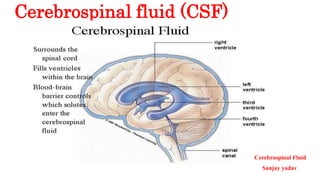
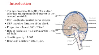
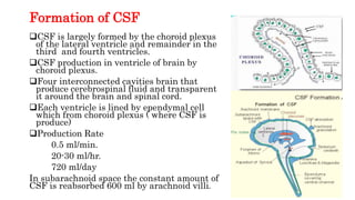
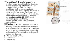
2. Content
Introduction
Formation of CSF
Function of CSF
Composition of CSF
Collection of CSF
Physical examination
Chemical examination
Microscopic examination
Cause of CSF
Symptoms of CSF
Diagnosis of CSF
Treatment of CSF
Prevention
References
3. Introduction
• The cerebrospinal fluid (CSF) is a clear,
color less transparent fluid present in the
cerebral ventricle.
• CSF is a fluid of central nerve system
• CSF is a ultra filtration of the blood.
• Properties volume:- 100 - 200 ml
• Rate of formation :- 0.3 ml/ min/ 600 – 700
ml daily
• Specific gravity:- 1.005
• Reaction:- alkaline 7.3 to 7.4 ph.
4. Formation of CSF
CSF is largely formed by the choroid plexus
of the lateral ventricle and remainder in the
third and fourth ventricles.
CSF production in ventricle of brain by
choroid plexus.
Four interconnected cavities brain that
produce cerebrospinal fluid and transparent
it around the brain and spinal cord.
Each ventricle is lined by ependymal cell
which from choroid plexus ( where CSF is
produce)
Production Rate
0.5 ml/min.
20-30 ml/hr.
720 ml/day
In subarachnoid space the constant amount of
CSF is reabsorbed 600 ml by arachnoid villi.
5. Function of CSF
• The major Function of CSF is to provide basic mechanism
sport and immunological protection of the brain.
• Provide fluid cushion for protection and support the brain
the spinal cord. ( by act like a fluid buffer)
• CSF supplies nutrients to the nervous system tissue (carries
nutrient and CSF removes waste products from cerebral
metabolism.
• Act as shock absorbed for brain and Spinal cord.
• Maintain the constant pressure inside the brain and around
the spinal cord.
• It act as a medium for the transfer of substance from tissue
of the brain spinal cord to the blood stream.
6. Composition of CSF
Water – 99%
Solids – 1%
Solids present in two forms
• Organic substance
• Inorganic substance
Organic substances present
• Protein – 15-47 mg/dl
• Sugar – 40-80 mg/dl
• Amino acid – few amount
• Cholesterol – 0.2-0.5 mg/dl
• Urea – 6-16 mg%
• Uric acid – 0.5-4.5 mg%
• Creatine – 0.5-1.2 mg%
• Lactic acid – few amount
Few – 0.8 lymphocyte/ per cumm
WBC cell – Neutrophils absent
Inorganic substance
• Na – 144-154 meg/l
• K – 2.0 to 3.5 meg/l
• Chloride – 118-132meg/l
• Bicarbonate
• Calcium etc.
CSF normal pressure – 5-15 mm hg
CSF pressure measurement machine
hydromenometer.
7. Collection of csf
Cerebrospinal fluid is collected by lumber puncture (LP) under
strict aseptic condition similar to any surgical collection.
An experience physician or a specially trained nurse dose the
LP and specimen collection.
After properly positioning the patient the sterile LP needle
with the stylet is inserted b/w the third –fourth (L3-L4) and
(L4-L5) a depth of 4 to 5 cm.
The stylet is with drown and the fluid flows freely in to a
sterile test tube through the needle.
A total between 6-7 ml of CSF is collected in 3 sterile tubes
which are chemically cleaned and washed with distilled and
deionized water.
The needle is remove the area is cleaned and bandage over the
needle site.
3 sterile tubes used
• Bacterial examination 1ml
• Chemical examination 2.5 ml
• Microscopic examination 2.5 ml
After collection test sterile with hr.
Do not refrigerate sample if bacteriology examination
Sone bacterial kill in 2-8 C
8. Physical examination
Absorbed the specimen and note down.
Color – color less
Red color – presence of blood
Yellow color – presence of bilirubin (known as
xanthochromia) jaundice condition
Appearance – clear
Cloudy – due to pus condition (bacterial infection)
Specific gravity – 1.003 to 1.008
PH – 7.3 to 7.4
Clot formation normal CSF does not clot
Clot formation is the presence of fibrinogen fibrin to
produce clot ( when BBB is disturb.
9. Chemical examination
Total solids - counting
• Protein – 15 to 45 mg/dl
• Albumin – 50-70%
• Globulin – 30-50%
Some method used to increased in globulin
in pondy’s method
• Glucose – 40-80 meg/l
• Chloride – 118-132 meg/l
• Sodium – 144-154meg/l
• Potassium – 2.0-3.5 meg/l
• Creatine – 0.5-1.2 mg/dl
• Urea – 6-16 mg/dl
• Uric acid – 0.5-4.5 mg/dl
Pandy’s method
Globulin are precipitated by a saturated
solution of phenol in water and cause fine
turbidity.
Reagent – (pandy’s reagent)
Phenol – 10 g
Distilled water – 150 ml
Procedure –
• Take 1 ml of pandy’s reagent in to a small
test tube.
• Add 2-3 drop of CSF (specimen)
• Examine the tube after addition of each
drop (do not mix)
• Read result immediately.
• Result - depending upon the degree of
precipitation of globulin.
• Trace – 1+, 2+, 3+, 4+
10. Microscopic examination
1. Cell counting - TLC, DLC
• Normal CSF contain few 0.8 lymphocytes / cumm
• Lymphocyte present normally in few amount about 0.5
WBC / cumm increased cell count in CSF is know as
pleocytosis.
• Used Neubauer chamber.
• Increased leucocyte – infective meningitis
• Lymphocyte increase – viral infection
• Neutrophils – increase bacterial infection
High count DLC – pyogenic infection
High lymphocyte count – mycotic infection, TB, viral
11. 2. Gram staining used for gram+ve, gram-ve,
morphology, cocci, bacilli, size etc.
Organism that commonly cause meningitis.
• Neisseria meningitides (-ve)
• Streptococcus pneumonia (+ve)
• Hemophilus influenzas (-ve)
3. Acid Fast staining
For tuberculosis meningitis.
4. (i) Wet mount of CSF
performed case of sleeping sickness trypanosomiasis
Take 1 drop of CSF top of slide and cover with cover slip
after that examine in 40x .
(ii) Indian Ink preparation
Specimen - CSF sediment used cryptococcus infection
Take 1 drop of CSF top of slide and 1 drop of Indian ink
preparation after that examine in microscope. Result –
round budding spores (surrounded by colorless capsule)
12. 5. Culture of CSF
Culturing the specimen :- (if CSF contain cell and total protein are
raised.
Inoculate chocolate agar – incubate in carbon dioxide
Incoculate – MacConkey agar and blood agar in the case of specimen
collected from a new bornt infant.
Incoculate sabouraud agar if cryptococci meningitis is suspected
1. Examine the chocolate agar plate For
• Neisseria meningitis's
• Hemophilus influenzae
• Streptococcus pneumonia
2. Examine the MacConkey agar and blood agar plates for.
• Escherichia coli
• Staphylococcus aureus
• Streptococcus agalactia
Note:- whenever passible perform biochemical test to confirm the result.
13. Causes of CSF
CSF (Cerebrospinal Fluid) can be caused by a variety of factors,
including:
• Normal production and circulation of CSF
• Infections or inflammation of the brain or spinal cord
• Head injury or trauma
• Brain tumors or other abnormalities
• Hydrocephalus (excess CSF accumulation in the brain)Bleeding in the brain or
spinal cord
• Certain medications or medical procedures
• Autoimmune disorders affecting the nervous system.
14. Symptoms
The symptoms of CSF (Cerebrospinal Fluid) vary depending
on the underlying cause and can range from mild to severe.
Some common symptoms of CSF include:
• Headaches
• Nausea and vomiting
• Stiff neck or back
• Vision problems
• Sensitivity to light
• Hearing problems
• Cognitive difficulties
• seizures
• Loss of balance and coordination
• Incontinence or urinary problems
• Sleep disturbances
• Fatigue
It's important to note that some cases of CSF may not
produce any noticeable symptoms. If you're experiencing any
of the above symptoms, it's important to seek medical
attention promptly as they may indicate a serious
underlying condition.
15. Diagnosis of CSF
CSF Chemical Tests
Protein level: This test measures the amount of protein in the CSF. Elevated
levels of protein can be a sign of inflammation or damage to the nervous
system.
Glucose level: This test measures the amount of glucose in the CSF.
Decreased levels of glucose can be a sign of bacterial meningitis or other
infections.
CSF lactate dehydrogenase (LD, LDH)—used to differentiate between
bacterial and viral meningitis; the level is usually increased with bacterial
meningitis and not with viral meningitis; may also be elevated with leukemia
or stroke.
Tumor markers—carcinoembryonic antigen (CEA), alpha-fetoprotein (AFP),
and human chorionic gonadotropin (hCG) may be increased in cancers that
have spread from other sites in the body (metastatic)).
16. Cell Counts, Differential and Microscopic Exam
• CSF total cell counts
• Red blood cell (RBC) count—normally no red blood cells are present in the CSF. The
presence of red blood cells may indicate bleeding into the CSF or may indicate a “traumatic
tap” – blood that leaked into the CSF sample during collection.
• White blood cell (WBC) count—normally very few white blood cells are present. A
significant increase in white blood cells in the CSF can be caused by infection or
inflammation of the central nervous system.
• CSF white blood cell (WBC) differential—identifies and counts the different types of WBCs that
are present. Small numbers of lymphocytes, monocytes (and, in neonates, neutrophils) are
normal in a sample of CSF.
• CSF Gram stain—this test is always performed on CSF when infection is suspected. A sample of
CSF is centrifuged and the concentrated portion is placed on a slide and treated with a special
stain. A laboratory professional examines the slide using a microscope to look for bacteria or
fungi, which can indicate bacterial or fungal meningitis.
• CSF culture—culture is used to detect any bacteria or fungi in the sample. A negative culture
does not rule out an infection because the microbes may be present in small numbers or unable
to grow in culture due to prior antibiotic therapy.
• CSF AFB testing—used to detect infection with mycobacteria, such as Mycobacterium
tuberculosis; molecular tests specific for M. tuberculosis may be performed when tuberculosis is
suspected.
17.
18. MRI (magnetic resonance imaging)
- CSF can indicate conditions such
as hydrocephalus, which is an
abnormal buildup of CSF in the
brain. In addition, the appearance
of CSF in the brain and spinal cord
can provide important information
for the diagnosis of conditions such
as multiple sclerosis or other
demyelinating disorders.
In combination with other
diagnostic tests, such as CSF
analysis and clinical examination,
CSF MRI can provide important
information for the diagnosis and
management of various
neurological conditions.
19. CT scan – This imaging test uses
a combination of X-rays and
computer technology to create
detailed images of any part of the
body, including bones, muscles, fat,
and organs. CT scans are more
detailed than general X-rays. They
are used to diagnose disorders of
the brain, spine, or other parts of
the nervous system.
Electroencephalogram (EEG). This
test records the brain's continuous
electrical activity through
electrodes attached to the scalp.
Ultrasound (sonography). This
imaging test uses high-frequency
sound waves and a computer to
make images of blood vessels,
tissues, and organs. Ultrasounds
are used to view internal organs as
they function. They also assess
blood flow through various vessels.
20. Treatment
Lumbar puncture: This is a
procedure where a needle is
inserted into the lower back to
remove a small amount of CSF
for diagnostic or therapeutic
purposes.
CSF replacement: In rare cases, CSF
may need to be replaced due to a condition
known as hydrocephalus, in which there is
an excess buildup of CSF in the brain.
This can be done through a procedure
known as a ventriculoperitoneal shunt,
which involves redirecting the flow of CSF
from the brain to the abdomen.
21. Intrathecal drug delivery: This
involves using a small catheter to deliver
medication directly into the CSF. This
can be an effective way to treat
conditions such as chronic pain or
spasticity. Intrathecal administration is a
route of administration for drugs via an
injection into the spinal canal, or into the
subarachnoid space so that it reaches
the cerebrospinal fluid (CSF) and is
useful in spinal anesthesia,
chemotherapy, or pain management
applications
Medication - As a hormone produced by
the body that stimulates the bone marrow to
produce more white blood cells, G-CSF can be
taken as a drug that can reduce the severity
and duration of neutropenia in patients with
some types of cancer.
1. Acetazolamide (ACTZ)
2. Ceftriaxone
3. cefotaxime
22. Prevention
Preventing CSF-related neurological conditions often involves reducing the risk
factors that can lead to these conditions. Here are some measures that can help
prevent CSF-related neurological conditions:
Vaccination: Vaccines can prevent several infections that can lead to meningitis and
other neurological conditions, such as Haemophiles influenzae type b, meningococcal
disease, and pneumococcal disease.
Practice good hygiene: Practicing good hygiene, such as washing your hands
regularly, can help prevent the spread of infections that can lead to neurological
conditions.
Wear protective gear: If you are participating in sports or other activities that can
lead to head injuries, wearing protective gear, such as helmets, can reduce the risk
of a head injury that can lead to CSF-related neurological conditions.
Avoid risky behavior: Avoiding risky behavior, such as using illicit drugs or engaging
in unprotected sex, can reduce the risk of infections that can lead to neurological
conditions.
Manage chronic conditions: If you have a chronic condition, such as diabetes or
hypertension, managing it effectively can reduce the risk of complications that can
lead to CSF-related neurological conditions.
Seek prompt medical attention: If you experience symptoms of a neurological
condition, such as headaches, neck stiffness, or changes in vision, seek prompt
medical attention to prevent the condition from worsening.
23. References
Cerebrospinal Fluid (CSF) Testing - Testing.com
Hydrocephalus - Symptoms and causes - Mayo Clinic
Cerebrospinal Fluid (CSF) Leak: Symptoms & Treatment (clevelandclinic.org)
Cerebrospinal fluid - Wikipedia
Cerebral Spinal Fluid (CSF) Analysis (healthline.com)
V. H. Talib book
Praful B. Godkar, Darshan P. Godkar book