Downloaded 409 times
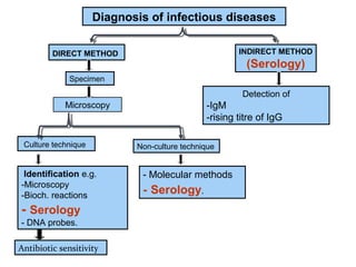


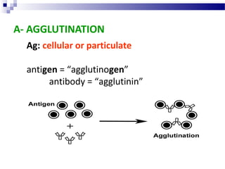
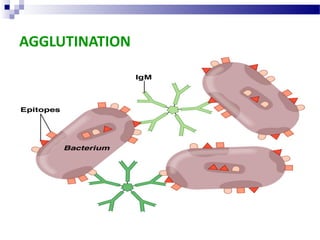


This document discusses various direct and indirect methods used for the diagnosis of infectious diseases. Direct methods involve detecting the pathogen itself through microscopy, culture techniques, or molecular methods on a specimen. Indirect methods involve detecting the immune response to the pathogen through serology techniques like detecting IgM or rising titers of IgG antibodies. It then discusses various antigen-antibody reactions that can be used for serological diagnosis, including agglutination, precipitation, complement fixation, viral neutralization, immunofluorescence, ELISA, and radioimmunoassay.