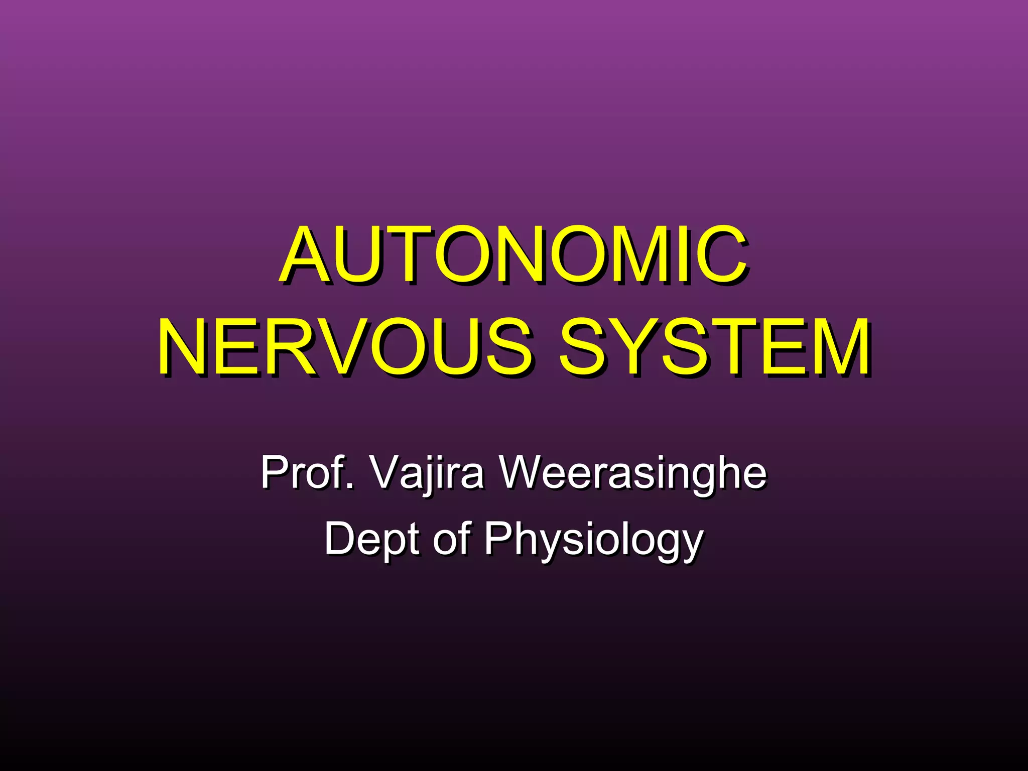
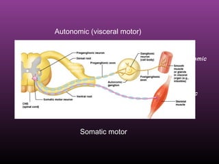
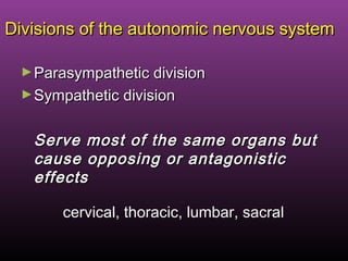
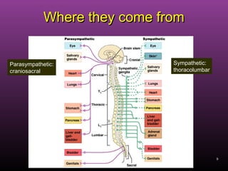
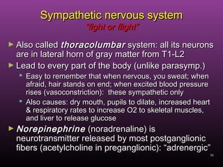
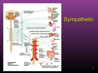
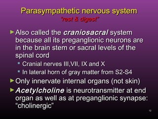
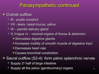
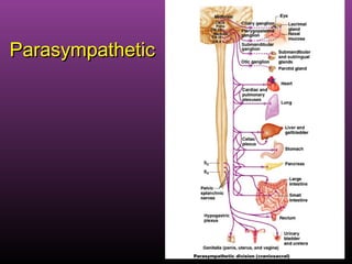
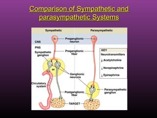
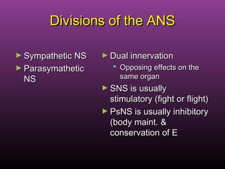
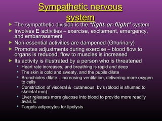
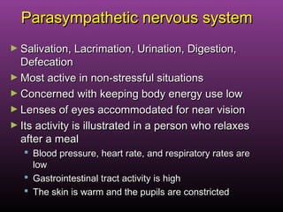
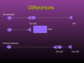
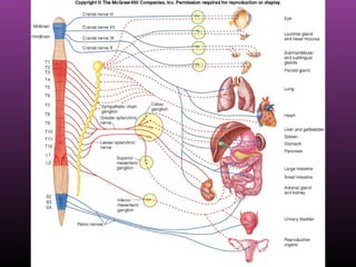
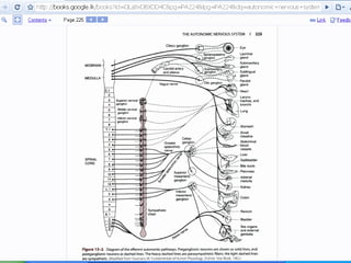
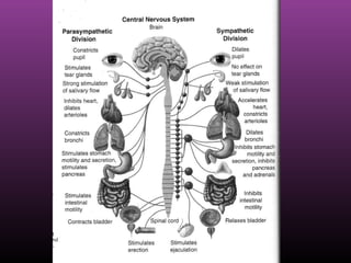
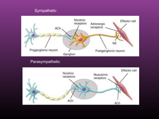
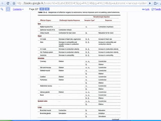
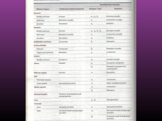
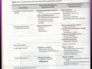
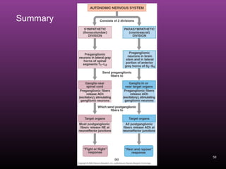
The autonomic nervous system regulates involuntary body functions and is divided into the sympathetic and parasympathetic nervous systems. The sympathetic system activates the body's fight or flight response and increases heart rate and respiration. The parasympathetic system calms the body and increases digestion. Both systems generally have opposing effects on organs with the sympathetic usually stimulating functions and the parasympathetic inhibiting them. Acetylcholine and norepinephrine act as neurotransmitters depending on the type of receptor in the target organ.
Anatomy & physiology of the Autonomic nervous systemRafid Rashid
Provides a good description of the anatomy & physiology of the autonomic nervous system for undergraduate medical students. It goes over the parts & functions of the sympathetic & parasympathetic nervous system respectively & compares the differences between them.
This presentation gives a detailed description of the physiology of Neural Regulation of Respiration in Human. The role played by the Medullary and Pontine Centers in respiration is highlighted. A detailed diagram and the reflexes involved in controlling Respiration are described for the understanding of the student. Standard reference books have been used to describe the topic.
Dr. Bataa Autonmic nervous system
The Indian Dental Academy is the Leader in continuing dental education , training dentists in all aspects of dentistry and
offering a wide range of dental certified courses in different formats.for more details please visit
www.indiandentalacademy.com
Basavarajeeyam is a Sreshta Sangraha grantha (Compiled book ), written by Neelkanta kotturu Basavaraja Virachita. It contains 25 Prakaranas, First 24 Chapters related to Rogas& 25th to Rasadravyas.
Title: Sense of Smell
Presenter: Dr. Faiza, Assistant Professor of Physiology
Qualifications:
MBBS (Best Graduate, AIMC Lahore)
FCPS Physiology
ICMT, CHPE, DHPE (STMU)
MPH (GC University, Faisalabad)
MBA (Virtual University of Pakistan)
Learning Objectives:
Describe the primary categories of smells and the concept of odor blindness.
Explain the structure and location of the olfactory membrane and mucosa, including the types and roles of cells involved in olfaction.
Describe the pathway and mechanisms of olfactory signal transmission from the olfactory receptors to the brain.
Illustrate the biochemical cascade triggered by odorant binding to olfactory receptors, including the role of G-proteins and second messengers in generating an action potential.
Identify different types of olfactory disorders such as anosmia, hyposmia, hyperosmia, and dysosmia, including their potential causes.
Key Topics:
Olfactory Genes:
3% of the human genome accounts for olfactory genes.
400 genes for odorant receptors.
Olfactory Membrane:
Located in the superior part of the nasal cavity.
Medially: Folds downward along the superior septum.
Laterally: Folds over the superior turbinate and upper surface of the middle turbinate.
Total surface area: 5-10 square centimeters.
Olfactory Mucosa:
Olfactory Cells: Bipolar nerve cells derived from the CNS (100 million), with 4-25 olfactory cilia per cell.
Sustentacular Cells: Produce mucus and maintain ionic and molecular environment.
Basal Cells: Replace worn-out olfactory cells with an average lifespan of 1-2 months.
Bowman’s Gland: Secretes mucus.
Stimulation of Olfactory Cells:
Odorant dissolves in mucus and attaches to receptors on olfactory cilia.
Involves a cascade effect through G-proteins and second messengers, leading to depolarization and action potential generation in the olfactory nerve.
Quality of a Good Odorant:
Small (3-20 Carbon atoms), volatile, water-soluble, and lipid-soluble.
Facilitated by odorant-binding proteins in mucus.
Membrane Potential and Action Potential:
Resting membrane potential: -55mV.
Action potential frequency in the olfactory nerve increases with odorant strength.
Adaptation Towards the Sense of Smell:
Rapid adaptation within the first second, with further slow adaptation.
Psychological adaptation greater than receptor adaptation, involving feedback inhibition from the central nervous system.
Primary Sensations of Smell:
Camphoraceous, Musky, Floral, Pepperminty, Ethereal, Pungent, Putrid.
Odor Detection Threshold:
Examples: Hydrogen sulfide (0.0005 ppm), Methyl-mercaptan (0.002 ppm).
Some toxic substances are odorless at lethal concentrations.
Characteristics of Smell:
Odor blindness for single substances due to lack of appropriate receptor protein.
Behavioral and emotional influences of smell.
Transmission of Olfactory Signals:
From olfactory cells to glomeruli in the olfactory bulb, involving lateral inhibition.
Primitive, less old, and new olfactory systems with different path
Ozempic: Preoperative Management of Patients on GLP-1 Receptor Agonists Saeid Safari
Preoperative Management of Patients on GLP-1 Receptor Agonists like Ozempic and Semiglutide
ASA GUIDELINE
NYSORA Guideline
2 Case Reports of Gastric Ultrasound
These lecture slides, by Dr Sidra Arshad, offer a quick overview of the physiological basis of a normal electrocardiogram.
Learning objectives:
1. Define an electrocardiogram (ECG) and electrocardiography
2. Describe how dipoles generated by the heart produce the waveforms of the ECG
3. Describe the components of a normal electrocardiogram of a typical bipolar lead (limb II)
4. Differentiate between intervals and segments
5. Enlist some common indications for obtaining an ECG
6. Describe the flow of current around the heart during the cardiac cycle
7. Discuss the placement and polarity of the leads of electrocardiograph
8. Describe the normal electrocardiograms recorded from the limb leads and explain the physiological basis of the different records that are obtained
9. Define mean electrical vector (axis) of the heart and give the normal range
10. Define the mean QRS vector
11. Describe the axes of leads (hexagonal reference system)
12. Comprehend the vectorial analysis of the normal ECG
13. Determine the mean electrical axis of the ventricular QRS and appreciate the mean axis deviation
14. Explain the concepts of current of injury, J point, and their significance
Study Resources:
1. Chapter 11, Guyton and Hall Textbook of Medical Physiology, 14th edition
2. Chapter 9, Human Physiology - From Cells to Systems, Lauralee Sherwood, 9th edition
3. Chapter 29, Ganong’s Review of Medical Physiology, 26th edition
4. Electrocardiogram, StatPearls - https://www.ncbi.nlm.nih.gov/books/NBK549803/
5. ECG in Medical Practice by ABM Abdullah, 4th edition
6. Chapter 3, Cardiology Explained, https://www.ncbi.nlm.nih.gov/books/NBK2214/
7. ECG Basics, http://www.nataliescasebook.com/tag/e-c-g-basics
Basavarajeeyam is an important text for ayurvedic physician belonging to andhra pradehs. It is a popular compendium in various parts of our country as well as in andhra pradesh. The content of the text was presented in sanskrit and telugu language (Bilingual). One of the most famous book in ayurvedic pharmaceutics and therapeutics. This book contains 25 chapters called as prakaranas. Many rasaoushadis were explained, pioneer of dhatu druti, nadi pareeksha, mutra pareeksha etc. Belongs to the period of 15-16 century. New diseases like upadamsha, phiranga rogas are explained.
Here is the updated list of Top Best Ayurvedic medicine for Gas and Indigestion and those are Gas-O-Go Syp for Dyspepsia | Lavizyme Syrup for Acidity | Yumzyme Hepatoprotective Capsules etc
Knee anatomy and clinical tests 2024.pdfvimalpl1234
This includes all relevant anatomy and clinical tests compiled from standard textbooks, Campbell,netter etc..It is comprehensive and best suited for orthopaedicians and orthopaedic residents.
4. 4
Autonomic nervous systemAutonomic nervous system
The autonomic nervous system is theThe autonomic nervous system is the
subdivision of the peripheral nervoussubdivision of the peripheral nervous
system that regulates body activities thatsystem that regulates body activities that
are generally not under conscious controlare generally not under conscious control
6. Difference between somaticDifference between somatic
motor and autonomic motormotor and autonomic motor
► Somatic motor:Somatic motor:
Cell bodies of motor neurons reside in CNS (brain orCell bodies of motor neurons reside in CNS (brain or
spinal cord)spinal cord)
Their axons (sheathed in spinal nerves) extend all theTheir axons (sheathed in spinal nerves) extend all the
way to their skeletal musclesway to their skeletal muscles
Fast neural transmissionFast neural transmission
► Autonomic system: chains of two neuronsAutonomic system: chains of two neurons
11stst
= preganglionic neuron (in brain or cord)= preganglionic neuron (in brain or cord)
22ndnd
= postgangionic neuron (cell body in ganglion outside= postgangionic neuron (cell body in ganglion outside
CNS)CNS)
Slower because lightly or unmyelinatedSlower because lightly or unmyelinated
6
7. 7
►Axon of 1Axon of 1stst
((preganglionicpreganglionic) neuron leaves) neuron leaves
CNS to synapse with the 2CNS to synapse with the 2ndnd
((postganglionicpostganglionic) neuron) neuron
►Axon of 2Axon of 2ndnd
((postganglionicpostganglionic) neuron) neuron
extends to the organ it servesextends to the organ it serves
Diagram contrasts somatic (lower) and autonomic:
autonomic
somatic
Note: the autonomic ganglion is motor
this dorsal
root ganglion
is sensory
8. Divisions of the autonomic nervous systemDivisions of the autonomic nervous system
►Parasympathetic divisionParasympathetic division
►Sympathetic divisionSympathetic division
Serve most of the same organs butServe most of the same organs but
cause opposing or antagonisticcause opposing or antagonistic
effectseffects
cervical, thoracic, lumbar, sacral
9. 9
Where they come fromWhere they come from
Parasympathetic:
craniosacral
Sympathetic:
thoracolumbar
10. 10
Sympathetic nervous systemSympathetic nervous system
“fight or flight”“fight or flight”
► Also calledAlso called thoracolumbarthoracolumbar system: all its neuronssystem: all its neurons
are in lateral horn of gray matter from T1-L2are in lateral horn of gray matter from T1-L2
► Lead to every part of the body (unlike parasymp.)Lead to every part of the body (unlike parasymp.)
Easy to remember that when nervous, you sweat; whenEasy to remember that when nervous, you sweat; when
afraid, hair stands on end; when excited blood pressureafraid, hair stands on end; when excited blood pressure
rises (vasoconstriction): these sympathetic onlyrises (vasoconstriction): these sympathetic only
Also causes: dry mouth, pupils to dilate, increased heartAlso causes: dry mouth, pupils to dilate, increased heart
& respiratory rates to increase O2 to skeletal muscles,& respiratory rates to increase O2 to skeletal muscles,
and liver to release glucoseand liver to release glucose
► NorepinephrineNorepinephrine (noradrenaline) is(noradrenaline) is
neurotransmitter released by most postganglionicneurotransmitter released by most postganglionic
fibers (acetylcholine in preganglionic): “adrenergic”fibers (acetylcholine in preganglionic): “adrenergic”
12. 12
Parasympathetic nervous systemParasympathetic nervous system
“rest & digest”“rest & digest”
►Also called theAlso called the craniosacralcraniosacral systemsystem
because all its preganglionic neurons arebecause all its preganglionic neurons are
in the brain stem or sacral levels of thein the brain stem or sacral levels of the
spinal cordspinal cord
Cranial nerves III,VII, IX and XCranial nerves III,VII, IX and X
In lateral horn of gray matter from S2-S4In lateral horn of gray matter from S2-S4
►Only innervate internal organs (not skin)Only innervate internal organs (not skin)
►AcetylcholineAcetylcholine is neurotransmitter at endis neurotransmitter at end
organ as well as at preganglionic synapse:organ as well as at preganglionic synapse:
“cholinergic”“cholinergic”
13. 13
Parasympathetic continuedParasympathetic continued
► Cranial outflowCranial outflow
III - pupils constrictIII - pupils constrict
VII - tears, nasal mucus, salivaVII - tears, nasal mucus, saliva
IX – parotid salivary glandIX – parotid salivary gland
X (Vagus n) – visceral organs of thorax & abdomen:X (Vagus n) – visceral organs of thorax & abdomen:
►Stimulates digestive glandsStimulates digestive glands
►Increases motility of smooth muscle of digestive tractIncreases motility of smooth muscle of digestive tract
►Decreases heart rateDecreases heart rate
►Causes bronchial constrictionCauses bronchial constriction
► Sacral outflow (S2-4): form pelvic splanchnic nervesSacral outflow (S2-4): form pelvic splanchnic nerves
Supply 2Supply 2ndnd
half of large intestinehalf of large intestine
Supply all the pelvic (genitourinary) organsSupply all the pelvic (genitourinary) organs
15. NeurotransmittersNeurotransmitters
►Somatic = all motor neurons release ACHSomatic = all motor neurons release ACH
which is always stimulatorywhich is always stimulatory
►Visceral = ACH & norepinephrineVisceral = ACH & norepinephrine
AllAll prepreganglionicganglionic fibers release ACHfibers release ACH
AllAll postpostganglionicganglionic PsNSPsNS fibers release ACHfibers release ACH
Most postganglionic SNS fibers releaseMost postganglionic SNS fibers release
norepinorepi
Can be stimulatory or inhibitory basedCan be stimulatory or inhibitory based
on receptor typeson receptor types
16. Comparison of Sympathetic andComparison of Sympathetic and
parasympathetic Systemsparasympathetic Systems
17. Divisions of the ANSDivisions of the ANS
► Sympathetic NSSympathetic NS
► ParasymatheticParasymathetic
NSNS
► Dual innervationDual innervation
Opposing effects on theOpposing effects on the
same organsame organ
► SNS is usuallySNS is usually
stimulatory (fight or flight)stimulatory (fight or flight)
► PsNS is usually inhibitoryPsNS is usually inhibitory
(body maint. &(body maint. &
conservation of Econservation of E
18. Sympathetic nervousSympathetic nervous
systemsystem
► The sympathetic division is theThe sympathetic division is the “fight-or-flight”“fight-or-flight” systemsystem
► InvolvesInvolves EE activities – exercise, excitement, emergency,activities – exercise, excitement, emergency,
and embarrassmentand embarrassment
► Non-essential activities are dampened (GI/urinary)Non-essential activities are dampened (GI/urinary)
► Promotes adjustments during exercise – blood flow toPromotes adjustments during exercise – blood flow to
organs is reduced, flow to muscles is increasedorgans is reduced, flow to muscles is increased
► Its activity is illustrated by a person who is threatenedIts activity is illustrated by a person who is threatened
Heart rate increases, and breathing is rapid and deepHeart rate increases, and breathing is rapid and deep
The skin is cold and sweaty, and the pupils dilateThe skin is cold and sweaty, and the pupils dilate
Bronchioles dilate…increasing ventilation, delivering more oxygenBronchioles dilate…increasing ventilation, delivering more oxygen
to cellsto cells
Constriction of visceral & cutaneous bv’s (blood is shunted toConstriction of visceral & cutaneous bv’s (blood is shunted to
skeletal mm)skeletal mm)
Liver releases more glucose into blood to provide more readilyLiver releases more glucose into blood to provide more readily
avail. Eavail. E
Targets adipocytes for lipolysisTargets adipocytes for lipolysis
19. Parasympathetic nervous systemParasympathetic nervous system
► Salivation, Lacrimation, Urination, Digestion,Salivation, Lacrimation, Urination, Digestion,
DefecationDefecation
► Most active in non-stressful situationsMost active in non-stressful situations
► Concerned with keeping body energy use lowConcerned with keeping body energy use low
► Lenses of eyes accommodated for near visionLenses of eyes accommodated for near vision
► Its activity is illustrated in a person who relaxesIts activity is illustrated in a person who relaxes
after a mealafter a meal
Blood pressure, heart rate, and respiratory rates areBlood pressure, heart rate, and respiratory rates are
lowlow
Gastrointestinal tract activity is highGastrointestinal tract activity is high
The skin is warm and the pupils are constrictedThe skin is warm and the pupils are constricted
21. Neurotransmitters and ReceptorsNeurotransmitters and Receptors
► Acetylcholine (ACh) and norepinephrine (NE) are theAcetylcholine (ACh) and norepinephrine (NE) are the
two major neurotransmitters of the ANStwo major neurotransmitters of the ANS
► ACh is released by all preganglionic axons and allACh is released by all preganglionic axons and all
parasympathetic postganglionic axonsparasympathetic postganglionic axons
► Cholinergic fibersCholinergic fibers – ACh-releasing fibers– ACh-releasing fibers
► Adrenergic fibersAdrenergic fibers – sympathetic postganglionic– sympathetic postganglionic
axons that release NEaxons that release NE
► Neurotransmitter effects can be excitatory or inhibitoryNeurotransmitter effects can be excitatory or inhibitory
depending upon the receptor typedepending upon the receptor type
22. Cholinergic ReceptorsCholinergic Receptors
►The two types of receptors that bindThe two types of receptors that bind
ACh areACh are nicotinicnicotinic andand muscarinicmuscarinic
►These are named after drugs that bindThese are named after drugs that bind
to them and mimic ACh effectsto them and mimic ACh effects
23. Nicotinic Receptors (cholinergic)Nicotinic Receptors (cholinergic)
►Nicotinic receptors are found on:Nicotinic receptors are found on:
Motor end plates (somatic targets)Motor end plates (somatic targets)
All ganglionic neurons of both sympatheticAll ganglionic neurons of both sympathetic
and parasympathetic divisionsand parasympathetic divisions
The hormone-producing cells of theThe hormone-producing cells of the
adrenal medullaadrenal medulla
►The effect of ACh binding to nicotinicThe effect of ACh binding to nicotinic
receptors isreceptors is always stimulatoryalways stimulatory
24. Muscarinic ReceptorsMuscarinic Receptors
(cholinergic)(cholinergic)
►Muscarinic receptors occur on all effectorMuscarinic receptors occur on all effector
cells stimulated by postganglioniccells stimulated by postganglionic
cholinergic fiberscholinergic fibers
►The effect of ACh binding:The effect of ACh binding:
Can be either inhibitory or excitatoryCan be either inhibitory or excitatory
Depends on the receptor typeDepends on the receptor type of theof the
target organtarget organ
25. Adrenergic ReceptorsAdrenergic Receptors
► The two types of adrenergic receptors are alphaThe two types of adrenergic receptors are alpha
and betaand beta
► Each type has two or three subclassesEach type has two or three subclasses
((αα11,, αα22,, ββ11,, ββ22 ,, ββ33))
► Effects of NE binding to:Effects of NE binding to:
αα receptors is generally stimulatoryreceptors is generally stimulatory
ββ receptors is generally inhibitoryreceptors is generally inhibitory
► A notable exception – NE binding toA notable exception – NE binding to ββ receptors ofreceptors of
the heart is stimulatorythe heart is stimulatory
28. Alpha ReceptorsAlpha Receptors
►Alpha 1: adrenergic receptors located onAlpha 1: adrenergic receptors located on
postsynaptic effector cells.postsynaptic effector cells.
Smooth muscles of blood vessels: ConstrictionSmooth muscles of blood vessels: Constriction
►Arteriolar constrictionArteriolar constriction
Bladder sphincterBladder sphincter
PenisPenis
UterusUterus
Pupillary muscles of irisPupillary muscles of iris
29. Alpha 2Alpha 2
►Same as the Alpha 1 but are located in theSame as the Alpha 1 but are located in the
presynaptic nerve terminals.presynaptic nerve terminals.
30. Adrenergic ReceptorAdrenergic Receptor
►Beta 1Beta 1
►CardiovascularCardiovascular
Cardiac muscle: increased contractilityCardiac muscle: increased contractility
increased force of contractionincreased force of contraction
Atrioventricular node: increased heart rateAtrioventricular node: increased heart rate
Sinoatrial node: increase in heart rateSinoatrial node: increase in heart rate
►EndocrineEndocrine
PancreasPancreas
31. Beta 1 DrugsBeta 1 Drugs
►Predominately works on vascular smoothPredominately works on vascular smooth
muscle of the heart.muscle of the heart.
32. Adrenergic ReceptorAdrenergic Receptor
►Beta 2Beta 2
►CardiovascularCardiovascular
Dilation of blood vesselsDilation of blood vessels
►EndocrineEndocrine
►Uterine relaxationUterine relaxation
►Respiratory: dilation of bronchial musclesRespiratory: dilation of bronchial muscles
33. HeartHeart
►Direct stimulation of receptorsDirect stimulation of receptors
Alpha 1 –Alpha 1 –
►Vasoconstriction of blood vessels which increasesVasoconstriction of blood vessels which increases
blood pressureblood pressure
►Pressor or vasopressor effect to maintain bloodPressor or vasopressor effect to maintain blood
pressurepressure
Beta 1Beta 1
►Increased force of myocardial contractionIncreased force of myocardial contraction
►Increased speed of electrical conduction in the heart.Increased speed of electrical conduction in the heart.
34. Sympathetic vs parasympatheticSympathetic vs parasympathetic
►Distribution on different organsDistribution on different organs
►Actions on different organsActions on different organs
►Drugs acting on receptorsDrugs acting on receptors
►Reflexes in different systemsReflexes in different systems
37. Sympathetic (Thoracolumbar)Sympathetic (Thoracolumbar)
DivisionDivision
► Preganglionic cell bodies in lateralPreganglionic cell bodies in lateral
horns of spinal cord T1-L2:horns of spinal cord T1-L2:
thoracolumbarthoracolumbar
► Preganglionic axons pass throughPreganglionic axons pass through
ventral roots toventral roots to whitewhite ramirami
communicantescommunicantes to theto the
retroperitonealretroperitoneal sympatheticsympathetic
chain gangliachain ganglia
38. Parasympathetic (Craniosacral)Parasympathetic (Craniosacral)
DivisionDivision
► Preganglionic cell bodies inPreganglionic cell bodies in
nuclei of brainstem or lateralnuclei of brainstem or lateral
parts of spinal cord gray matterparts of spinal cord gray matter
from S2-S4from S2-S4
Preganglionic axons fromPreganglionic axons from
brain pass to terminalbrain pass to terminal
ganglia through cranialganglia through cranial
nerves III, VII, IX and Xnerves III, VII, IX and X
Preganglionic axons fromPreganglionic axons from
sacral region pass throughsacral region pass through
pelvic nervespelvic nerves to terminalto terminal
gangliaganglia
► Terminal gangliaTerminal ganglia locatedlocated
near organ innervated ornear organ innervated or
embedded in wall of organembedded in wall of organ
52. 52
Adrenal glandsAdrenal glands
On top of kidneysOn top of kidneys
Adrenal medullaAdrenal medulla
(inside part) is a(inside part) is a
major organ ofmajor organ of
the sympatheticthe sympathetic
nervous systemnervous system
53. 53
Adrenal gland is an exceptionAdrenal gland is an exception
►Synapse in glandSynapse in gland
►Can cause body-wideCan cause body-wide
release of epinephrinerelease of epinephrine
(adrenaline) and(adrenaline) and
norepinephrinenorepinephrine
(noradrenaline) in an(noradrenaline) in an
extreme emergencyextreme emergency
(adrenaline “rush” or(adrenaline “rush” or
surge)surge)
55. Another exception - SweatAnother exception - Sweat
glandsglands
► The sweat glands are innervated by the sympatheticThe sweat glands are innervated by the sympathetic
nervous system (part of the fight or flight responsenervous system (part of the fight or flight response
system)system)
► Preganglionic neuron is short, originates from thePreganglionic neuron is short, originates from the
thoracolumbar region of the spinal cord, usesthoracolumbar region of the spinal cord, uses
acetylcholine as its neurotransmitter (nicotinic )acetylcholine as its neurotransmitter (nicotinic )
► Postganglionic neuron for sweat gland innervationPostganglionic neuron for sweat gland innervation
differs from other sympathetic postganglionic neuronsdiffers from other sympathetic postganglionic neurons
in that it releases acetylcholine to act on muscarinicin that it releases acetylcholine to act on muscarinic
receptorsreceptors
56.
57. Another exception – skeletalAnother exception – skeletal
muscle blood vesselsmuscle blood vessels
►These are also innervated by sympatheticThese are also innervated by sympathetic
cholinergic fibrescholinergic fibres