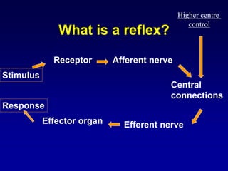
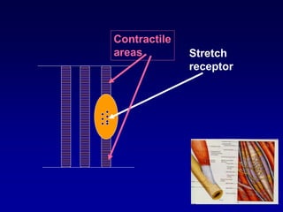
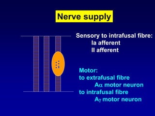
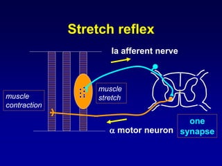
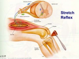
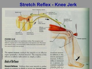
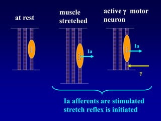
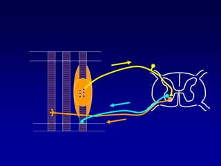
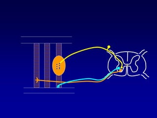
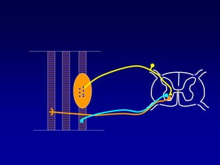
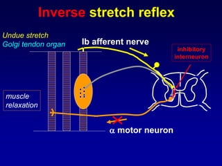
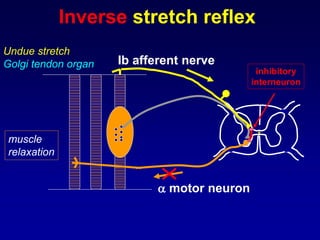
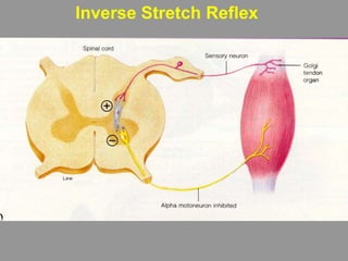
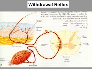
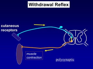
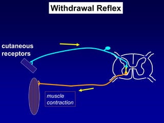
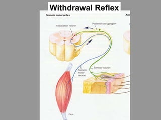
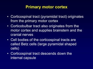
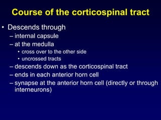
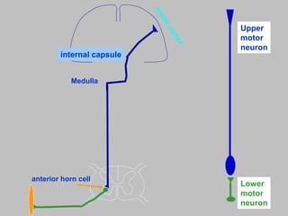
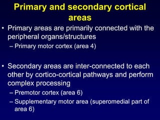
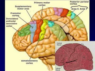
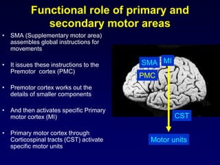
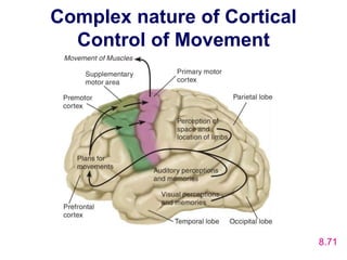
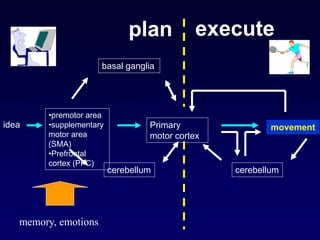
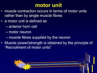
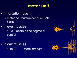
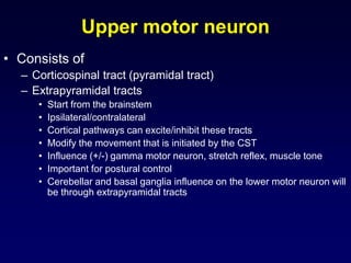
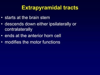
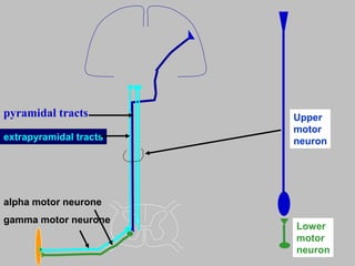
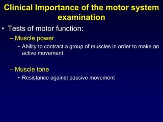
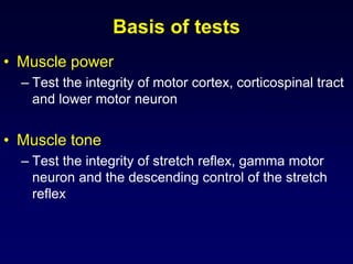
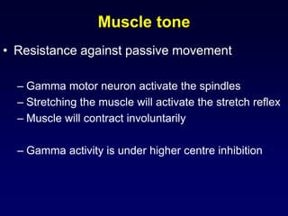
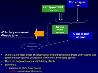
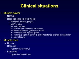
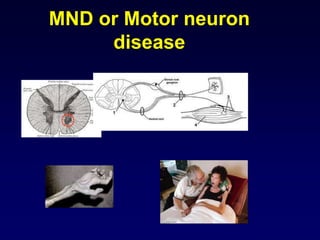
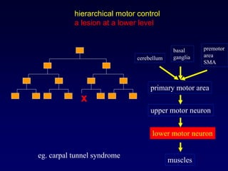
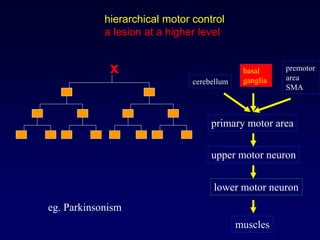
Motor system
Prof. Vajira Weerasinghe
Professor of Physiology
Faculty of Medicine
Motor Functions
• 1. Voluntary motor functions
– Voluntary movement
• 2. Involuntary motor functions
– Reflexes
What is a reflex?
• Response to a stimulus
• Involuntary, without significant
involvement of the brain
• Stimulus Response
Task:
Write down 3 reflexes
What is a reflex?
Stimulus
Effector organ
Response
Central
connections
Efferent nerve
Afferent nerveReceptor
Higher centre
control
Stretch reflex
• This is a basic reflex present in the
spinal cord
• Stimulus: muscle stretch
• Response: contraction of the muscle
• Receptors: stretch receptors located
in the muscle spindle
skeletal muscle
• two types of muscle fibres
– extrafusal
• normally contracting fibres
– Intrafusal
• fibres present inside the muscle spindle
• lie parallel to extrafusal fibres
• either end of the fibre contractile
• central part contains
stretch receptors
Extrafusal
fibre
Intrafusal
fibre
Contractile
areas Stretch
receptor
Nerve supply
Sensory to intrafusal fibre:
Ia afferent
II afferent
Motor:
to extrafusal fibre
A motor neuron
to intrafusal fibre
A motor neuron
Ia afferent nerve
 motor neuron
one
synapse
muscle
stretchmuscle
contraction
Stretch reflex
• When a muscle is stretched
• stretch receptors in the intrafusal fibres
are stimulated
• via type Ia afferent impulse is transmitted
to the spinal cord
•  motor neuron is stimulated
• muscle is contracted
• Monosynaptic
• Neurotransmitter is glutamate
Stretch
Reflex
Stretch Reflex - Knee Jerk
– nuclear bag fibre
• primary (Ia) afferent
– supplies annulospiral ending in the centre
– provide information on muscle length and velocity
(phasic response) fast stretch reflex
– nuclear chain fibre
• primary (Ia) and secondary (II) afferent
– supplies flower spray ending
– monitor the length of the muscle (tonic response) –
slow stretch reflex
Two types of intrafusal fibres
Ia afferent fibre
II afferent fibre
nuclear bag fibre
nuclear chain fibre
 motor
neuron
 motor
neuron
Importance of stretch reflex
• detects muscle length and changes
in muscle length
• Phasic stretch reflex
– Stretching the quadriceps muscle quickly (e.g. by tapping
the patellar tendon) evokes a discharge in the primary
afferent (Ia) fibres
– These form monosynaptic excitatory connections with motor
neurons supplying physiological extensors of the knee,
which contract briefly
• Tonic stretch reflex
– Passive bending of the joint elicits a discharge from the
group II afferents that increases the tone of physiological
extensor (antigravity) muscles
– Tonic stretch reflex is important for maintaining erect body
posture
 motor neuron
• cell body is located in the anterior
horn
• motor neuron travels through the
motor nerve
• supplies the intrafusal fibres
(contractile elements at either end)
 motor neuron
 motor
neuron
 motor neuron
• When  motor neuron is active
– extrafusal fibres are contracted
– muscle contracts
• when  motor neuron is active
– intrafusal fibres are contracted
– stretch receptors are stimulated
– stretch reflex is activated
– impulses will travel through Ia
afferents
– alpha motor neuron is activated
– muscle contracts
at rest
muscle
stretched
active  motor
neuron
Ia
Ia

Ia afferents are stimulated
stretch reflex is initiated
 motor neuron activity
• active all the time - mild contraction
• Maintain the sensitivity of the muscle
spindle to stretch
• modified by the descending pathways
• descending excitatory and inhibitory
influences
• sum effect is generally inhibitory in nature
Alpha gamma co-activation
• gamma motoneurons are activated in parallel
with alpha motoneurons to maintain the firing
of spindle afferents when the extrafusal
muscles shorten
• Activity from brain centres often causes
simultaneous contraction of both extra- and
intrafusal fibres, thereby ensuring that the
spindle is sensitive to stretch at all muscle
lengths
Inverse stretch reflex
• When the muscle is strongly
stretched -> muscle is relaxed
• Golgi tendon organs are stimulated
• Via type Ib afferents impulse is
transmitted to the spinal cord
• inhibitory interneuron is stimulated
•  motor neuron is inhibited
• muscle is relaxed
 motor neuron
Undue stretch
Golgi tendon organ
muscle
relaxation
Ib afferent nerve
inhibitory
interneuron
Inverse stretch reflex
 motor neuron
Undue stretch
Golgi tendon organ
muscle
relaxation
Ib afferent nerve
inhibitory
interneuron
Inverse stretch reflex
Inverse Stretch Reflex
Importance of inverse
stretch reflex
• detects muscle tension
Deep tendon reflexes (DTR)
• Biceps jerk
• Triceps jerk
• Knee jerk
• Ankle jerk
• reflex level
• biceps jerk C56
• triceps jerk C78
• knee jerk L34
• ankle jerk S12
Spinal cord level of stretch
reflexes (tendon jerks)
Superficial reflexes
• Withdrawal reflex
• Superficial abdominal reflex
• Flexor plantar reflex
Withdrawal Reflex
• Stimulus:
– cutaneous stimulation (usually noxious)
• Response:
– withdrawal of the hand
• Polysynaptic reflex
Withdrawal Reflex
muscle
contraction
cutaneous
receptors
polysynaptic
Withdrawal Reflex
muscle
contraction
cutaneous
receptors
Withdrawal Reflex
Withdrawal Reflex
Agonist and antagonist
• Elbow flexion, extension
• Wrist flexion, extension
• Shoulder adduction, abduction
• Hip flexion, extension
• Thigh adduction, abduction
• Knee flexion, extension
• Ankle dorsiflexion, plantar flexion
Reciprocal innervation
• inside the spinal cord
– Agonist and antagonistic muscles are
reciprocally innervated
– stimulation of flexor muscles
– inhibition of extensor muscles
– excitatory neurotransmitter is glutamate
– inhibitory neurotransmitter is glycine
flexor
extensor
+++
----
Reciprocal Innervation
Withdrawal Reflex
Flexor & Crossed extensor reflex
Withdrawal Reflex
Superficial abdominal
reflexes
• light scratch of the abdominal skin
• brisk unilateral contraction of the
abdominal wall
Flexor plantar reflex
• Scratching the sole of foot
• Plantar flexion
• Normal response
Primitive reflexes
• These are reflexes present in
newborn babies but disappear as the
child develops
• They were evolutionarily primitive in
origin
• In adults these reflexes are inhibited
by the higher centres
Other primitive reflexes
• Moro reflex: startle reaction
• Walking/stepping reflex
• Sucking reflex
• Tonic neck reflex
• Palmar grasp reflex
Grasp reflexTonic neck reflex
Walking/stepping reflexMoro reflex: startle reaction
Babinski sign
• when outer border of the sole of the foot is
scratched
• upward movement of big toe (dorsiflexion)
• fanning out of other toes
• also called extensor plantar reflex
• feature of
• upper motor neuron lesion
• seen in infants during 1st year of life (because of
immature corticospinal tract)
positive Babinski sign
Clinical Importance of reflexes
(tendon jerks)
• Locate a lesion in the motor system
• To differentiate upper motor neuron
lesion from a lower motor neuron
lesion
Motor System
• Starts at the motor cortex
• Motor cortex is located at the frontal lobe
– precentral cortex
Motor homunculus
First discovered
by
Penfield
Brodmann areas Primary motor cortex
Area 4
Motor cortex
• different areas of the body are
represented in different cortical areas in
the motor cortex
• Motor homunculus
– somatotopic representation
– not proportionate to structures but
proportionate to function
– distorted map
– upside down map
Motor cortical areas
• primary motor cortex (MI)
– precentral gyrus
• Movements are executed
• secondary motor cortex (MII)
– premotor cortex
– supplementary motor area (SMA)
• Movements are planned together with cerebellum, basal
ganglia and other cortical areas
Primary motor cortex
• Corticospinal tract (pyramidal tract) originates
from the primary motor cortex
• Corticobulbar tract also originates from the
motor cortex and supplies brainstem and the
cranial nerves
• Cell bodies of the corticospinal tracts are
called Betz cells (large pyramidal shaped
cells)
• Corticospinal tract descends down the
internal capsule
Course of the corticospinal tract
• Descends through
– internal capsule
– at the medulla
• cross over to the other side
• uncrossed tracts
– descends down as the corticospinal tract
– ends in each anterior horn cell
– synapse at the anterior horn cell (directly or through
interneurons)
Medulla
internal capsule
Upper
motor
neuron
Lower
motor
neuron
anterior horn cell
Primary and secondary cortical
areas
• Primary areas are primarily connected with the
peripheral organs/structures
– Primary motor cortex (area 4)
• Secondary areas are inter-connected to each
other by cortico-cortical pathways and perform
complex processing
– Premotor cortex (area 6)
– Supplementary motor area (superomedial part of
area 6)
Functional role of primary and
secondary motor areas
• SMA (Supplementary motor area)
assembles global instructions for
movements
• It issues these instructions to the
Premotor cortex (PMC)
• Premotor cortex works out the
details of smaller components
• And then activates specific Primary
motor cortex (MI)
• Primary motor cortex through
Corticospinal tracts (CST) activate
specific motor units
SMA
PMC
MI
CST
Motor units
Complex nature of Cortical
Control of Movement
8.71
idea
•premotor area
•supplementary
motor area
(SMA)
•Prefrontal
cortex (PFC)
Primary
motor cortex
movement
basal ganglia
cerebellum cerebellum
plan execute
memory, emotions
Motor system
• Consists of
– Upper motor neuron
– Lower motor neuron
Lower motor neuron
• consists of mainly
• alpha motor neuron
– and also gamma motor neuron
alpha motor neuron
gamma motor neuron
alpha motor neuron
gamma motor neuron
corticospinal tract
Arrangement at the
anterior horn cell
alpha motor neuron
• this is also called the final common pathway
• Contraction of the muscle occurs through this
whether
– voluntary contraction through corticospinal tract
or
– involuntary contraction through gamma motor
neuron - stretch reflex - Ia afferent
motor unit
• muscle contraction occurs in terms of motor units
rather than by single muscle fibres
• a motor unit is defined as
– anterior horn cell
– motor neuron
– muscle fibres supplied by the neuron
• Muscle power/strength is obtained by the principle of
“Recruitment of motor units”
motor unit
• Innervation ratio
– motor neuron:number of muscle
fibres
• in eye muscles
– 1:23 offers a fine degree of
control
• in calf muscles
– 1:1000 more strength
Upper motor neuron
• Consists of
– Corticospinal tract (pyramidal tract)
– Extrapyramidal tracts
• Start from the brainstem
• Ipsilateral/contralateral
• Cortical pathways can excite/inhibit these tracts
• Modify the movement that is initiated by the CST
• Influence (+/-) gamma motor neuron, stretch reflex, muscle tone
• Important for postural control
• Cerebellar and basal ganglia influence on the lower motor neuron will
be through extrapyramidal tracts
Extrapyramidal tracts
• starts at the brain stem
• descends down either ipsilaterally or
contralaterally
• ends at the anterior horn cell
• modifies the motor functions
Extrapyramidal tracts
• there are 4 tracts
– reticulospinal tracts
– vestibulospinal tracts
– rubrospinal tracts
– tectospinal tracts
reticulospinal tract
• relay station for descending motor impulses
except pyramidal tracts
• receives & modifies motor commands to the
proximal & axial muscles
• maintain normal postural tone
• excitatory to alpha & gamma motorneurons
• end on interneurons too
• this effect is inhibited by cerebral influence
• mainly ipsilateral
midbrain
pons
medulla
spinal cord
reticulospinal tract
• pontine reticular formation
– medial reticulospinal tracts
• controls proximal muscles (axial), excitatory to flexor
• medullary reticular formation
– lateral reticulospinal tracts (also medial)
• excitatory or inhibitory to axial muscles
Reticular formation
• A set of network of interconnected
neurons located in the central
core of the brainstem
• It is made up of ascending and
descending fibers
• It plays a big role in filtering
incoming stimuli to discriminate
irrelevant background stimuli
• There are a large number of
neurons with great degree of
convergence and divergence
Functions
• Maintain consciousness, sleep and arousal
• Motor functions (postural and muscle tone
control)
– Reticulospinal pathways are part of the
extrapyramidal tracts
• Pain modulation (inhibition)
– Several nuclei (PAG, NRM) are part of the
descending pain modulatory (inhibitory) pathway
vestibular nuclei & tracts
• responsible for maintaining tone in antigravity
muscles & for coordinating the postural
adjustments in limbs & eyes
• connections with vestibular receptors (otolith
organs) & cerebellum
• mainly ipsilateral
• supplies extensors
midbrain
pons
medulla
spinal cord
vestibulospinal tract
mainly extensors
• vestibulospinal tracts
– lateral vestibulospinal tract
– medial vestibulospinal tract
– excitatory to antigravity alpha motor neurons &
supplies interneurons too
– lateral tract
• excitation of extensor muscles & relaxation of flexor
muscles
– medial tract
• inhibition of neck & axial muscles
red nucleus
• present in the midbrain
• rubrospinal tract originates from the red nucleus
• ends on interneurons
• control the distal muscles of limbs
• excite limb flexors & inhibit extensors
• higher centre influence (cerebral cortex)
• mainly contralateral
• supplies flexors
• Functionally this tract is not important in human motor
system
midbrain
pons
medulla
spinal cord
rubrospinal tract
mainly flexors
tectospinal tract
• tectospinal tract originates from the tectum of
the midbrain
• ends on interneurons
• mainly contralateral
• supplies cervical segments only
• Functionally this tract is not important in human
motor system
midbrain
pons
medulla
spinal cord
tectospinal tract
cervical segments
inferior olivary nucleus
• present in the medulla
• function:
– motor coordination
• via projections to the cerebellum
• sole source of climbing fibres to the cerebellum
– motor learning
– Functionally this nucleus is not important in human
motor system
Upper
motor
neuron
Lower
motor
neuron
extrapyramidal tracts
pyramidal tracts
alpha motor neurone
gamma motor neurone
Clinical Importance of the motor system
examination
• Tests of motor function:
– Muscle power
• Ability to contract a group of muscles in order to make an
active movement
– Muscle tone
• Resistance against passive movement
Basis of tests
• Muscle power
– Test the integrity of motor cortex, corticospinal tract
and lower motor neuron
• Muscle tone
– Test the integrity of stretch reflex, gamma motor
neuron and the descending control of the stretch
reflex
Muscle tone
• Resistance against passive movement
– Gamma motor neuron activate the spindles
– Stretching the muscle will activate the stretch reflex
– Muscle will contract involuntarily
– Gamma activity is under higher centre inhibition
• There is a complex effect of corticospinal and extrapyramidal tracts on the alpha and
gamma motor neurons (in addition to the effect by muscle spindle)
• There are both excitatory and inhibitory effects
• Sum effect
– excitatory on alpha motor neuron
– Inhibitory on gamma motor neuron
Corticospinal
tract
Extrapyramidal
tracts
Alpha motor
neuron
Gamma
motor
neuron•Voluntary movement
•Muscle tone
Muscle spindle
Clinical situations
• Muscle power
– Normal
– Reduced (muscle weakness)
• Paralysis, paresis, plegia
• MRC grades
0 - no movement
1 - flicker is perceptible in the muscle
2 - movement only if gravity eliminated
3 - can move limb against gravity
4 - can move against gravity & some resistance exerted by examiner
5 - normal power
• Muscle tone
– Normal
– Reduced
• Hypotonia (Flaccidity)
– Increased
• Hypertonia (Spasticity)
Main abnormalities
• Muscle Weakness / paralysis
– Reduced muscle power
• Flaccidity
– Reduced muscle tone
• Spasticity
– Increased muscle tone
• Lower motor neuron lesion causes
– flaccid paralysis (flaccid weakness)
• Upper motor neuron lesion causes
– spastic paralysis (spastic weakness)
Lower motor neuron lesion
• muscle weakness
• flaccid paralysis
• muscle wasting (disuse atrophy)
• reduced muscle tone (hypotonia)
• reflexes: reduced or absent (hyporeflexia or areflexia)
• spontaneous muscle contractions (fasciculations)
• plantar reflex: flexor
• superficial abdominal reflexes: present
• eg. Brachial plexus damage
Muscle wasting
Fasciculations
Upper motor neuron lesion
• muscle weakness
• spastic paralysis
• increased muscle tone (hypertonia)
• reflexes: exaggerated (hyperreflexia)
• Babinski sign: positive
• superficial abdominal reflexes: absent
• muscle wasting is very rare
• clonus can be seen:
– rhythmical series of contractions in response to sudden stretch
• clasp knife effect can be seen
– passive stretch causing initial increased resistance which is released
later
• eg. Stroke
Clasp knife effect
Clonus
Stroke patient walking
Babinski sign
• when outer border of the sole of the foot is
scratched
• upward movement of big toe
• fanning out of other toes
• feature of upper motor neuron lesion
• extensor plantar reflex
• seen in infants during 1st year of life (because
of immature corticospinal tract)
positive Babinski sign
• Observation
• When the spinal cord is suddenly transected, essentially all
cord functions, including spinal cord reflexes, immediately
become depressed
• This is called “spinal shock”
• Period of spinal shock is about 2 weeks in humans
• It may vary depending on the level spinal cord injury
• Higher the animal in evolution greater is the spinal shock
period
Spinal cord transection and spinal shock
During spinal shock period
• complete loss of all reflexes
• no tone
• paralysis
• complete anaesthesia
• no peristalsis
• bladder and rectal reflexes absent
• no sweating
• arterial blood pressure decreases
Possible mechanism of spinal shock
• Normal activity of the spinal cord reflexes depends to a great
extent on continual tonic excitation from higher centers
(pyramidal and extrapyramidal tracts)
• Spinal shock may be due to the sudden cessation of tonic
bombardment of spinal cord interneuron pool by descending
influences
• During recovery from spinal shock, the excitability of spinal cord
reflexes increase due to the lack of descending inhibition and
possible denervation hypersensitivity
• After the spinal shock period typical upper motor neuron
features appear
after the spinal shock
• reflexes will reappear, mostly exaggerated
• bladder become reflex
• mass reflex will appear
– afferent stimuli irradiate to several reflex centres
– noxious stimulus causes: withdrawal response,
evacuation of bladder, rectum, sweating, pallor
Site of lesions
Cortex
Internal capsule
Brain stem
Spinal cord
Anterior horn cell
Motor nerve
Neuromuscular junction
Muscle
Neurological diseases
Disease Involvement
• Stroke UMN
• Peripheral neuropathy LMN
– Mononeuropathy
– Polyneuropathy
• Plexopathy LMN
• Radiculopathy LMN
• Myelopathy LMN, UMN
• Motor neuron disease LMN, UMN
• Monoplegia (monoparesis)
• Hemiplegia (hemiparesis)
• Paraplegia (paraparesis)
• Quadriplegia (quadriparesis)
Site of lesions
monoplegia
only 1 limb is affected either UL or LL,
lower motor neuron lesion
hemiplegia
one half of the body including
UL and LL
lesion in the Internal capsule
paraplegia
both lower limbs
thoracic cord lesion
quadriplegia (tetraplegia)
all 4 limbs are affected
cervical cord or brain stem lesion
Some common neurological
diseases
Stroke
• Cerebrovascular accident (CVA)
• A serious neurological disease
• Large number of deaths per year
• Cerebrovascular ischaemia causing
infarction or haemorrhage
• Sudden onset hemiplegia
• Hypertension, diabetes, obesity are
risk factors
Peripheral neuropathies
• Mononeuropathies
– Carpal tunnel syndrome (CTS)
– Ulnar neuropathy - claw hand
– Saturday night palsy (radial nerve lesion) – wrist drop
– Common peroneal nerve lesion – foot drop
– Posterior tibial nerve lesion – tarsal tunnel syndrome
– Sciatic nerve lesion
– Facial nerve lesion – Bell’s palsy
• Polyneuropathies
– Diabetic, vitamin deficiency, toxic
Median nerve compression
(Carpal tunnel syndrome)
Ulnar nerve lesion
(Ulnar tunnel syndrome)
Clawing of the hand
Radial nerve lesion
(Saturday night palsy)
Wrist drop
Wrist guard
Common peroneal nerve
lesion
Foot drop
Ankle guard
Posterior tibial nerve lesion
(Tarsal tunnel syndrome)
Sciatic nerve lesion
Facial nerve lesion
(Facial palsy or Bell’s
palsy)
Brachial plexopathy
(Erb’s palsy)
Cervical spondylosis
Sciatica
Cervical or thoracic
myelopathy
Paraplegia
Quadriplegia
MND or Motor neuron
disease
• Anterior horn cell disease
• MND: motor neuron disease
• ALS: Amyotrophic lateral sclerosis
• Weakness of lower limbs, upper limbs
• Speech defect: dysarthria
• Difficulty in swallowing: dysphagia
MND or Motor neuron
disease
OVERVIEW OF THE MOTOR
NERVOUS SYSTEM
Components of the motor system
•
Main motor system
– Primary motor area (MI)
– Corticospinal tract (corticobulbar tract)
– Lower motor neuron
– Peripheral nerve (cranial nerve)
– Neuromuscular junction
– Skeletal muscle
– Descending tracts (extrapyramidal tracts)
Secondary motor areas
– Premotor cortex
– Supplementary motor area (SMA)
Cerebellum
Basal ganglia
Vestibular system
Spinal level
• reflexes
• inhibited by the higher levels
Brain stem level
• maintenance of muscle tone
• vestibular functions and balance
Cortical level
• Corticospinal system controls the spinal
cord and the lower motor neuron
cerebellum
• coordinate motor activities
• planning motor commands
• Motor learning
Basal ganglia
• help cortex to execute patterns of
movements
• planning motor commands
• Purposeful movement
• Control of muscle tone and posture
Premotor cortex and SMA
• these are necessary to plan motor command
and also work as an intermediate centre
between cerebellum, basal ganglia and
motor cortex
• control complex movements, bimanual
tasks
Functional role of primary and
secondary motor areas
• SMA (Supplementary Motor Area)
initiate instructions for movements
• issues these instructions to the
PreMotor Area.
• PreMotor Cortex (PMC) works out
the details of smaller components
• activates specific Primary Motor
Cortex (MI)
• Primary Motor Cortex through
corticospinal tracts (CST) activate
specific motor units
SMA
PMC
MI
CST
Motor units
idea
•premotor area
•supplementary
motor area
motor cortex movement
basal ganglia
cerebellum cerebellum
plan execute
memory, emotions
hierarchical motor control
muscles
lower motor neuron
upper motor neuron
primary motor area
premotor
area
SMA
cerebellum
basal
ganglia
hierarchical motor control
a lesion at a lower level
muscles
lower motor neuron
upper motor neuron
primary motor area
premotor
area
SMA
cerebellum
basal
ganglia
x
eg. carpal tunnel syndrome
hierarchical motor control
a lesion at a higher level
muscles
lower motor neuron
upper motor neuron
primary motor area
premotor
area
SMA
cerebellum
basal
ganglia
x
eg. Parkinsonism
• But what finally drives us to
action???
•perhaps motivation
•motivation is controlled by limbic
system and hypothalamus

Y2 s1 motor system 2019

  • 1.
    Motor system Prof. VajiraWeerasinghe Professor of Physiology Faculty of Medicine
  • 2.
    Motor Functions • 1.Voluntary motor functions – Voluntary movement • 2. Involuntary motor functions – Reflexes
  • 3.
    What is areflex? • Response to a stimulus • Involuntary, without significant involvement of the brain • Stimulus Response Task: Write down 3 reflexes
  • 4.
    What is areflex? Stimulus Effector organ Response Central connections Efferent nerve Afferent nerveReceptor Higher centre control
  • 5.
    Stretch reflex • Thisis a basic reflex present in the spinal cord • Stimulus: muscle stretch • Response: contraction of the muscle • Receptors: stretch receptors located in the muscle spindle
  • 7.
    skeletal muscle • twotypes of muscle fibres – extrafusal • normally contracting fibres – Intrafusal • fibres present inside the muscle spindle • lie parallel to extrafusal fibres • either end of the fibre contractile • central part contains stretch receptors
  • 8.
  • 9.
  • 10.
    Nerve supply Sensory tointrafusal fibre: Ia afferent II afferent Motor: to extrafusal fibre A motor neuron to intrafusal fibre A motor neuron
  • 11.
    Ia afferent nerve motor neuron one synapse muscle stretchmuscle contraction Stretch reflex
  • 12.
    • When amuscle is stretched • stretch receptors in the intrafusal fibres are stimulated • via type Ia afferent impulse is transmitted to the spinal cord •  motor neuron is stimulated • muscle is contracted • Monosynaptic • Neurotransmitter is glutamate
  • 13.
  • 14.
  • 15.
    – nuclear bagfibre • primary (Ia) afferent – supplies annulospiral ending in the centre – provide information on muscle length and velocity (phasic response) fast stretch reflex – nuclear chain fibre • primary (Ia) and secondary (II) afferent – supplies flower spray ending – monitor the length of the muscle (tonic response) – slow stretch reflex Two types of intrafusal fibres
  • 16.
    Ia afferent fibre IIafferent fibre nuclear bag fibre nuclear chain fibre  motor neuron  motor neuron
  • 17.
    Importance of stretchreflex • detects muscle length and changes in muscle length
  • 18.
    • Phasic stretchreflex – Stretching the quadriceps muscle quickly (e.g. by tapping the patellar tendon) evokes a discharge in the primary afferent (Ia) fibres – These form monosynaptic excitatory connections with motor neurons supplying physiological extensors of the knee, which contract briefly • Tonic stretch reflex – Passive bending of the joint elicits a discharge from the group II afferents that increases the tone of physiological extensor (antigravity) muscles – Tonic stretch reflex is important for maintaining erect body posture
  • 19.
     motor neuron •cell body is located in the anterior horn • motor neuron travels through the motor nerve • supplies the intrafusal fibres (contractile elements at either end)
  • 20.
     motor neuron motor neuron  motor neuron
  • 21.
    • When motor neuron is active – extrafusal fibres are contracted – muscle contracts • when  motor neuron is active – intrafusal fibres are contracted – stretch receptors are stimulated – stretch reflex is activated – impulses will travel through Ia afferents – alpha motor neuron is activated – muscle contracts
  • 22.
    at rest muscle stretched active motor neuron Ia Ia  Ia afferents are stimulated stretch reflex is initiated
  • 28.
     motor neuronactivity • active all the time - mild contraction • Maintain the sensitivity of the muscle spindle to stretch • modified by the descending pathways • descending excitatory and inhibitory influences • sum effect is generally inhibitory in nature
  • 29.
    Alpha gamma co-activation •gamma motoneurons are activated in parallel with alpha motoneurons to maintain the firing of spindle afferents when the extrafusal muscles shorten • Activity from brain centres often causes simultaneous contraction of both extra- and intrafusal fibres, thereby ensuring that the spindle is sensitive to stretch at all muscle lengths
  • 31.
    Inverse stretch reflex •When the muscle is strongly stretched -> muscle is relaxed • Golgi tendon organs are stimulated • Via type Ib afferents impulse is transmitted to the spinal cord • inhibitory interneuron is stimulated •  motor neuron is inhibited • muscle is relaxed
  • 32.
     motor neuron Unduestretch Golgi tendon organ muscle relaxation Ib afferent nerve inhibitory interneuron Inverse stretch reflex
  • 33.
     motor neuron Unduestretch Golgi tendon organ muscle relaxation Ib afferent nerve inhibitory interneuron Inverse stretch reflex
  • 34.
  • 35.
    Importance of inverse stretchreflex • detects muscle tension
  • 36.
    Deep tendon reflexes(DTR) • Biceps jerk • Triceps jerk • Knee jerk • Ankle jerk
  • 37.
    • reflex level •biceps jerk C56 • triceps jerk C78 • knee jerk L34 • ankle jerk S12 Spinal cord level of stretch reflexes (tendon jerks)
  • 38.
    Superficial reflexes • Withdrawalreflex • Superficial abdominal reflex • Flexor plantar reflex
  • 39.
    Withdrawal Reflex • Stimulus: –cutaneous stimulation (usually noxious) • Response: – withdrawal of the hand • Polysynaptic reflex
  • 40.
  • 41.
  • 42.
  • 43.
  • 44.
    Agonist and antagonist •Elbow flexion, extension • Wrist flexion, extension • Shoulder adduction, abduction • Hip flexion, extension • Thigh adduction, abduction • Knee flexion, extension • Ankle dorsiflexion, plantar flexion
  • 45.
    Reciprocal innervation • insidethe spinal cord – Agonist and antagonistic muscles are reciprocally innervated – stimulation of flexor muscles – inhibition of extensor muscles – excitatory neurotransmitter is glutamate – inhibitory neurotransmitter is glycine flexor extensor +++ ----
  • 46.
  • 47.
    Withdrawal Reflex Flexor &Crossed extensor reflex
  • 48.
  • 49.
    Superficial abdominal reflexes • lightscratch of the abdominal skin • brisk unilateral contraction of the abdominal wall
  • 50.
    Flexor plantar reflex •Scratching the sole of foot • Plantar flexion • Normal response
  • 51.
    Primitive reflexes • Theseare reflexes present in newborn babies but disappear as the child develops • They were evolutionarily primitive in origin • In adults these reflexes are inhibited by the higher centres
  • 52.
    Other primitive reflexes •Moro reflex: startle reaction • Walking/stepping reflex • Sucking reflex • Tonic neck reflex • Palmar grasp reflex
  • 53.
    Grasp reflexTonic neckreflex Walking/stepping reflexMoro reflex: startle reaction
  • 54.
    Babinski sign • whenouter border of the sole of the foot is scratched • upward movement of big toe (dorsiflexion) • fanning out of other toes • also called extensor plantar reflex • feature of • upper motor neuron lesion • seen in infants during 1st year of life (because of immature corticospinal tract)
  • 55.
  • 56.
    Clinical Importance ofreflexes (tendon jerks) • Locate a lesion in the motor system • To differentiate upper motor neuron lesion from a lower motor neuron lesion
  • 57.
    Motor System • Startsat the motor cortex • Motor cortex is located at the frontal lobe – precentral cortex
  • 61.
  • 62.
    Brodmann areas Primarymotor cortex Area 4
  • 63.
    Motor cortex • differentareas of the body are represented in different cortical areas in the motor cortex • Motor homunculus – somatotopic representation – not proportionate to structures but proportionate to function – distorted map – upside down map
  • 64.
    Motor cortical areas •primary motor cortex (MI) – precentral gyrus • Movements are executed • secondary motor cortex (MII) – premotor cortex – supplementary motor area (SMA) • Movements are planned together with cerebellum, basal ganglia and other cortical areas
  • 65.
    Primary motor cortex •Corticospinal tract (pyramidal tract) originates from the primary motor cortex • Corticobulbar tract also originates from the motor cortex and supplies brainstem and the cranial nerves • Cell bodies of the corticospinal tracts are called Betz cells (large pyramidal shaped cells) • Corticospinal tract descends down the internal capsule
  • 66.
    Course of thecorticospinal tract • Descends through – internal capsule – at the medulla • cross over to the other side • uncrossed tracts – descends down as the corticospinal tract – ends in each anterior horn cell – synapse at the anterior horn cell (directly or through interneurons)
  • 67.
  • 68.
    Primary and secondarycortical areas • Primary areas are primarily connected with the peripheral organs/structures – Primary motor cortex (area 4) • Secondary areas are inter-connected to each other by cortico-cortical pathways and perform complex processing – Premotor cortex (area 6) – Supplementary motor area (superomedial part of area 6)
  • 70.
    Functional role ofprimary and secondary motor areas • SMA (Supplementary motor area) assembles global instructions for movements • It issues these instructions to the Premotor cortex (PMC) • Premotor cortex works out the details of smaller components • And then activates specific Primary motor cortex (MI) • Primary motor cortex through Corticospinal tracts (CST) activate specific motor units SMA PMC MI CST Motor units
  • 71.
    Complex nature ofCortical Control of Movement 8.71
  • 72.
    idea •premotor area •supplementary motor area (SMA) •Prefrontal cortex(PFC) Primary motor cortex movement basal ganglia cerebellum cerebellum plan execute memory, emotions
  • 73.
    Motor system • Consistsof – Upper motor neuron – Lower motor neuron
  • 74.
    Lower motor neuron •consists of mainly • alpha motor neuron – and also gamma motor neuron alpha motor neuron gamma motor neuron
  • 75.
    alpha motor neuron gammamotor neuron corticospinal tract Arrangement at the anterior horn cell
  • 76.
    alpha motor neuron •this is also called the final common pathway • Contraction of the muscle occurs through this whether – voluntary contraction through corticospinal tract or – involuntary contraction through gamma motor neuron - stretch reflex - Ia afferent
  • 77.
    motor unit • musclecontraction occurs in terms of motor units rather than by single muscle fibres • a motor unit is defined as – anterior horn cell – motor neuron – muscle fibres supplied by the neuron • Muscle power/strength is obtained by the principle of “Recruitment of motor units”
  • 78.
    motor unit • Innervationratio – motor neuron:number of muscle fibres • in eye muscles – 1:23 offers a fine degree of control • in calf muscles – 1:1000 more strength
  • 79.
    Upper motor neuron •Consists of – Corticospinal tract (pyramidal tract) – Extrapyramidal tracts • Start from the brainstem • Ipsilateral/contralateral • Cortical pathways can excite/inhibit these tracts • Modify the movement that is initiated by the CST • Influence (+/-) gamma motor neuron, stretch reflex, muscle tone • Important for postural control • Cerebellar and basal ganglia influence on the lower motor neuron will be through extrapyramidal tracts
  • 80.
    Extrapyramidal tracts • startsat the brain stem • descends down either ipsilaterally or contralaterally • ends at the anterior horn cell • modifies the motor functions
  • 81.
    Extrapyramidal tracts • thereare 4 tracts – reticulospinal tracts – vestibulospinal tracts – rubrospinal tracts – tectospinal tracts
  • 82.
    reticulospinal tract • relaystation for descending motor impulses except pyramidal tracts • receives & modifies motor commands to the proximal & axial muscles • maintain normal postural tone • excitatory to alpha & gamma motorneurons • end on interneurons too • this effect is inhibited by cerebral influence • mainly ipsilateral
  • 83.
  • 84.
    • pontine reticularformation – medial reticulospinal tracts • controls proximal muscles (axial), excitatory to flexor • medullary reticular formation – lateral reticulospinal tracts (also medial) • excitatory or inhibitory to axial muscles
  • 85.
    Reticular formation • Aset of network of interconnected neurons located in the central core of the brainstem • It is made up of ascending and descending fibers • It plays a big role in filtering incoming stimuli to discriminate irrelevant background stimuli • There are a large number of neurons with great degree of convergence and divergence
  • 86.
    Functions • Maintain consciousness,sleep and arousal • Motor functions (postural and muscle tone control) – Reticulospinal pathways are part of the extrapyramidal tracts • Pain modulation (inhibition) – Several nuclei (PAG, NRM) are part of the descending pain modulatory (inhibitory) pathway
  • 87.
    vestibular nuclei &tracts • responsible for maintaining tone in antigravity muscles & for coordinating the postural adjustments in limbs & eyes • connections with vestibular receptors (otolith organs) & cerebellum • mainly ipsilateral • supplies extensors
  • 88.
  • 89.
    • vestibulospinal tracts –lateral vestibulospinal tract – medial vestibulospinal tract – excitatory to antigravity alpha motor neurons & supplies interneurons too – lateral tract • excitation of extensor muscles & relaxation of flexor muscles – medial tract • inhibition of neck & axial muscles
  • 90.
    red nucleus • presentin the midbrain • rubrospinal tract originates from the red nucleus • ends on interneurons • control the distal muscles of limbs • excite limb flexors & inhibit extensors • higher centre influence (cerebral cortex) • mainly contralateral • supplies flexors • Functionally this tract is not important in human motor system
  • 91.
  • 92.
    tectospinal tract • tectospinaltract originates from the tectum of the midbrain • ends on interneurons • mainly contralateral • supplies cervical segments only • Functionally this tract is not important in human motor system
  • 93.
  • 94.
    inferior olivary nucleus •present in the medulla • function: – motor coordination • via projections to the cerebellum • sole source of climbing fibres to the cerebellum – motor learning – Functionally this nucleus is not important in human motor system
  • 95.
  • 96.
    Clinical Importance ofthe motor system examination • Tests of motor function: – Muscle power • Ability to contract a group of muscles in order to make an active movement – Muscle tone • Resistance against passive movement
  • 97.
    Basis of tests •Muscle power – Test the integrity of motor cortex, corticospinal tract and lower motor neuron • Muscle tone – Test the integrity of stretch reflex, gamma motor neuron and the descending control of the stretch reflex
  • 98.
    Muscle tone • Resistanceagainst passive movement – Gamma motor neuron activate the spindles – Stretching the muscle will activate the stretch reflex – Muscle will contract involuntarily – Gamma activity is under higher centre inhibition
  • 99.
    • There isa complex effect of corticospinal and extrapyramidal tracts on the alpha and gamma motor neurons (in addition to the effect by muscle spindle) • There are both excitatory and inhibitory effects • Sum effect – excitatory on alpha motor neuron – Inhibitory on gamma motor neuron Corticospinal tract Extrapyramidal tracts Alpha motor neuron Gamma motor neuron•Voluntary movement •Muscle tone Muscle spindle
  • 100.
    Clinical situations • Musclepower – Normal – Reduced (muscle weakness) • Paralysis, paresis, plegia • MRC grades 0 - no movement 1 - flicker is perceptible in the muscle 2 - movement only if gravity eliminated 3 - can move limb against gravity 4 - can move against gravity & some resistance exerted by examiner 5 - normal power • Muscle tone – Normal – Reduced • Hypotonia (Flaccidity) – Increased • Hypertonia (Spasticity)
  • 101.
    Main abnormalities • MuscleWeakness / paralysis – Reduced muscle power • Flaccidity – Reduced muscle tone • Spasticity – Increased muscle tone
  • 102.
    • Lower motorneuron lesion causes – flaccid paralysis (flaccid weakness) • Upper motor neuron lesion causes – spastic paralysis (spastic weakness)
  • 103.
    Lower motor neuronlesion • muscle weakness • flaccid paralysis • muscle wasting (disuse atrophy) • reduced muscle tone (hypotonia) • reflexes: reduced or absent (hyporeflexia or areflexia) • spontaneous muscle contractions (fasciculations) • plantar reflex: flexor • superficial abdominal reflexes: present • eg. Brachial plexus damage
  • 104.
  • 105.
  • 106.
    Upper motor neuronlesion • muscle weakness • spastic paralysis • increased muscle tone (hypertonia) • reflexes: exaggerated (hyperreflexia) • Babinski sign: positive • superficial abdominal reflexes: absent • muscle wasting is very rare • clonus can be seen: – rhythmical series of contractions in response to sudden stretch • clasp knife effect can be seen – passive stretch causing initial increased resistance which is released later • eg. Stroke
  • 107.
  • 108.
  • 109.
    Babinski sign • whenouter border of the sole of the foot is scratched • upward movement of big toe • fanning out of other toes • feature of upper motor neuron lesion • extensor plantar reflex • seen in infants during 1st year of life (because of immature corticospinal tract)
  • 110.
  • 111.
    • Observation • Whenthe spinal cord is suddenly transected, essentially all cord functions, including spinal cord reflexes, immediately become depressed • This is called “spinal shock” • Period of spinal shock is about 2 weeks in humans • It may vary depending on the level spinal cord injury • Higher the animal in evolution greater is the spinal shock period Spinal cord transection and spinal shock
  • 112.
    During spinal shockperiod • complete loss of all reflexes • no tone • paralysis • complete anaesthesia • no peristalsis • bladder and rectal reflexes absent • no sweating • arterial blood pressure decreases
  • 113.
    Possible mechanism ofspinal shock • Normal activity of the spinal cord reflexes depends to a great extent on continual tonic excitation from higher centers (pyramidal and extrapyramidal tracts) • Spinal shock may be due to the sudden cessation of tonic bombardment of spinal cord interneuron pool by descending influences • During recovery from spinal shock, the excitability of spinal cord reflexes increase due to the lack of descending inhibition and possible denervation hypersensitivity • After the spinal shock period typical upper motor neuron features appear
  • 114.
    after the spinalshock • reflexes will reappear, mostly exaggerated • bladder become reflex • mass reflex will appear – afferent stimuli irradiate to several reflex centres – noxious stimulus causes: withdrawal response, evacuation of bladder, rectum, sweating, pallor
  • 115.
    Site of lesions Cortex Internalcapsule Brain stem Spinal cord Anterior horn cell Motor nerve Neuromuscular junction Muscle
  • 116.
    Neurological diseases Disease Involvement •Stroke UMN • Peripheral neuropathy LMN – Mononeuropathy – Polyneuropathy • Plexopathy LMN • Radiculopathy LMN • Myelopathy LMN, UMN • Motor neuron disease LMN, UMN • Monoplegia (monoparesis) • Hemiplegia (hemiparesis) • Paraplegia (paraparesis) • Quadriplegia (quadriparesis)
  • 117.
    Site of lesions monoplegia only1 limb is affected either UL or LL, lower motor neuron lesion hemiplegia one half of the body including UL and LL lesion in the Internal capsule paraplegia both lower limbs thoracic cord lesion quadriplegia (tetraplegia) all 4 limbs are affected cervical cord or brain stem lesion
  • 118.
  • 119.
    Stroke • Cerebrovascular accident(CVA) • A serious neurological disease • Large number of deaths per year • Cerebrovascular ischaemia causing infarction or haemorrhage • Sudden onset hemiplegia • Hypertension, diabetes, obesity are risk factors
  • 120.
    Peripheral neuropathies • Mononeuropathies –Carpal tunnel syndrome (CTS) – Ulnar neuropathy - claw hand – Saturday night palsy (radial nerve lesion) – wrist drop – Common peroneal nerve lesion – foot drop – Posterior tibial nerve lesion – tarsal tunnel syndrome – Sciatic nerve lesion – Facial nerve lesion – Bell’s palsy • Polyneuropathies – Diabetic, vitamin deficiency, toxic
  • 121.
  • 122.
    Ulnar nerve lesion (Ulnartunnel syndrome) Clawing of the hand
  • 123.
    Radial nerve lesion (Saturdaynight palsy) Wrist drop Wrist guard
  • 124.
  • 125.
    Posterior tibial nervelesion (Tarsal tunnel syndrome)
  • 126.
  • 127.
    Facial nerve lesion (Facialpalsy or Bell’s palsy)
  • 128.
  • 129.
  • 130.
  • 131.
  • 132.
    MND or Motorneuron disease • Anterior horn cell disease • MND: motor neuron disease • ALS: Amyotrophic lateral sclerosis • Weakness of lower limbs, upper limbs • Speech defect: dysarthria • Difficulty in swallowing: dysphagia
  • 133.
    MND or Motorneuron disease
  • 134.
    OVERVIEW OF THEMOTOR NERVOUS SYSTEM
  • 135.
    Components of themotor system • Main motor system – Primary motor area (MI) – Corticospinal tract (corticobulbar tract) – Lower motor neuron – Peripheral nerve (cranial nerve) – Neuromuscular junction – Skeletal muscle – Descending tracts (extrapyramidal tracts) Secondary motor areas – Premotor cortex – Supplementary motor area (SMA) Cerebellum Basal ganglia Vestibular system
  • 136.
    Spinal level • reflexes •inhibited by the higher levels
  • 137.
    Brain stem level •maintenance of muscle tone • vestibular functions and balance
  • 138.
    Cortical level • Corticospinalsystem controls the spinal cord and the lower motor neuron
  • 139.
    cerebellum • coordinate motoractivities • planning motor commands • Motor learning
  • 140.
    Basal ganglia • helpcortex to execute patterns of movements • planning motor commands • Purposeful movement • Control of muscle tone and posture
  • 141.
    Premotor cortex andSMA • these are necessary to plan motor command and also work as an intermediate centre between cerebellum, basal ganglia and motor cortex • control complex movements, bimanual tasks
  • 142.
    Functional role ofprimary and secondary motor areas • SMA (Supplementary Motor Area) initiate instructions for movements • issues these instructions to the PreMotor Area. • PreMotor Cortex (PMC) works out the details of smaller components • activates specific Primary Motor Cortex (MI) • Primary Motor Cortex through corticospinal tracts (CST) activate specific motor units SMA PMC MI CST Motor units
  • 143.
    idea •premotor area •supplementary motor area motorcortex movement basal ganglia cerebellum cerebellum plan execute memory, emotions
  • 144.
    hierarchical motor control muscles lowermotor neuron upper motor neuron primary motor area premotor area SMA cerebellum basal ganglia
  • 145.
    hierarchical motor control alesion at a lower level muscles lower motor neuron upper motor neuron primary motor area premotor area SMA cerebellum basal ganglia x eg. carpal tunnel syndrome
  • 146.
    hierarchical motor control alesion at a higher level muscles lower motor neuron upper motor neuron primary motor area premotor area SMA cerebellum basal ganglia x eg. Parkinsonism
  • 147.
    • But whatfinally drives us to action??? •perhaps motivation •motivation is controlled by limbic system and hypothalamus