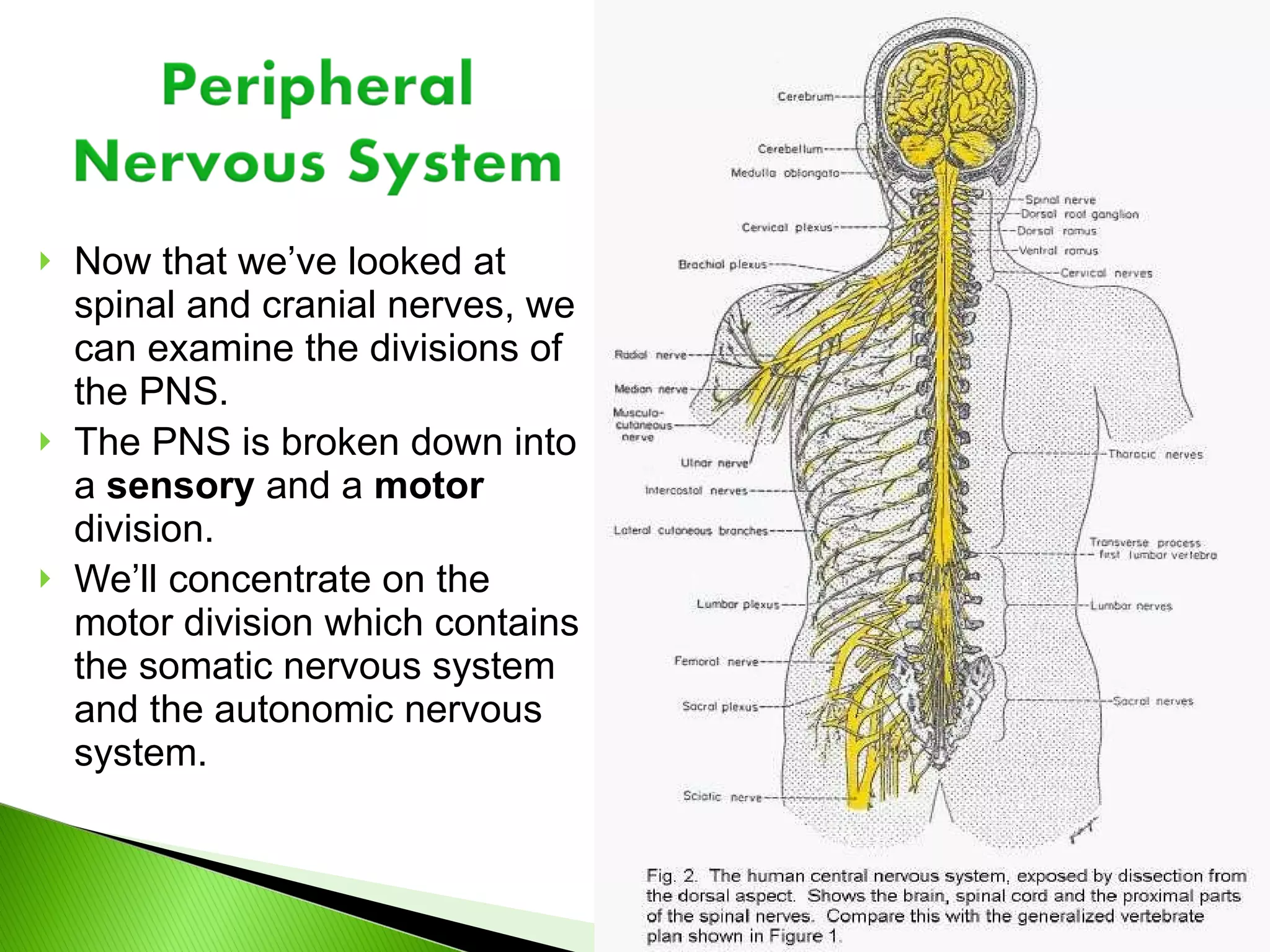
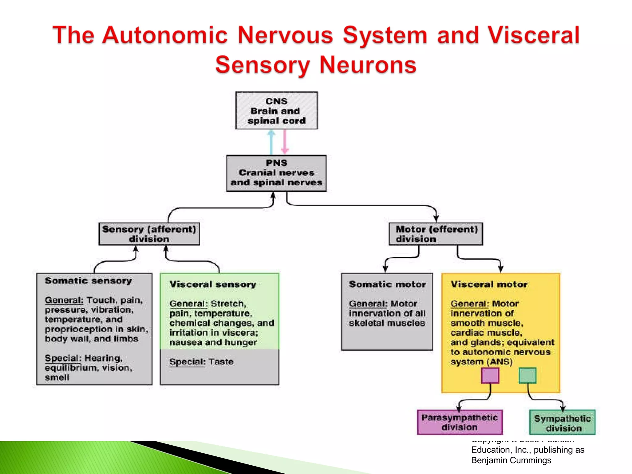
Now that we’velooked at spinal and cranial nerves, we can examine the divisions of the PNS. The PNS is broken down into a sensory and a motor division. We’ll concentrate on the motor division which contains the somatic nervous system and the autonomic nervous system.
3.
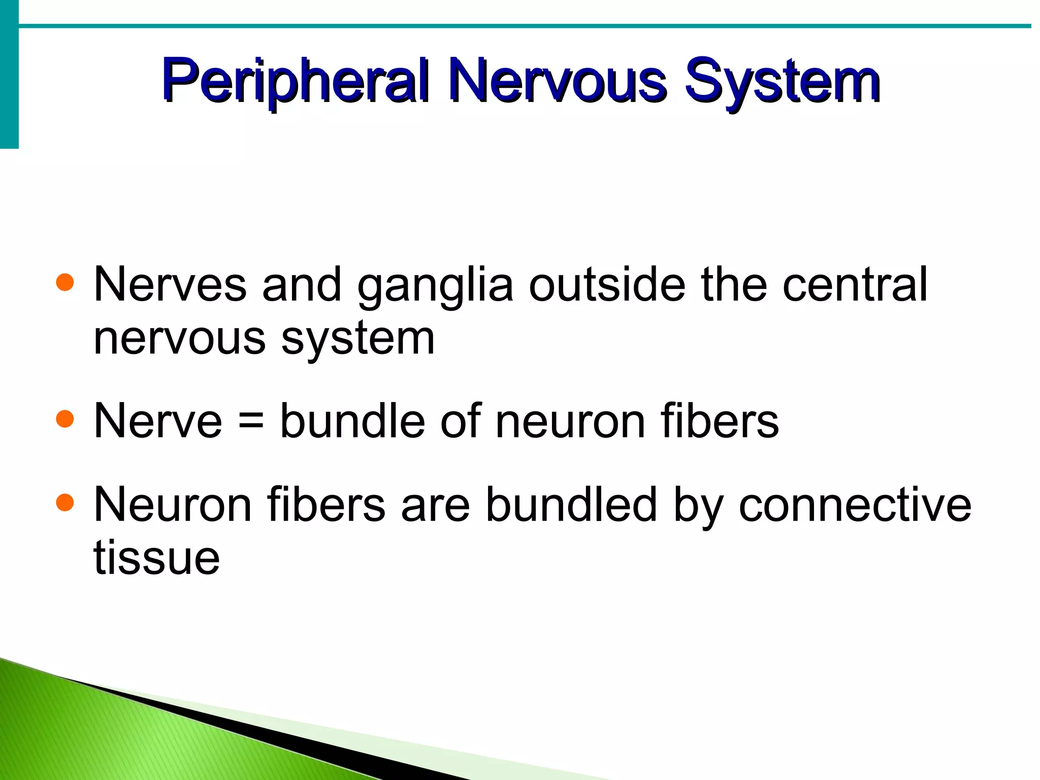
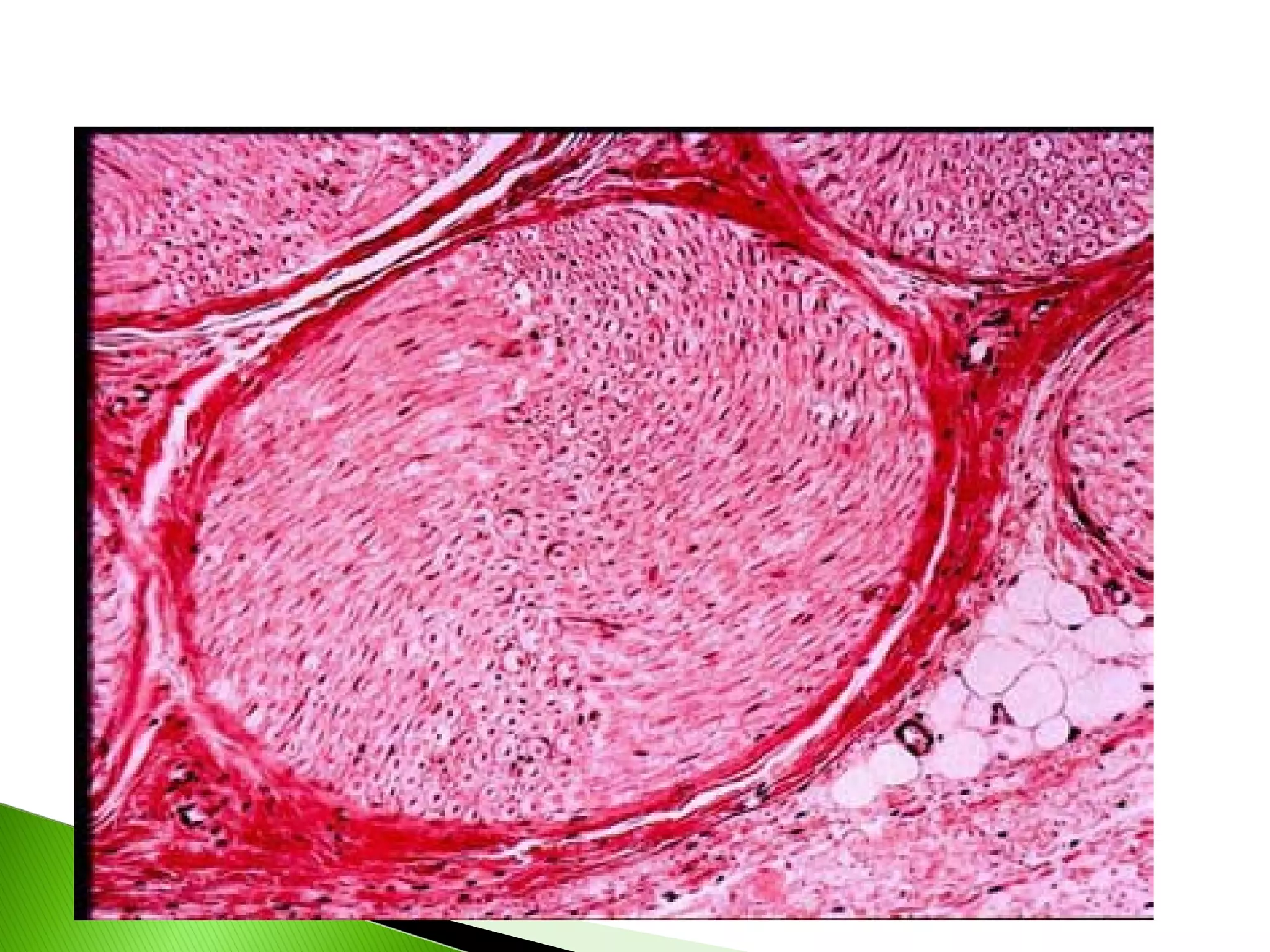
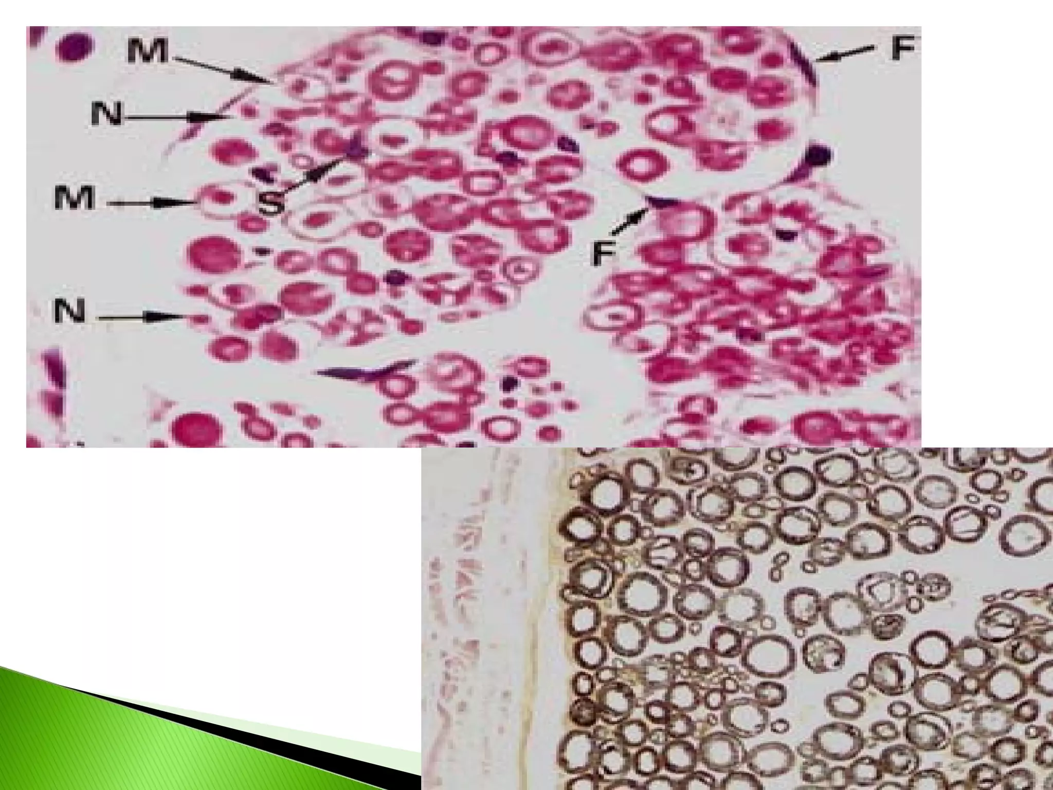
Peripheral Nervous SystemNerves and ganglia outside the central nervous system Nerve = bundle of neuron fibers Neuron fibers are bundled by connective tissue
4.
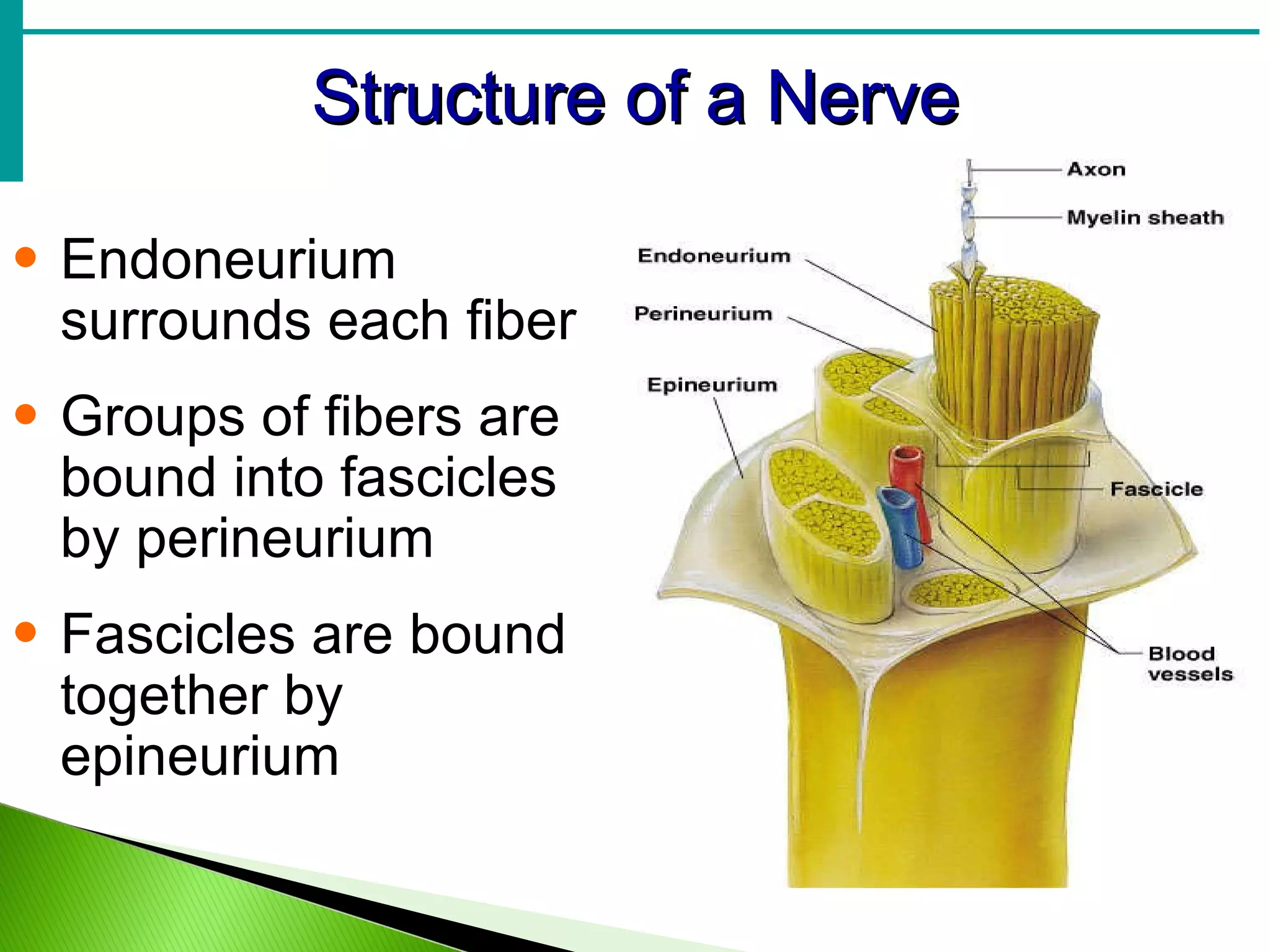
Structure of aNerve Endoneurium surrounds each fiber Groups of fibers are bound into fascicles by perineurium Fascicles are bound together by epineurium
Classification of NervesMixed nerves – both sensory and motor fibers Afferent (sensory) nerves – carry impulses toward the CNS Efferent (motor) nerves – carry impulses away from the CNS
9.
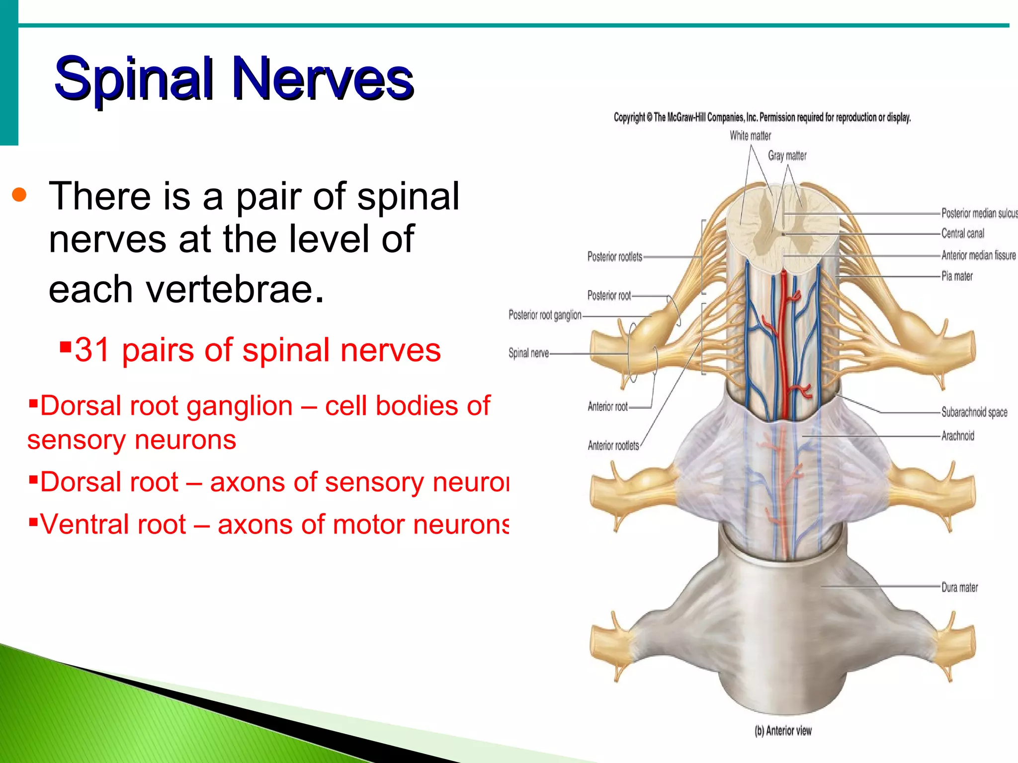
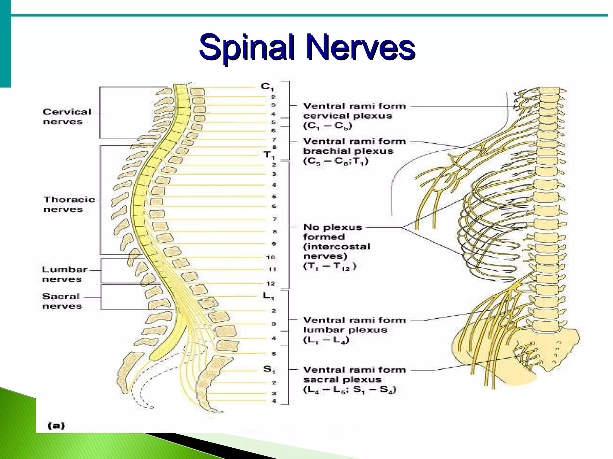
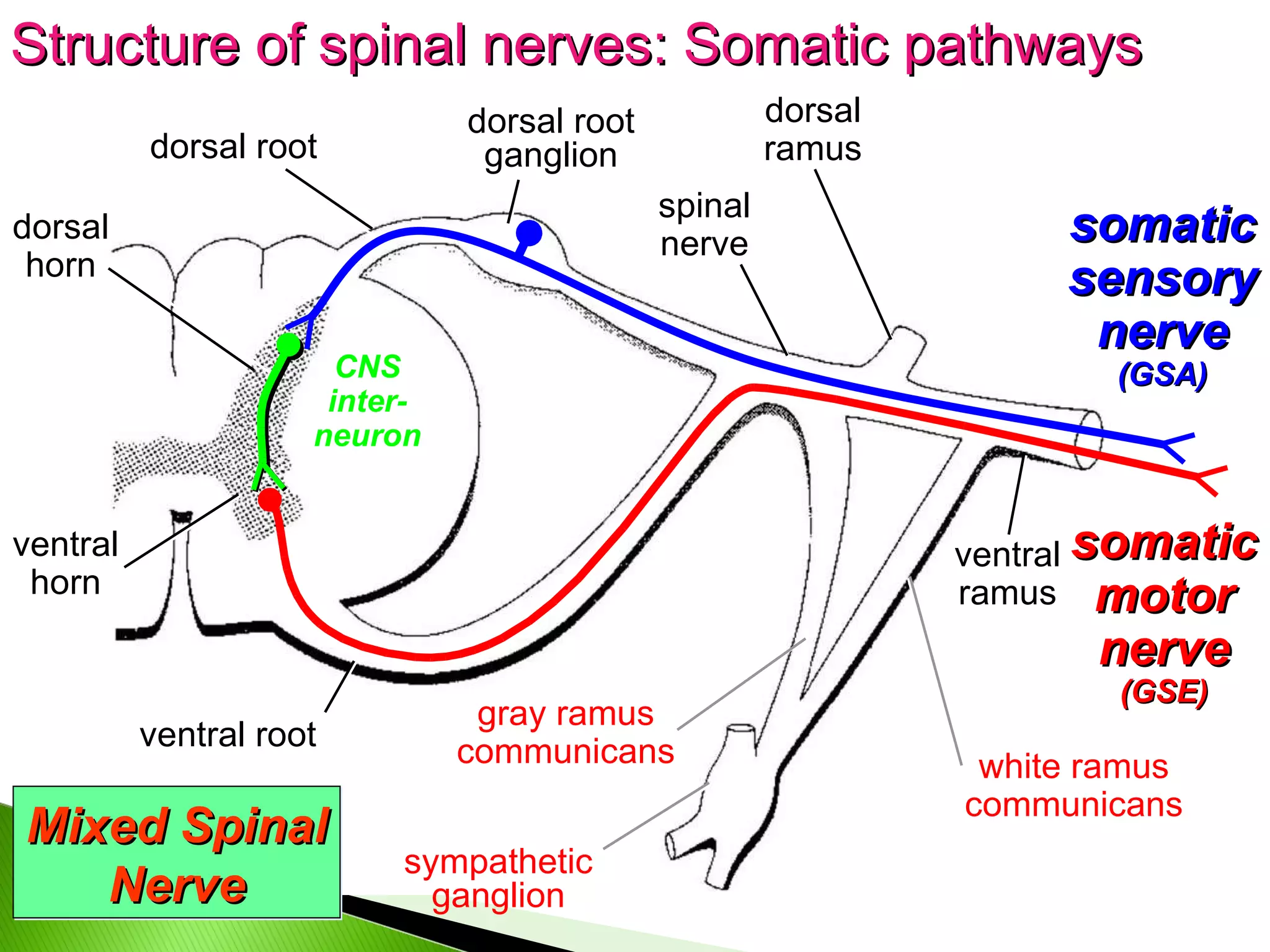
Spinal Nerves Thereis a pair of spinal nerves at the level of each vertebrae . Dorsal root ganglion – cell bodies of sensory neurons Dorsal root – axons of sensory neurons Ventral root – axons of motor neurons 31 pairs of spinal nerves
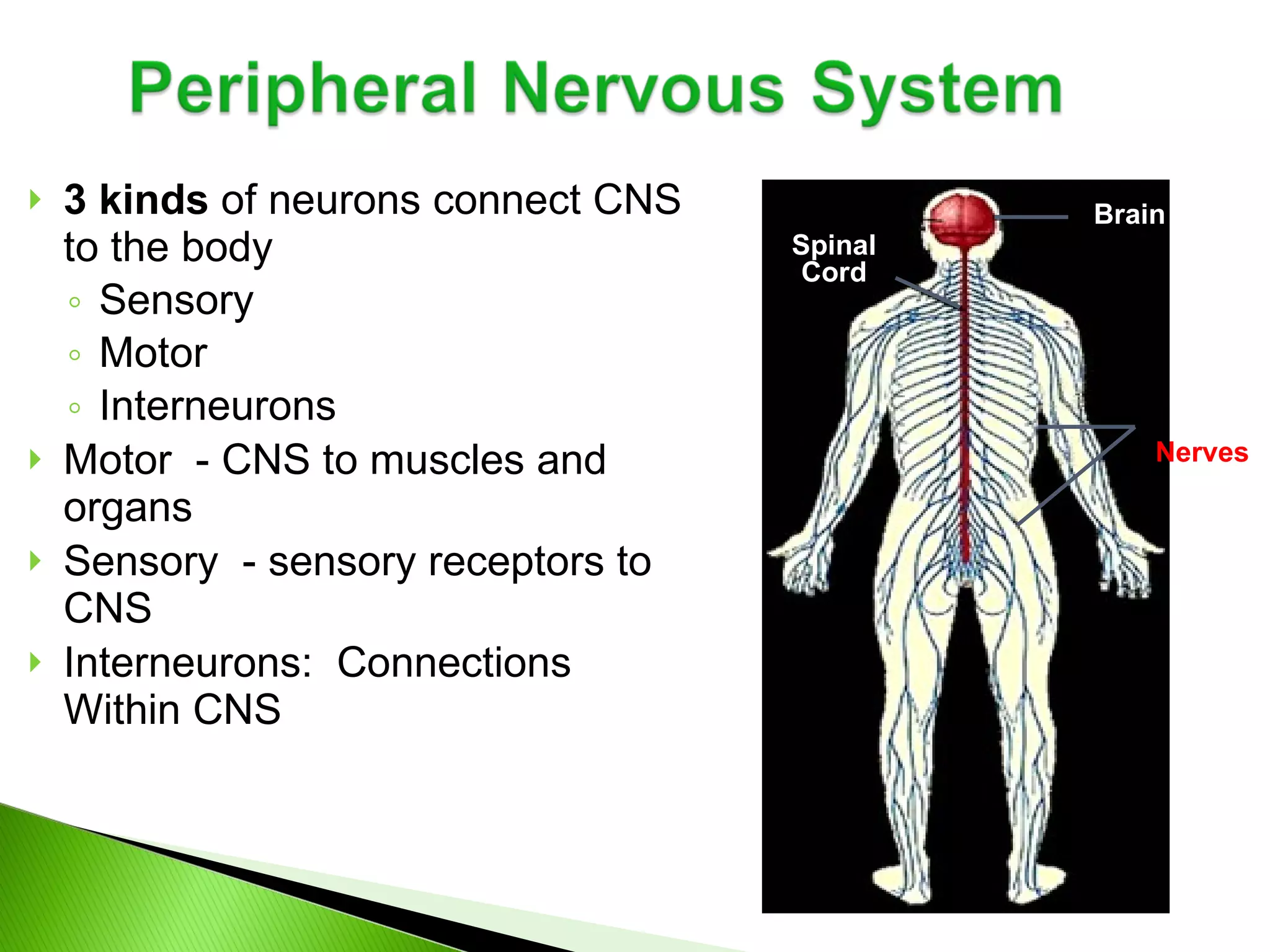
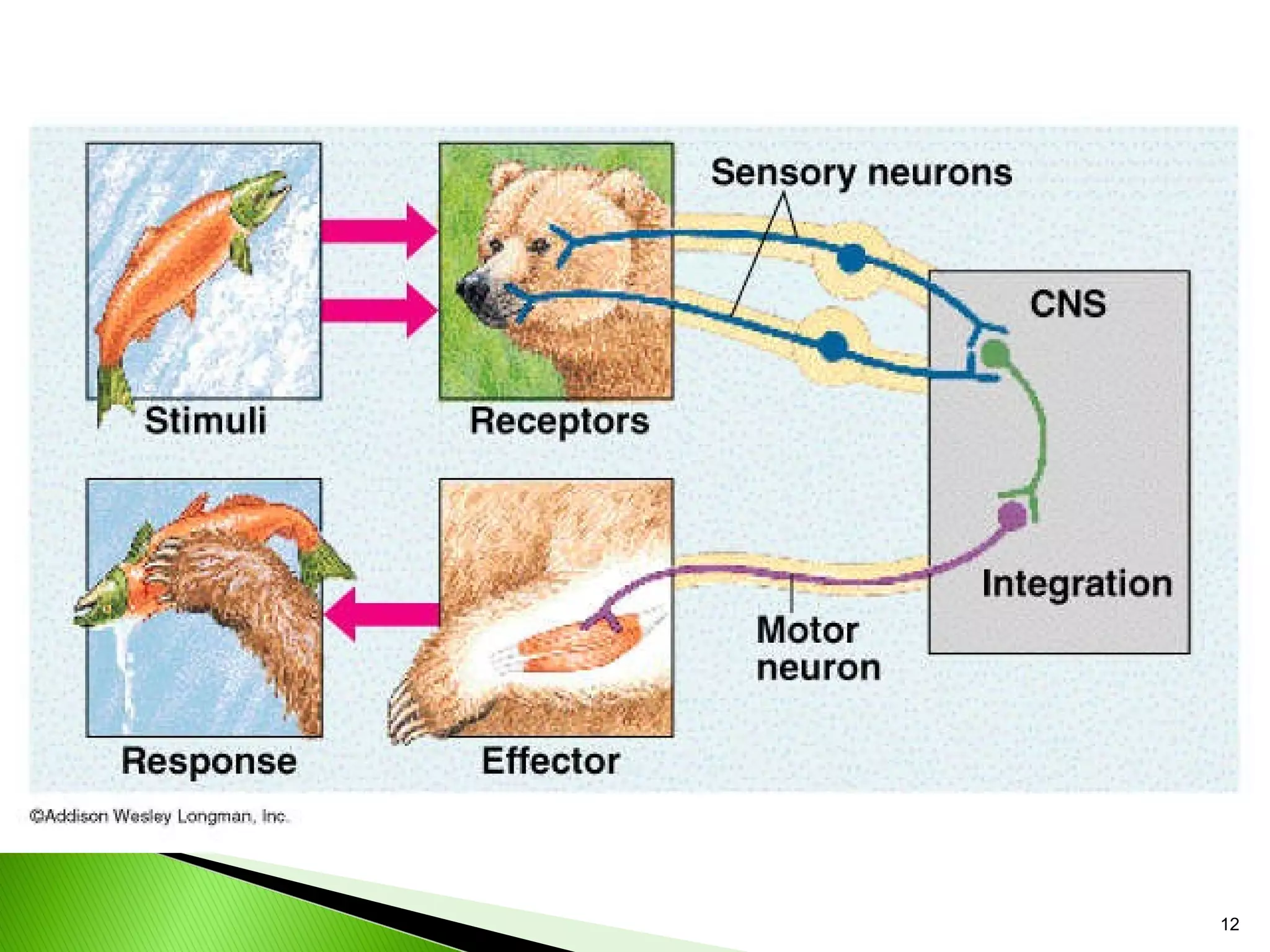
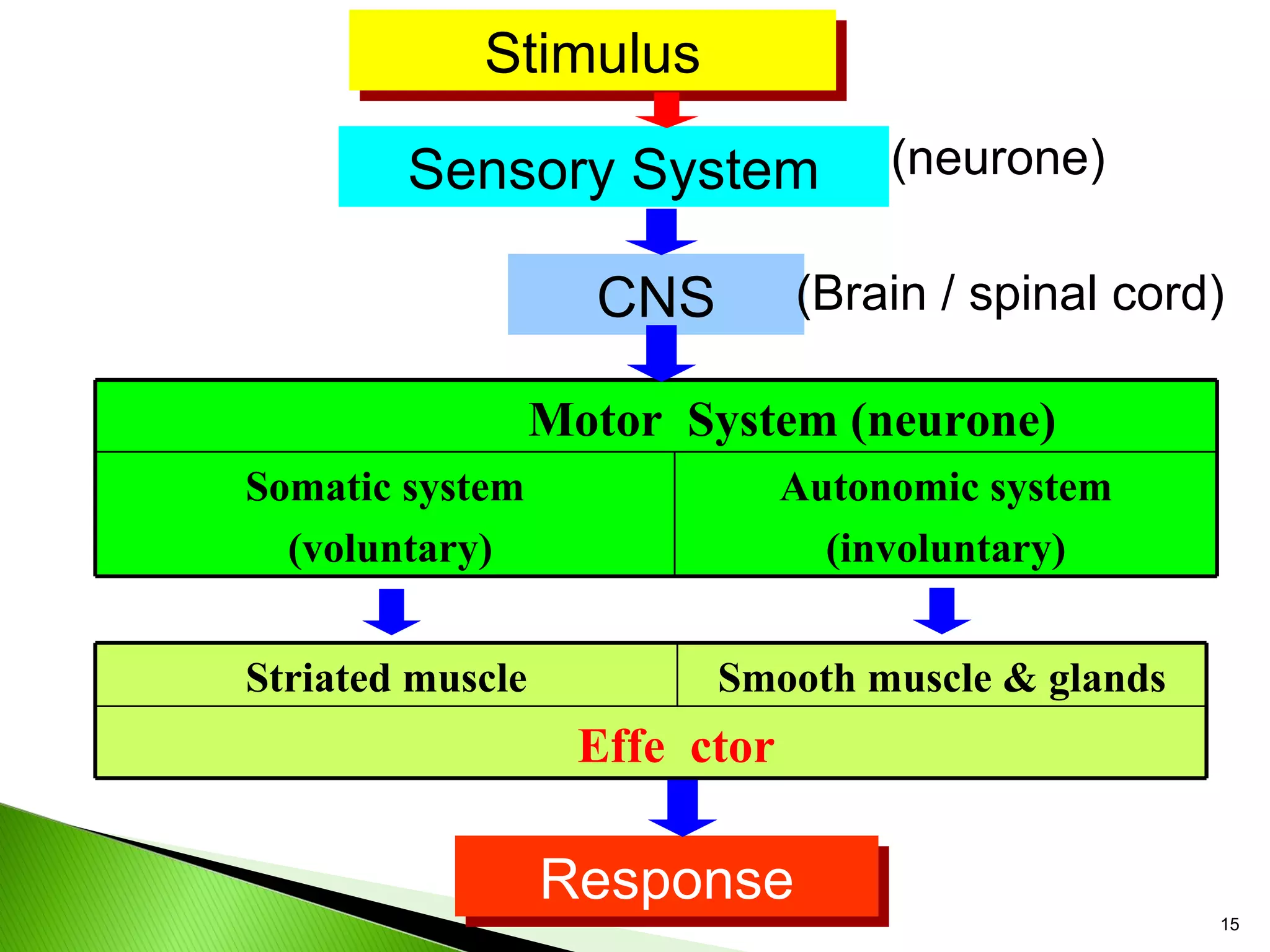
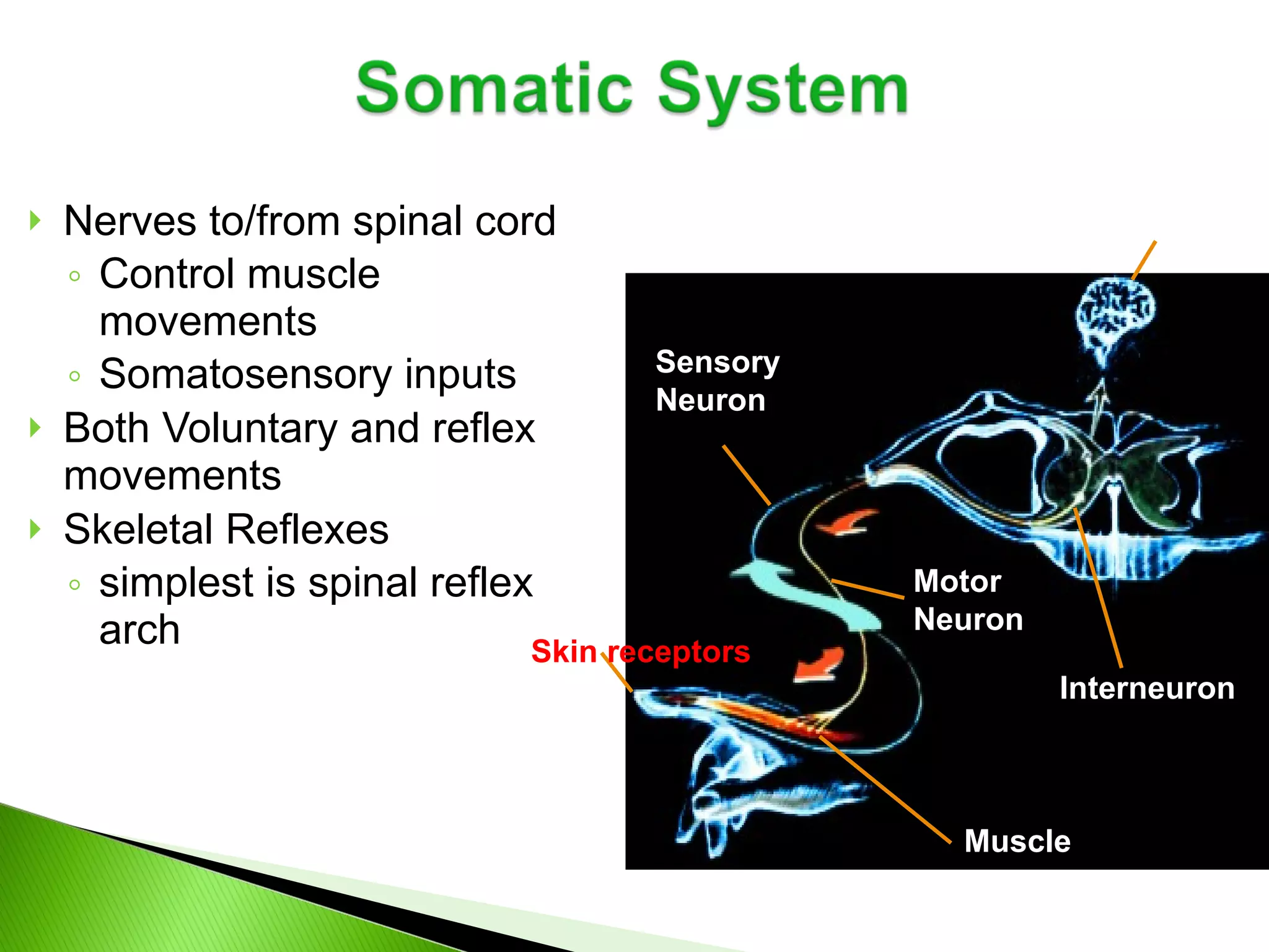
3 kinds of neurons connect CNS to the body Sensory Motor Interneurons Motor - CNS to muscles and organs Sensory - sensory receptors to CNS Interneurons: Connections Within CNS Spinal Cord Brain Nerves
13.
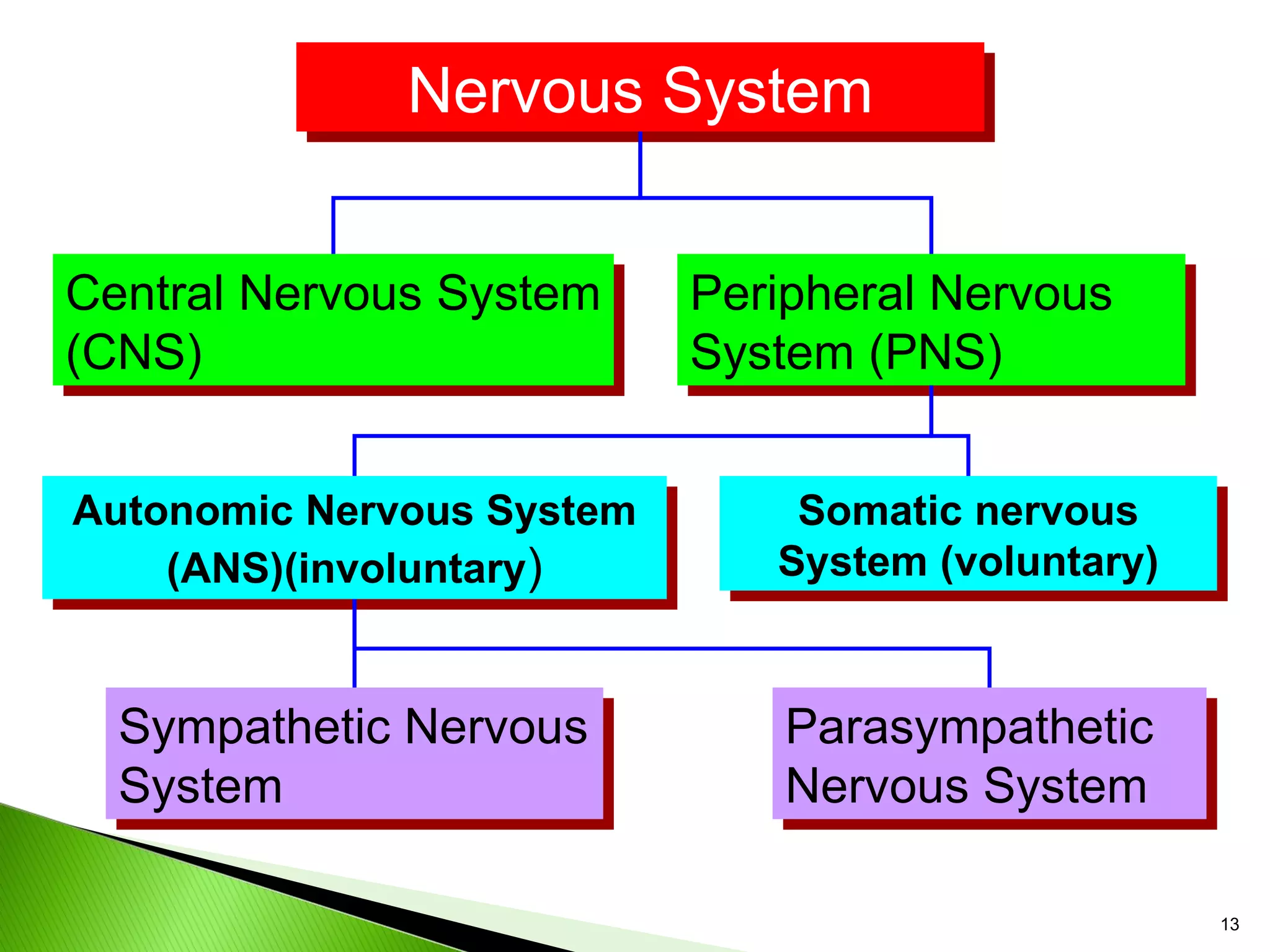
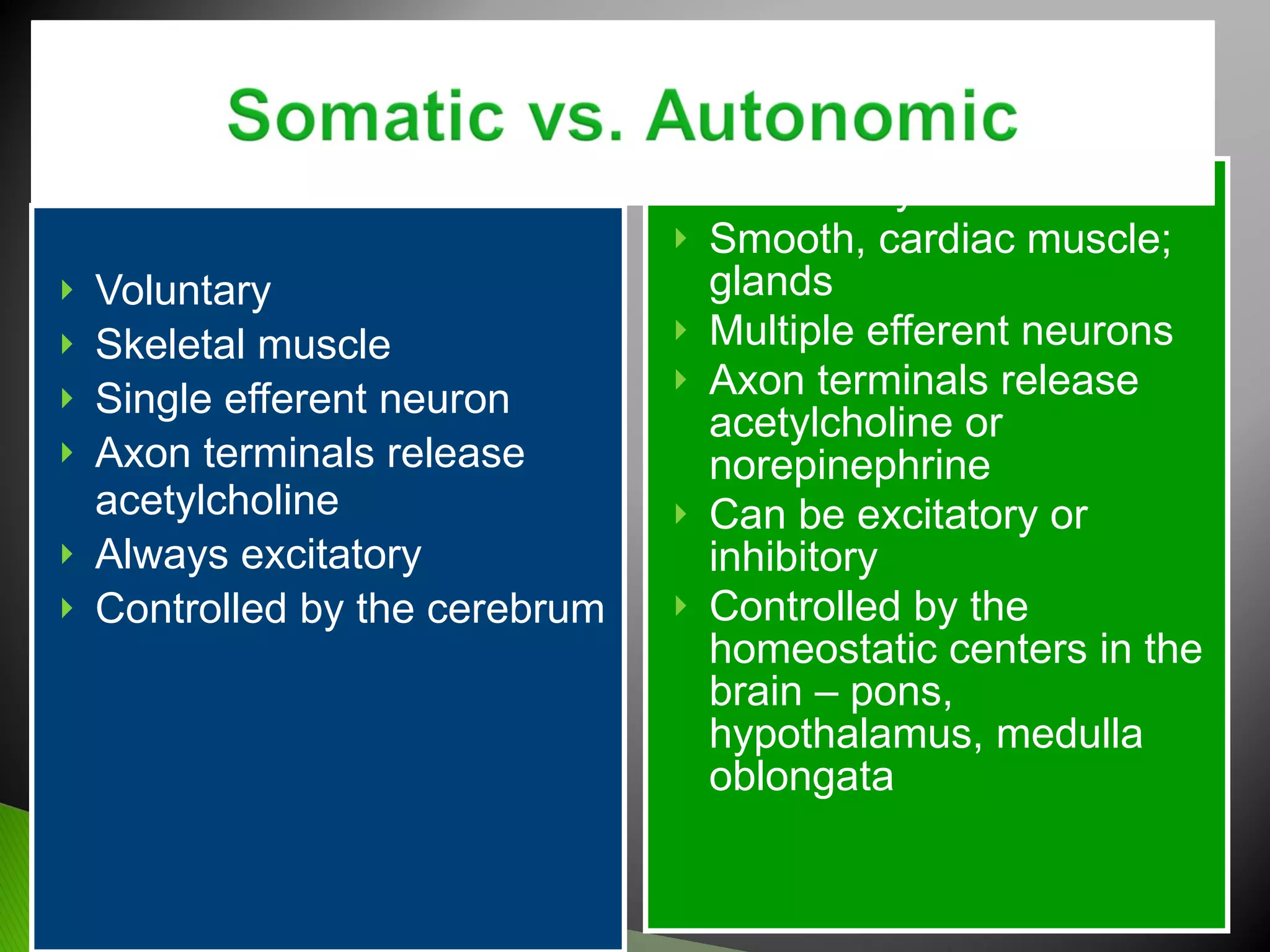
Nervous System CentralNervous System (CNS) Peripheral Nervous System (PNS) Autonomic Nervous System (ANS)(involuntary ) Somatic nervous System (voluntary) Sympathetic Nervous System Parasympathetic Nervous System
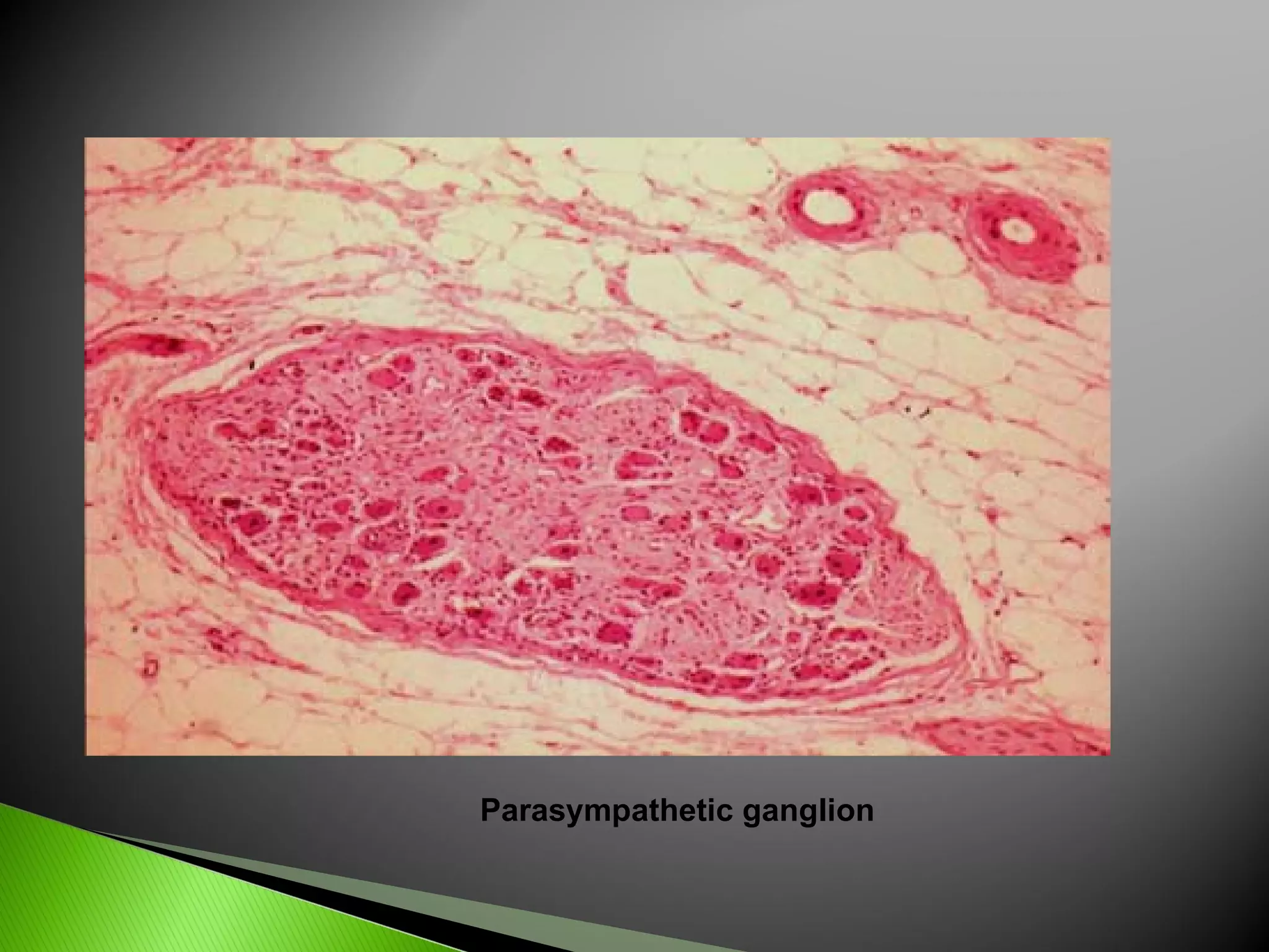
Autonomic Nervous SystemThe involuntary branch of the nervous system Consists of only motor nerves Divided into two divisions Sympathetic division Parasympathetic division
21.
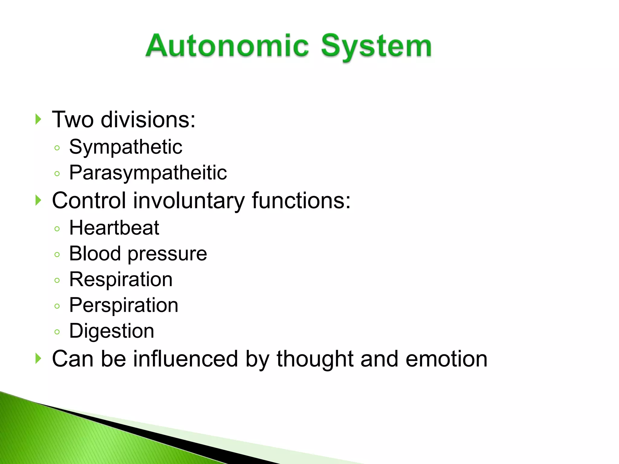
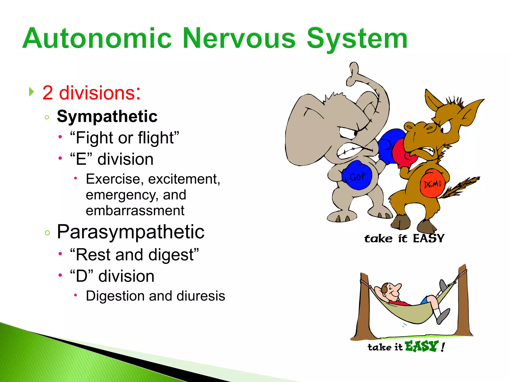
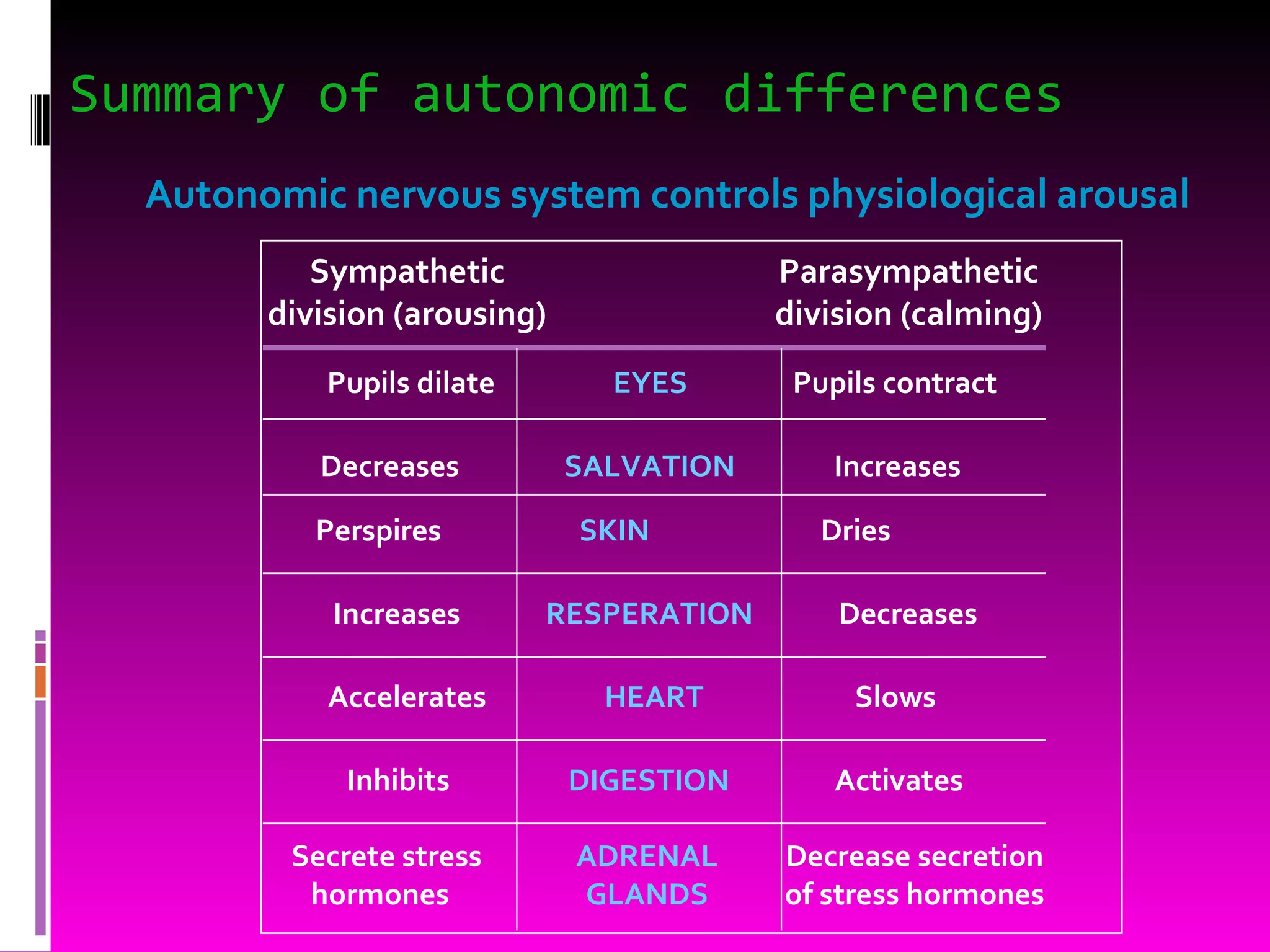
Two divisions: Sympathetic Parasympatheitic Control involuntary functions: Heartbeat Blood pressure Respiration Perspiration Digestion Can be influenced by thought and emotion
22.
2 divisions :Sympathetic “ Fight or flight” “ E” division Exercise, excitement, emergency, and embarrassment Parasympathetic “ Rest and digest” “ D” division Digestion and diuresis
23.
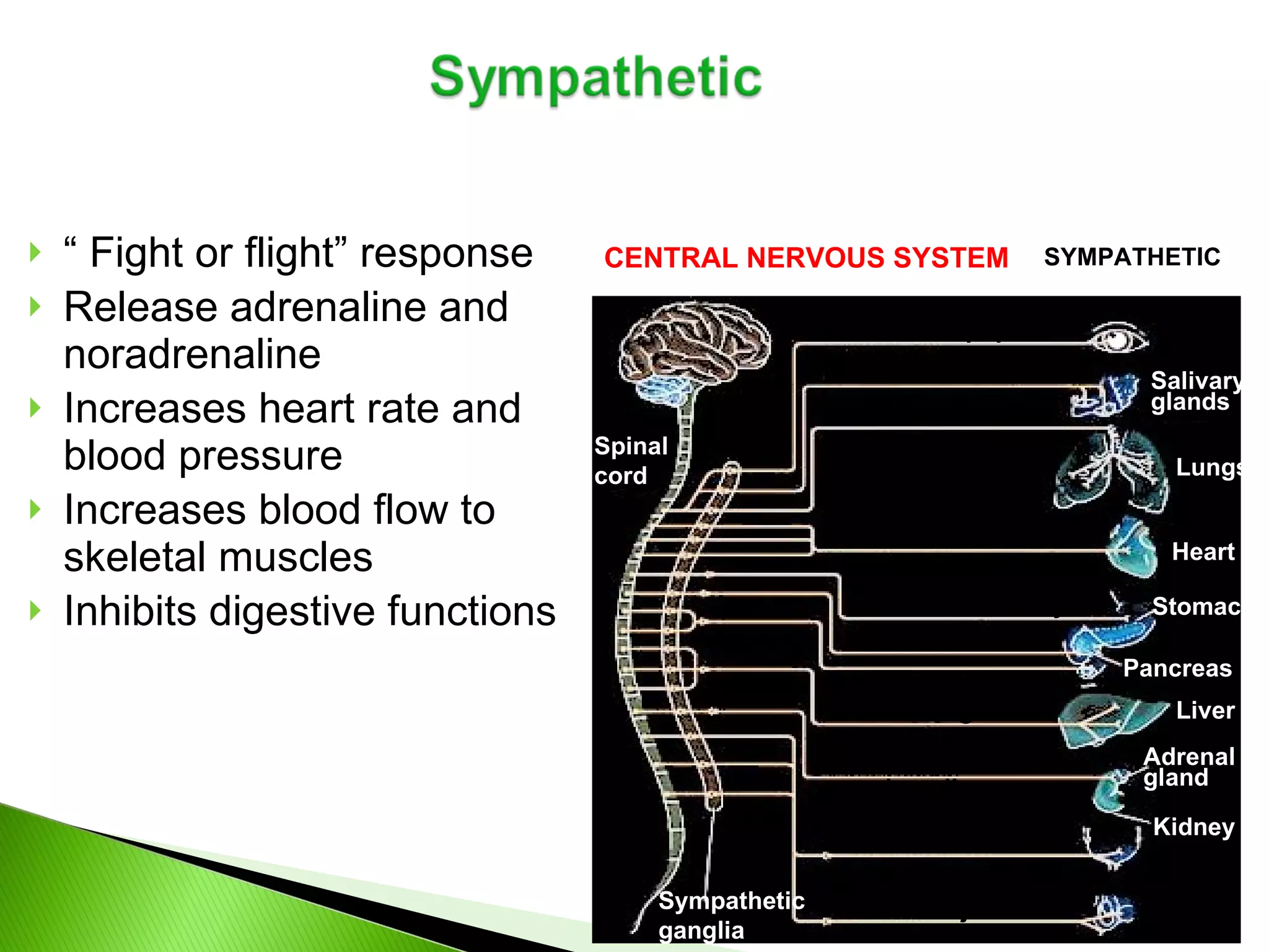
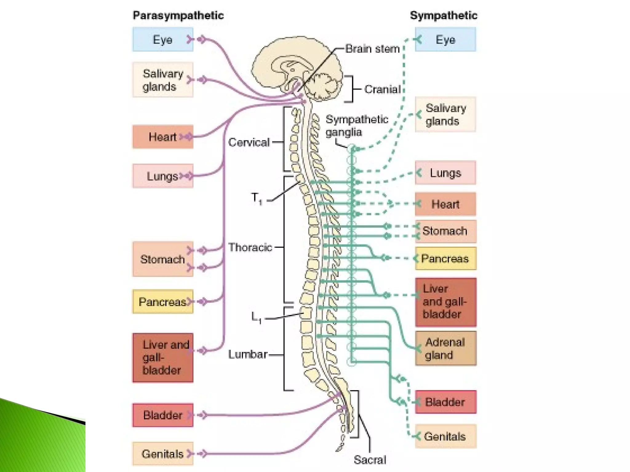
“ Fightor flight” response Release adrenaline and noradrenaline Increases heart rate and blood pressure Increases blood flow to skeletal muscles Inhibits digestive functions CENTRAL NERVOUS SYSTEM Brain Spinal cord SYMPATHETIC Dilates pupil Stimulates salivation Relaxes bronchi Accelerates heartbeat Inhibits activity Stimulates glucose Secretion of adrenaline, nonadrenaline Relaxes bladder Stimulates ejaculation in male Sympathetic ganglia Salivary glands Lungs Heart Stomach Pancreas Liver Adrenal gland Kidney
24.
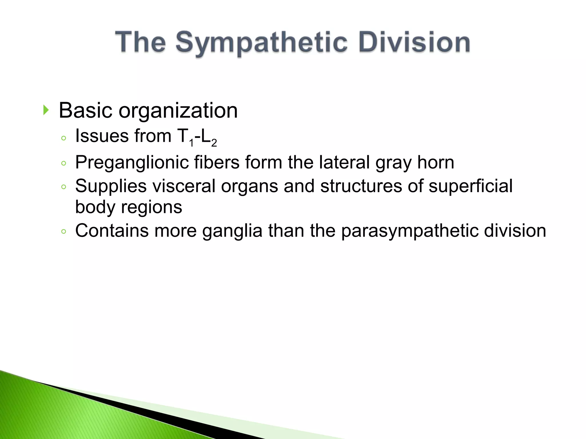
Basic organization Issuesfrom T 1 -L 2 Preganglionic fibers form the lateral gray horn Supplies visceral organs and structures of superficial body regions Contains more ganglia than the parasympathetic division
25.
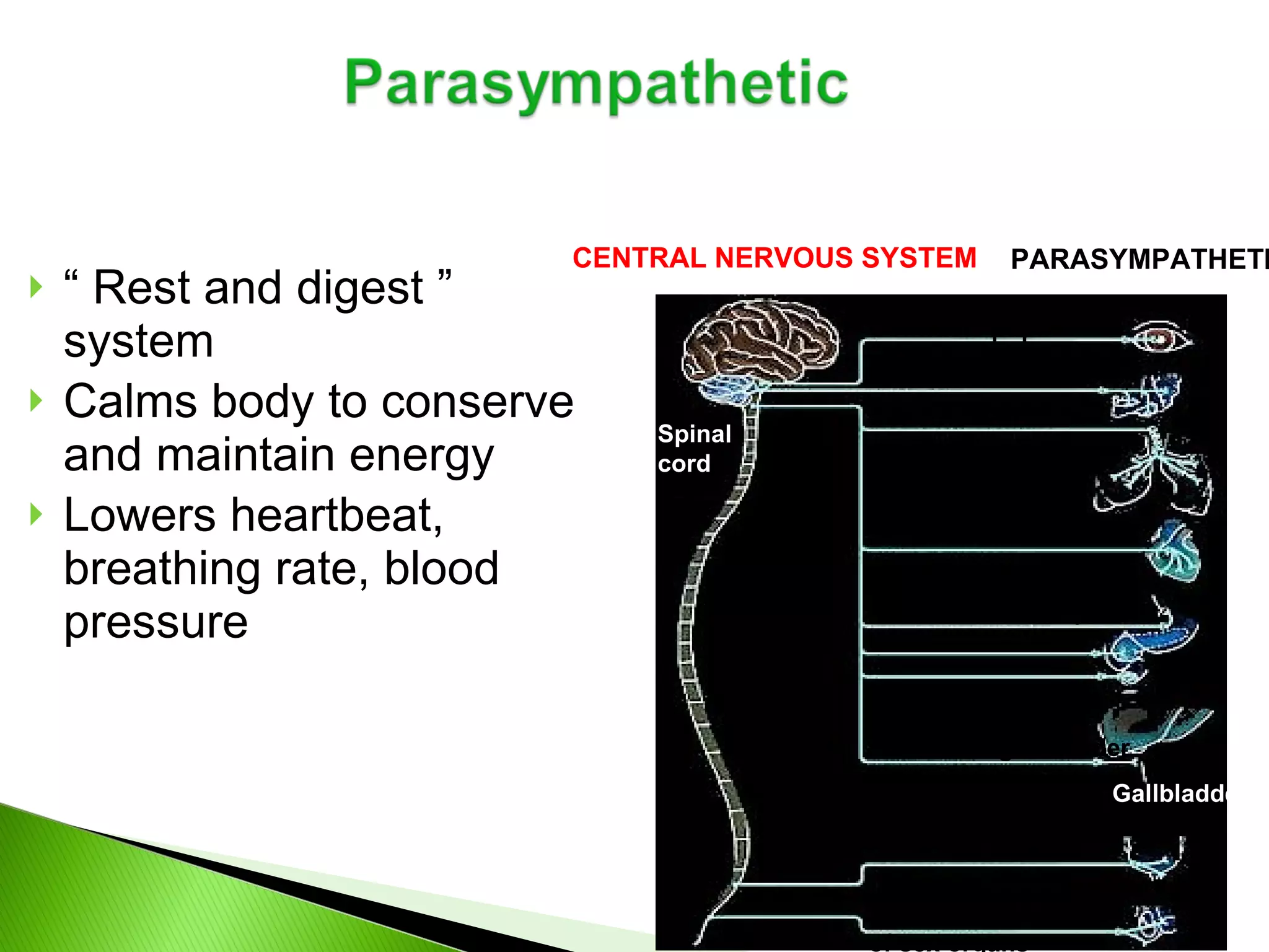
“ Restand digest ” system Calms body to conserve and maintain energy Lowers heartbeat, breathing rate, blood pressure CENTRAL NERVOUS SYSTEM Brain PARASYMPATHETIC Spinal cord Stimulates salivation Constricts bronchi Slows heartbeat Stimulates activity Contracts bladder Stimulates erection of sex organs Stimulates gallbladder Gallbladder Contracts pupil
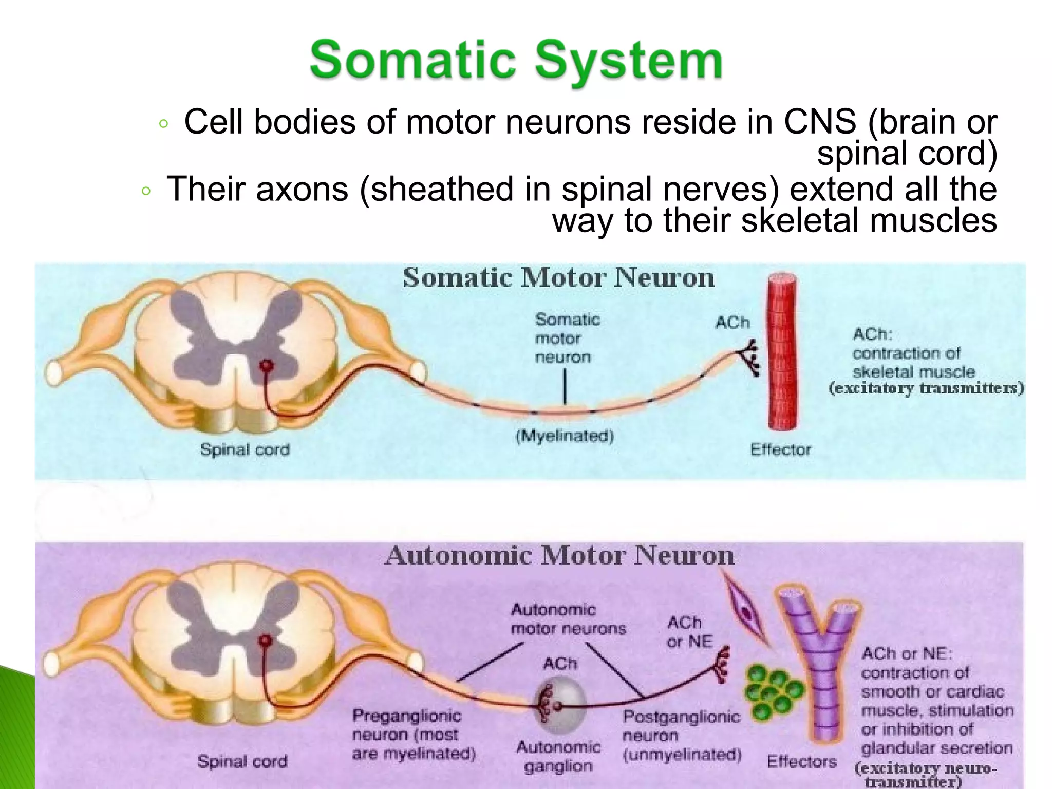
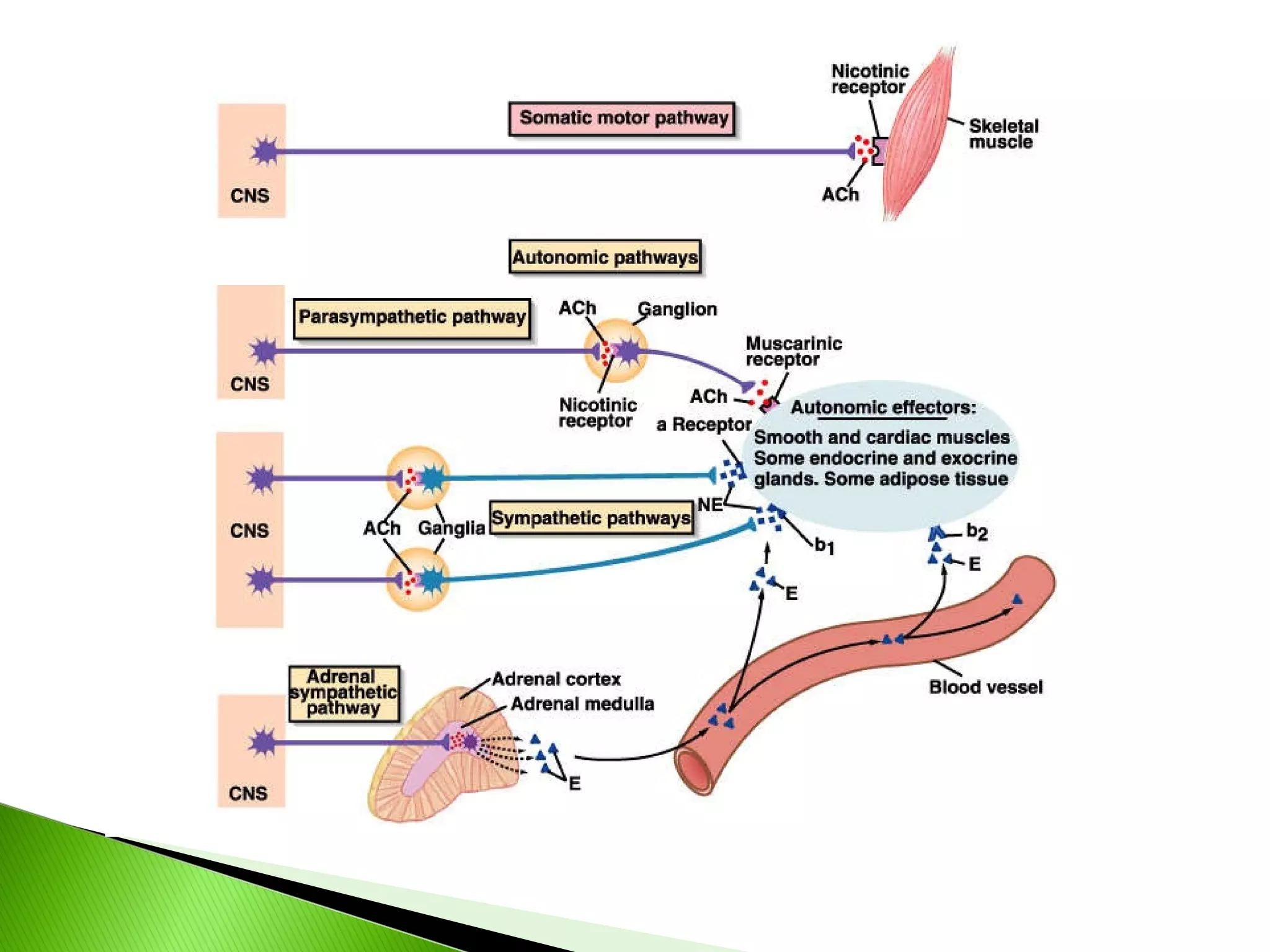
Voluntary Skeletal muscleSingle efferent neuron Axon terminals release acetylcholine Always excitatory Controlled by the cerebrum Involuntary Smooth, cardiac muscle; glands Multiple efferent neurons Axon terminals release acetylcholine or norepinephrine Can be excitatory or inhibitory Controlled by the homeostatic centers in the brain – pons, hypothalamus, medulla oblongata
29.
30.
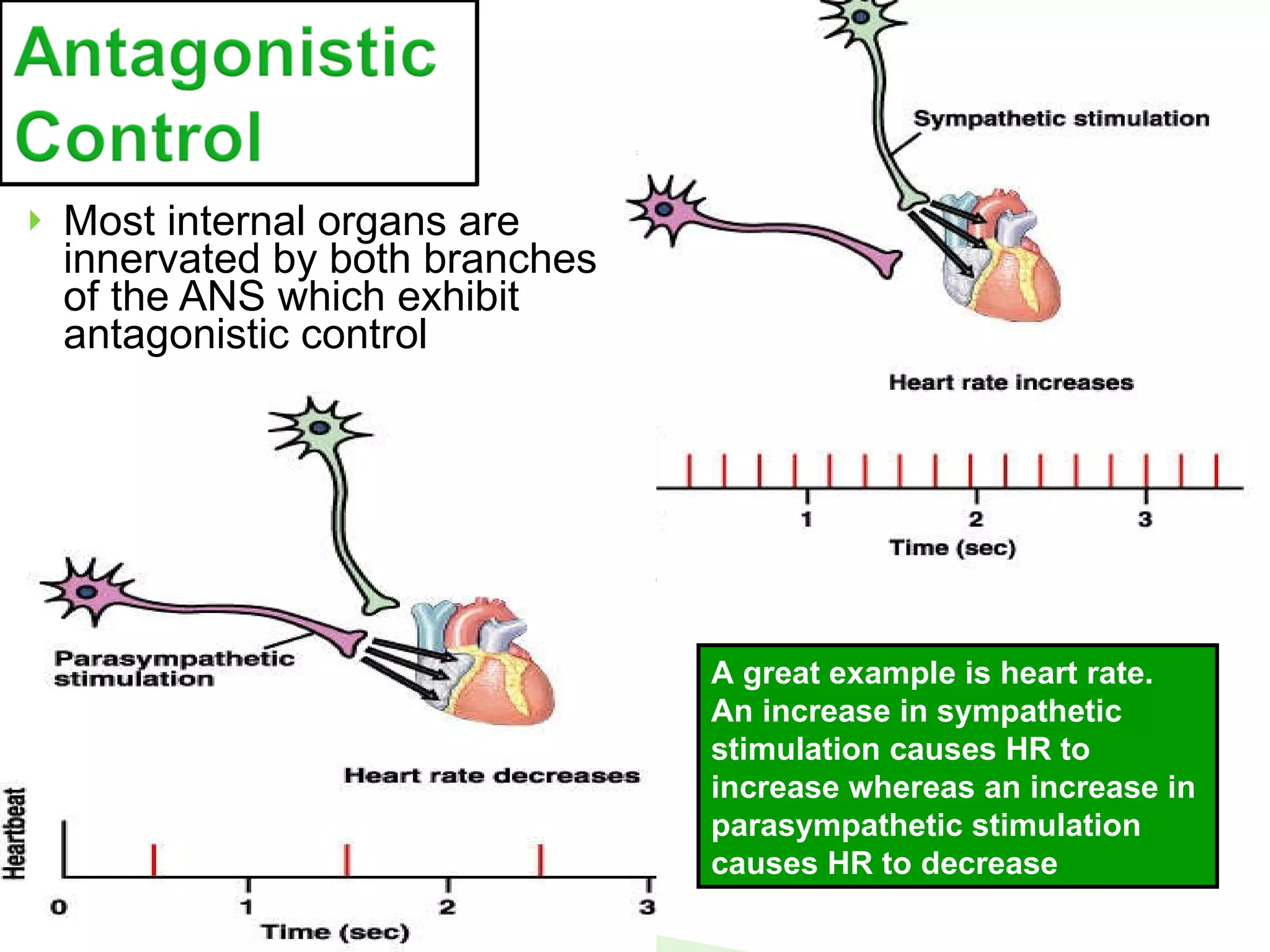
Most internal organsare innervated by both branches of the ANS which exhibit antagonistic control A great example is heart rate. An increase in sympathetic stimulation causes HR to increase whereas an increase in parasympathetic stimulation causes HR to decrease
31.
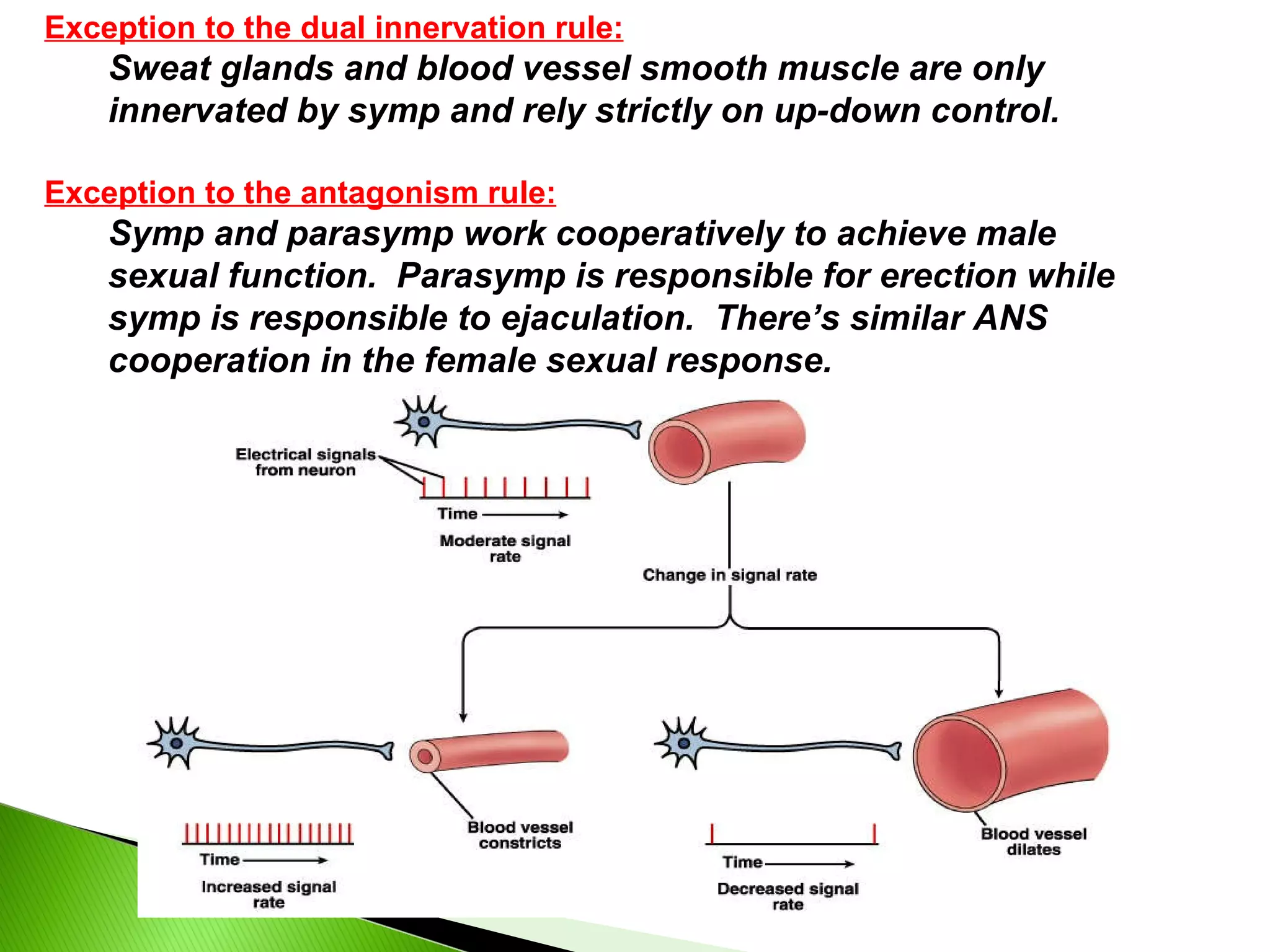
Exception to thedual innervation rule: Sweat glands and blood vessel smooth muscle are only innervated by symp and rely strictly on up-down control. Exception to the antagonism rule: Symp and parasymp work cooperatively to achieve male sexual function. Parasymp is responsible for erection while symp is responsible to ejaculation. There’s similar ANS cooperation in the female sexual response.
32.
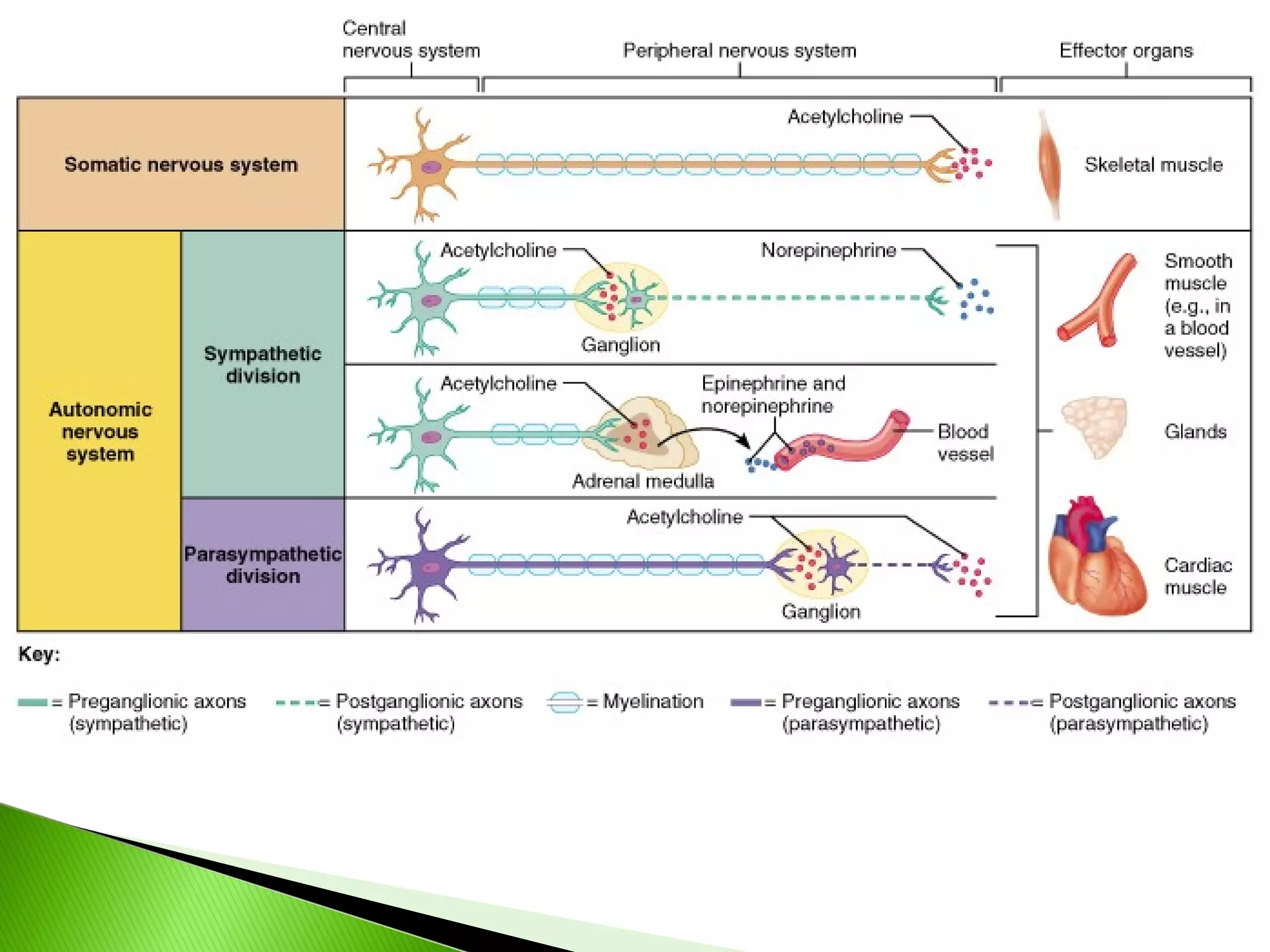
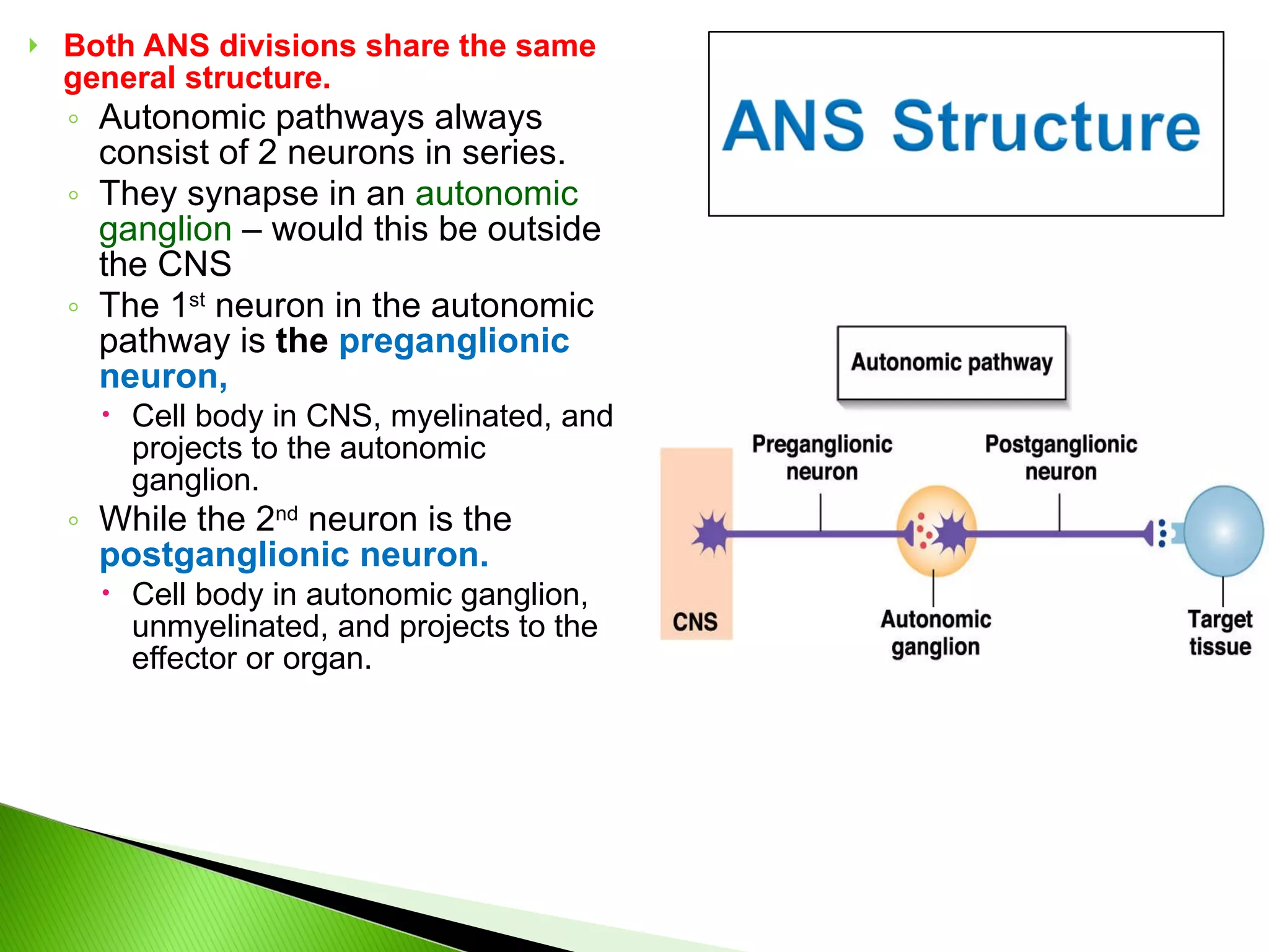
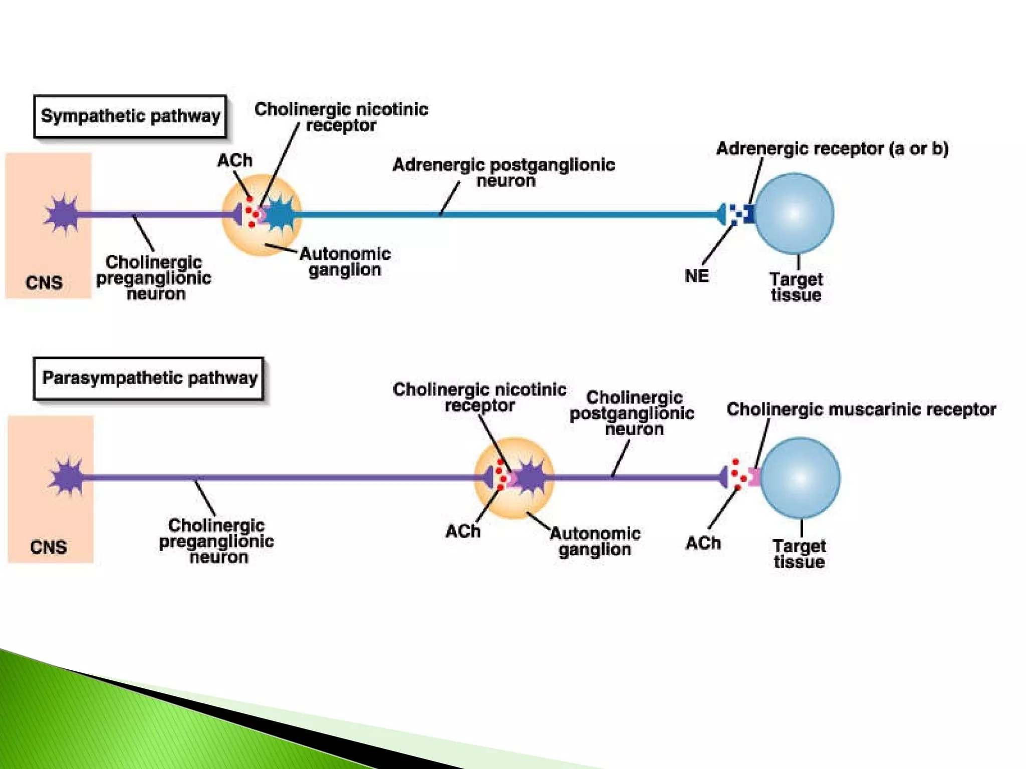
Both ANS divisionsshare the same general structure. Autonomic pathways always consist of 2 neurons in series. They synapse in an autonomic ganglion – would this be outside the CNS The 1 st neuron in the autonomic pathway is the preganglionic neuron, Cell body in CNS, myelinated, and projects to the autonomic ganglion. While the 2 nd neuron is the postganglionic neuron. Cell body in autonomic ganglion, unmyelinated, and projects to the effector or organ.
33.
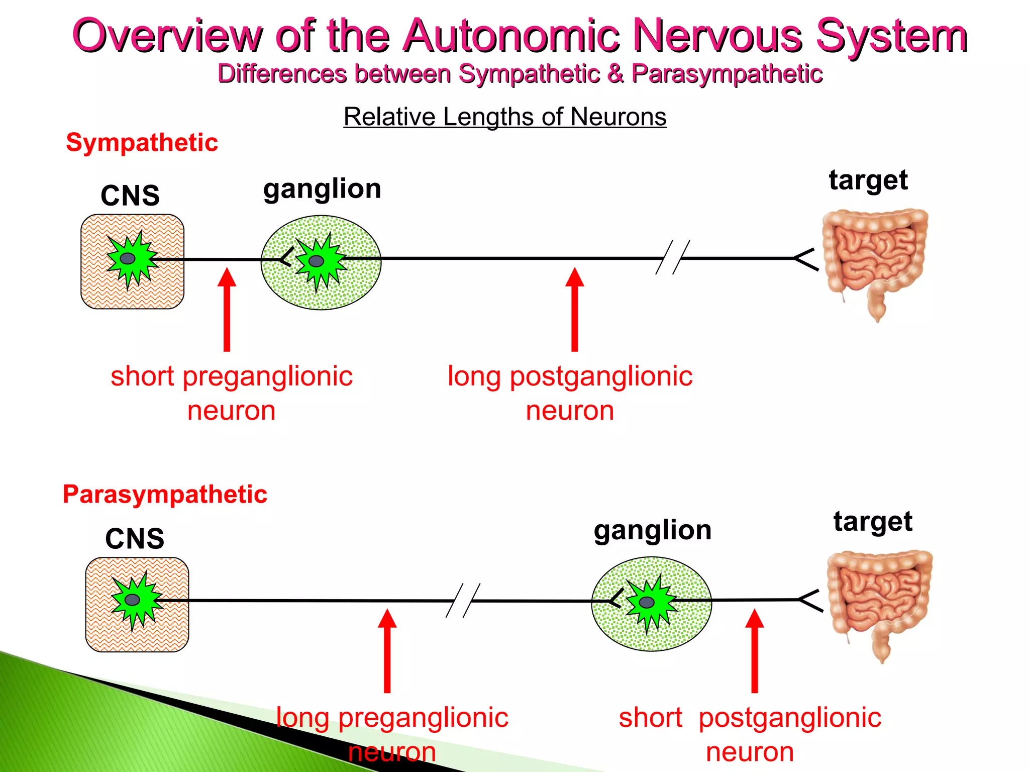
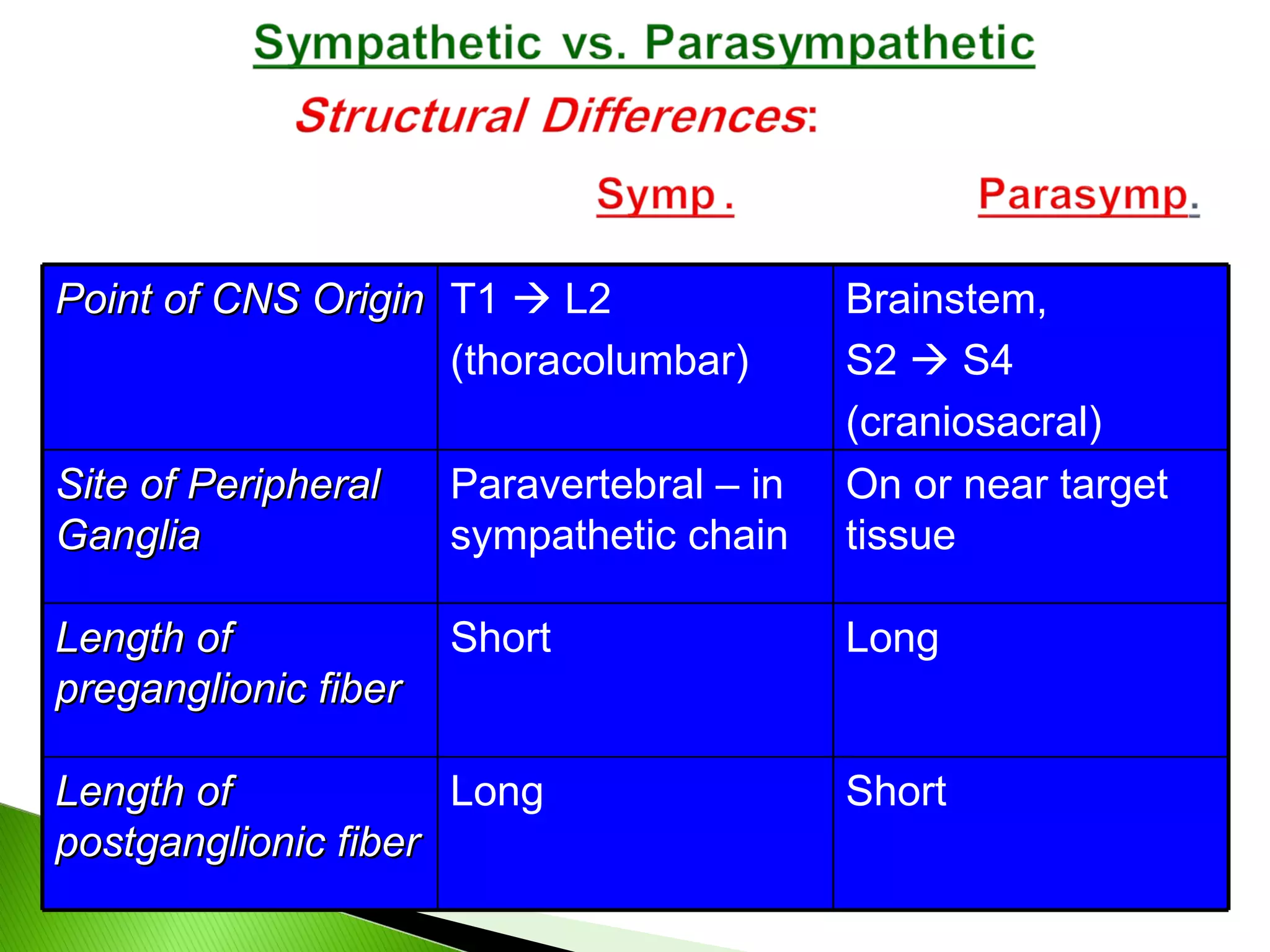
Sympathetic CNS ganglionshort preganglionic neuron long postganglionic neuron target Parasympathetic CNS ganglion long preganglionic neuron target short postganglionic neuron Overview of the Autonomic Nervous System Differences between Sympathetic & Parasympathetic Relative Lengths of Neurons
34.
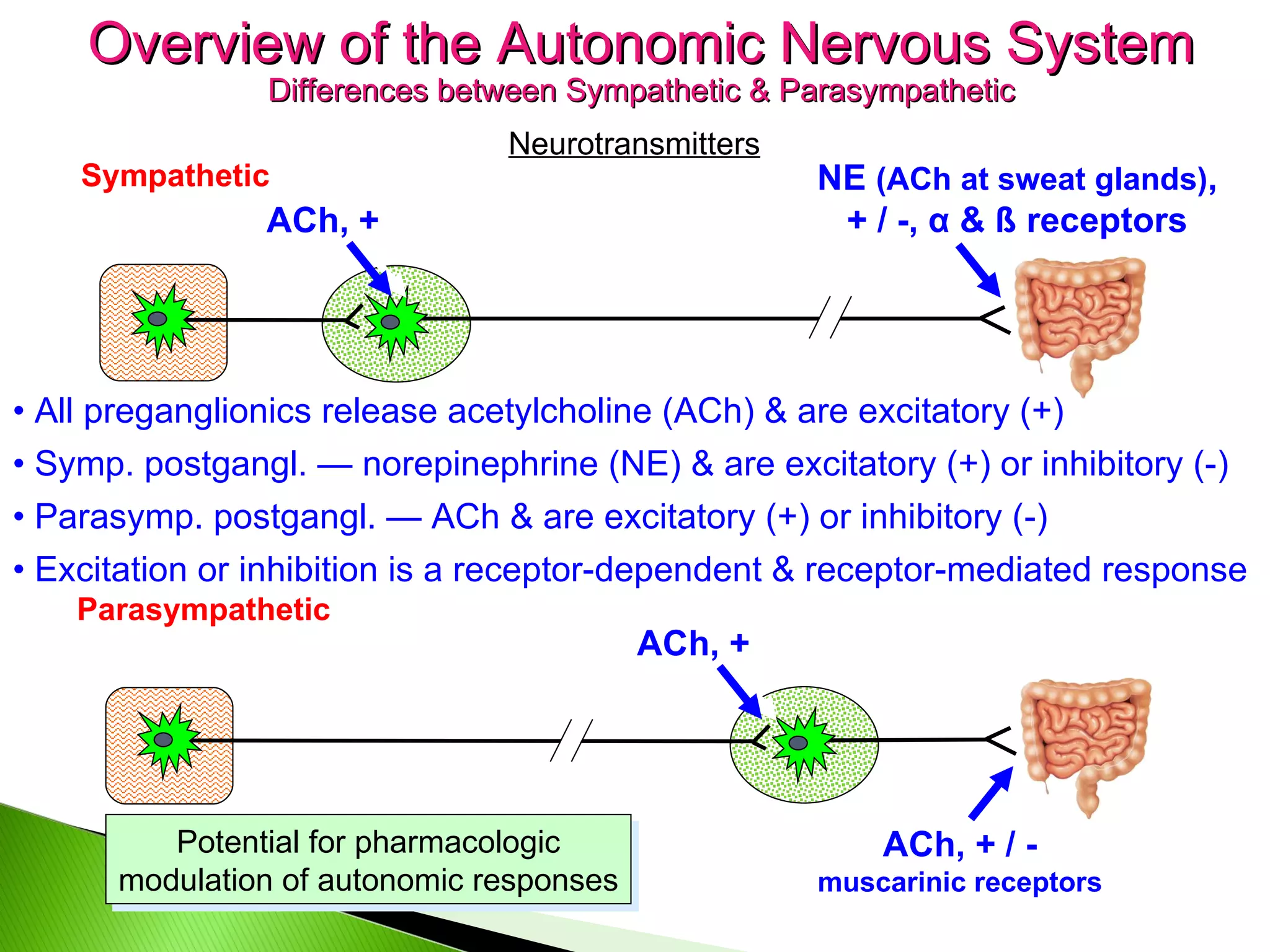
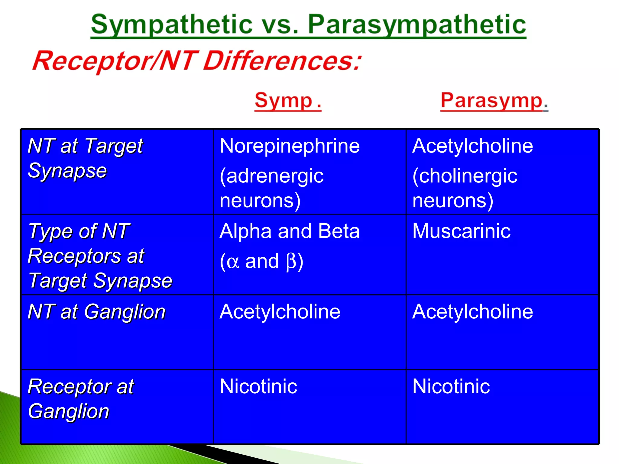
Parasympathetic Overview ofthe Autonomic Nervous System Differences between Sympathetic & Parasympathetic Neurotransmitters ACh, + NE (ACh at sweat glands) , + / -, α & ß receptors ACh, + / - muscarinic receptors • All preganglionics release acetylcholine (ACh) & are excitatory (+) • Symp. postgangl. — norepinephrine (NE) & are excitatory (+) or inhibitory (-) • Parasymp. postgangl. — ACh & are excitatory (+) or inhibitory (-) Sympathetic • Excitation or inhibition is a receptor-dependent & receptor-mediated response Potential for pharmacologic modulation of autonomic responses ACh, +
35.
Point of CNSOrigin T1 L2 (thoracolumbar) Brainstem, S2 S4 (craniosacral) Site of Peripheral Ganglia Paravertebral – in sympathetic chain On or near target tissue Length of preganglionic fiber Short Long Length of postganglionic fiber Long Short
36.
NT at TargetSynapse Norepinephrine (adrenergic neurons) Acetylcholine (cholinergic neurons) Type of NT Receptors at Target Synapse Alpha and Beta ( and ) Muscarinic NT at Ganglion Acetylcholine Acetylcholine Receptor at Ganglion Nicotinic Nicotinic
37.
38.
39.
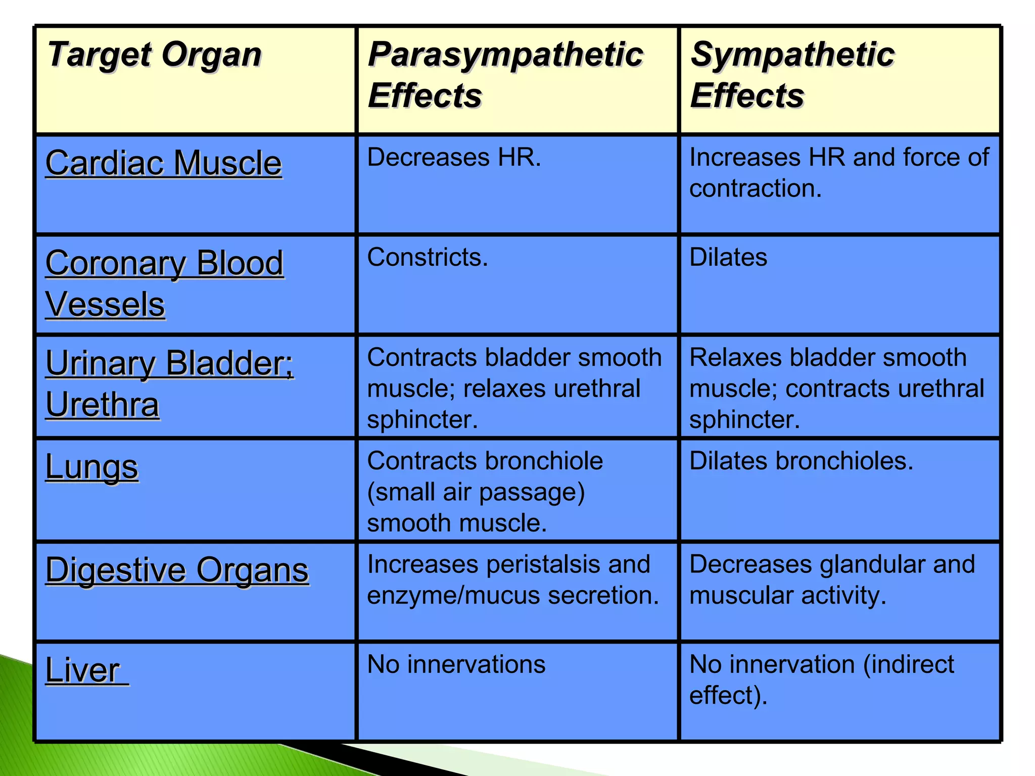
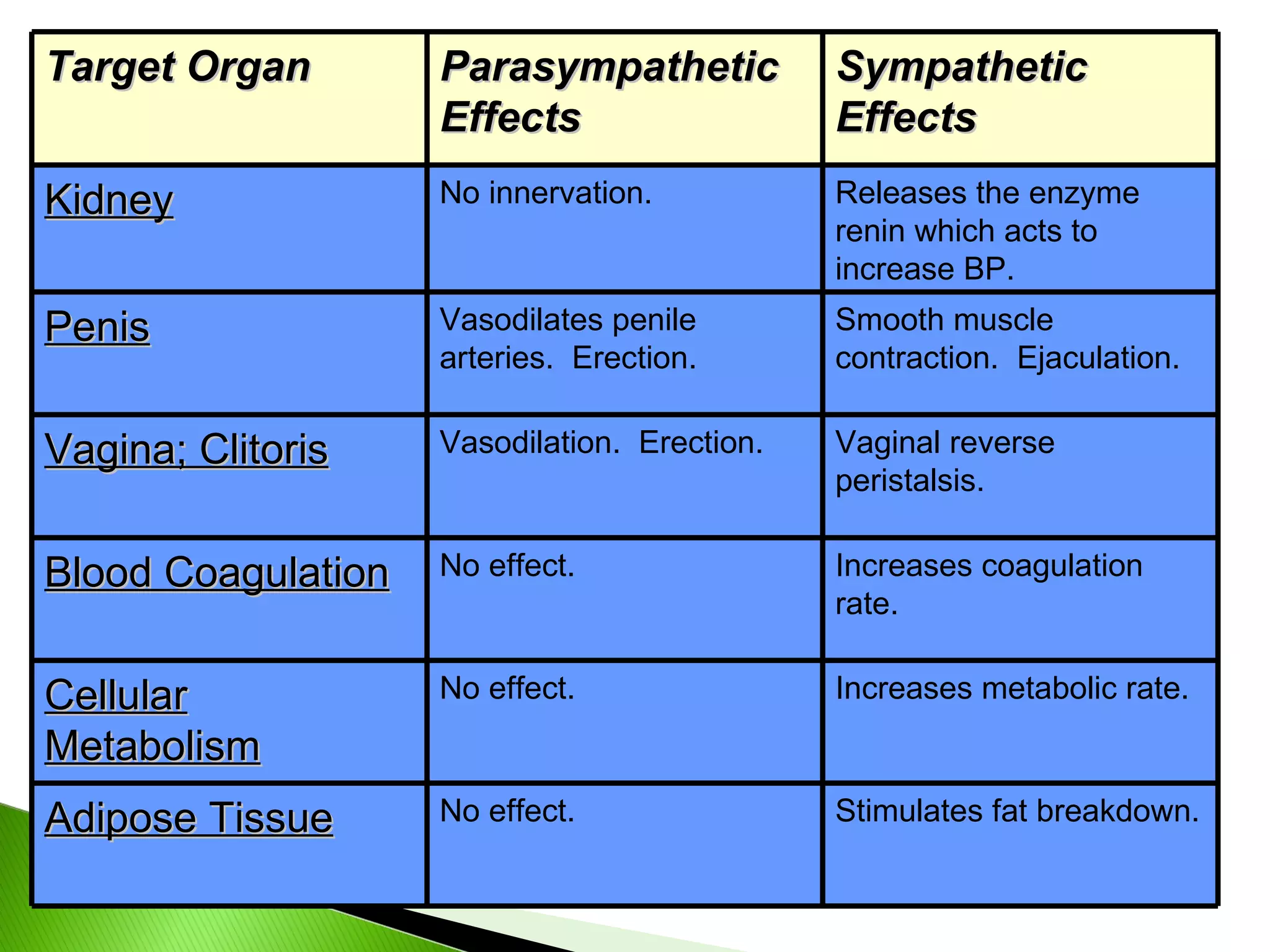
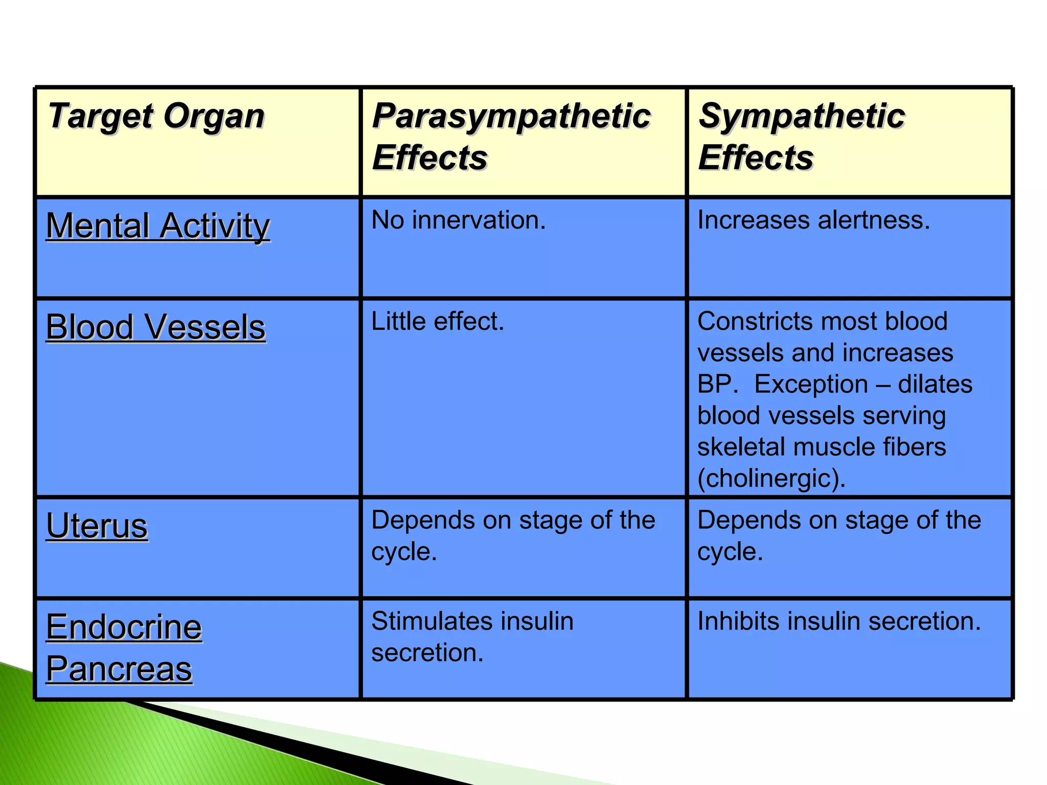
In the followingtables, note the effects of the sympathetic and parasympathetic nervous systems on various body organs.
40.
Target Organ ParasympatheticEffects Sympathetic Effects Eye (Iris) Stimulates constrictor muscles. Pupil constriction. Stimulates dilator muscles. Pupil dilates. Eye (Ciliary muscle) Stimulates. Lens accommodates – allows for close vision. No innervation. Salivary Glands Watery secretion. Mucous secretion. Sweat Glands No innervation. Stimulates sweating in large amounts. (Cholinergic) Gallbladder Stimulates smooth muscle to contract and expel bile. Inhibits gallbladder smooth muscle. Arrector Pili No innervation Stimulates contraction. Piloerection (Goosebumps)
41.
Target Organ ParasympatheticEffects Sympathetic Effects Cardiac Muscle Decreases HR. Increases HR and force of contraction. Coronary Blood Vessels Constricts. Dilates Urinary Bladder; Urethra Contracts bladder smooth muscle; relaxes urethral sphincter. Relaxes bladder smooth muscle; contracts urethral sphincter. Lungs Contracts bronchiole (small air passage) smooth muscle. Dilates bronchioles. Digestive Organs Increases peristalsis and enzyme/mucus secretion. Decreases glandular and muscular activity. Liver No innervations No innervation (indirect effect).
42.
Target Organ ParasympatheticEffects Sympathetic Effects Kidney No innervation. Releases the enzyme renin which acts to increase BP. Penis Vasodilates penile arteries. Erection. Smooth muscle contraction. Ejaculation. Vagina; Clitoris Vasodilation. Erection. Vaginal reverse peristalsis. Blood Coagulation No effect. Increases coagulation rate. Cellular Metabolism No effect. Increases metabolic rate. Adipose Tissue No effect. Stimulates fat breakdown.
43.
Target Organ ParasympatheticEffects Sympathetic Effects Mental Activity No innervation. Increases alertness. Blood Vessels Little effect. Constricts most blood vessels and increases BP. Exception – dilates blood vessels serving skeletal muscle fibers (cholinergic). Uterus Depends on stage of the cycle. Depends on stage of the cycle. Endocrine Pancreas Stimulates insulin secretion. Inhibits insulin secretion.
44.
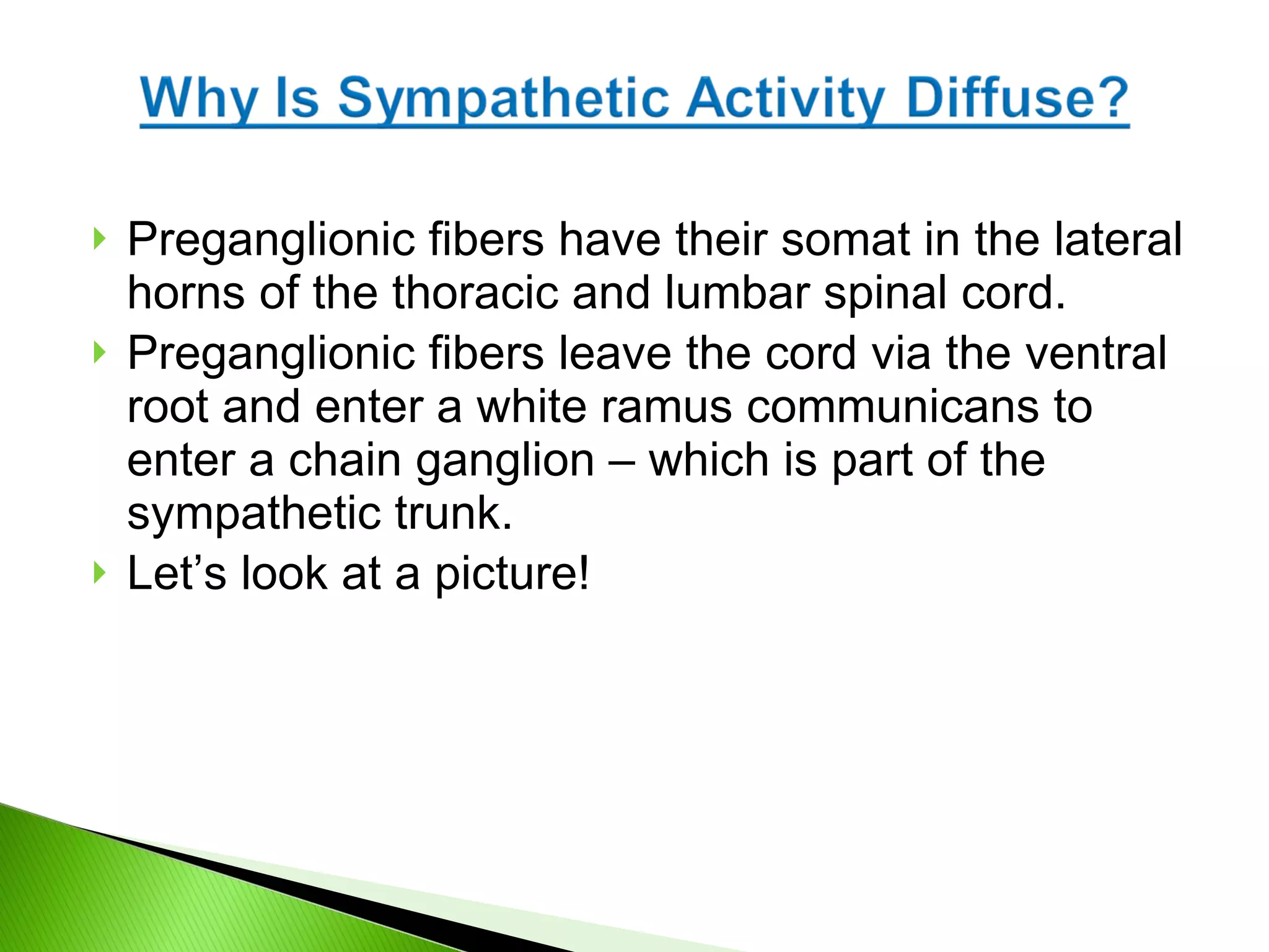
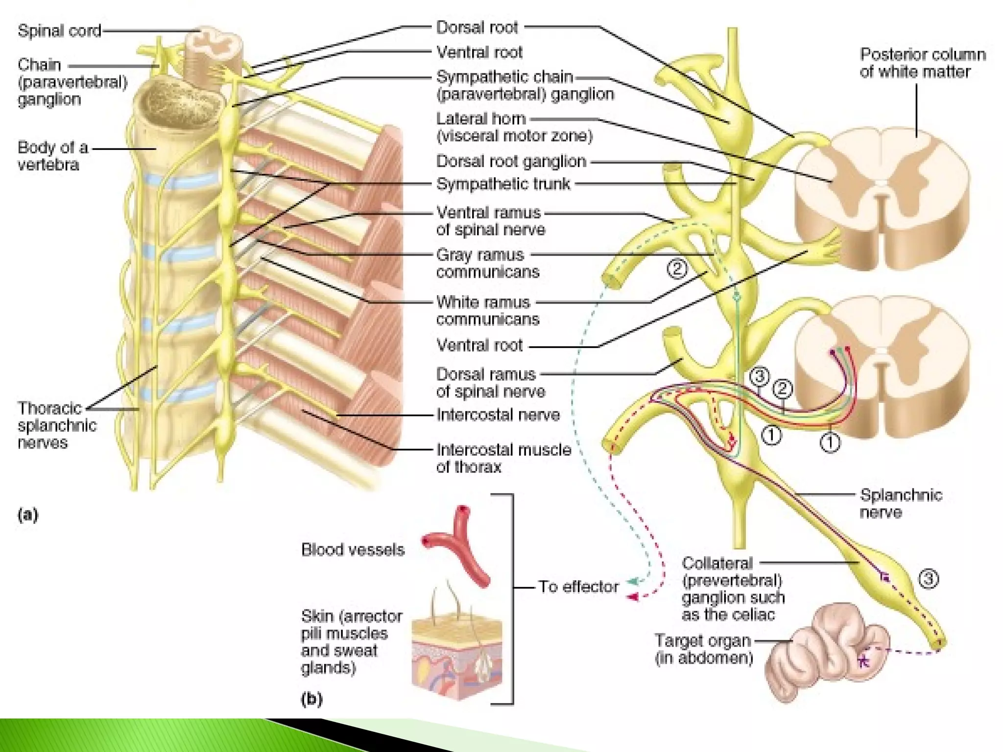
Preganglionic fibers havetheir somat in the lateral horns of the thoracic and lumbar spinal cord. Preganglionic fibers leave the cord via the ventral root and enter a white ramus communicans to enter a chain ganglion – which is part of the sympathetic trunk. Let’s look at a picture!
45.
46.
47.
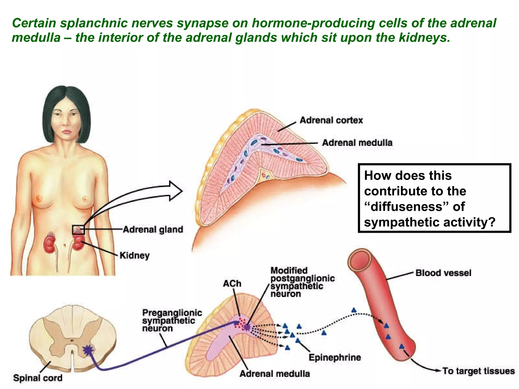
Certain splanchnic nervessynapse on hormone-producing cells of the adrenal medulla – the interior of the adrenal glands which sit upon the kidneys. How does this contribute to the “diffuseness” of sympathetic activity?
48.
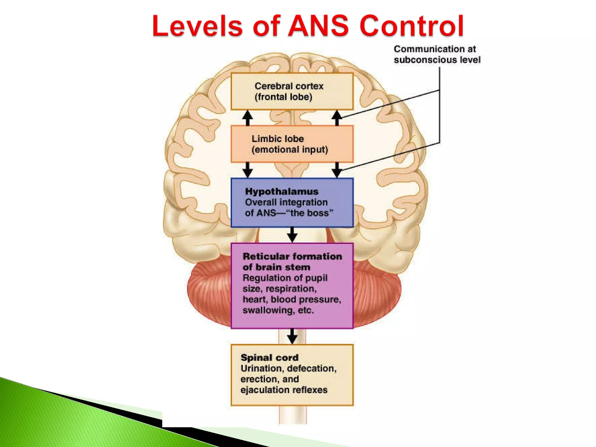
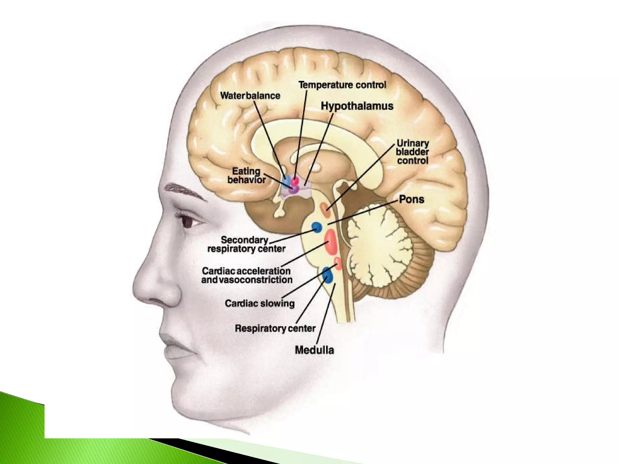
The Hypothalamus isthe Boss: Its anterior and medial regions direct parasympathetic function while its posterior and lateral regions direct sympathetic function These centers exert control directly and via nuclei in the reticular formation (e.g., the cardiovascular centers in the MO, respiratory centers in MO and pons, etc.) The connection of the limbic system to the hypothalamus mediates our “flight or flight” response to emotional situations.