Downloaded 185 times


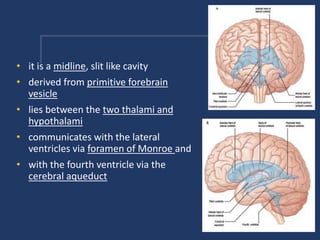
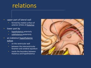
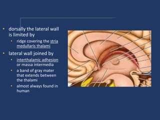
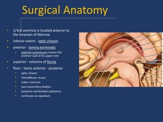
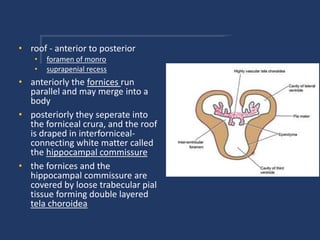
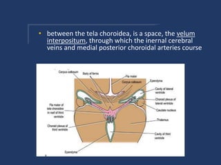
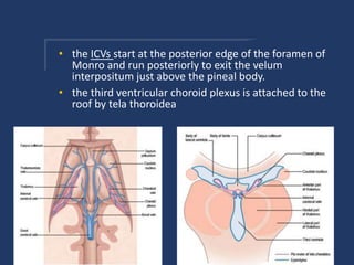
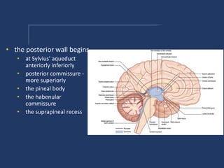
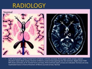
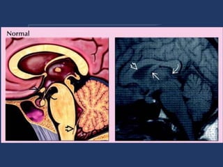

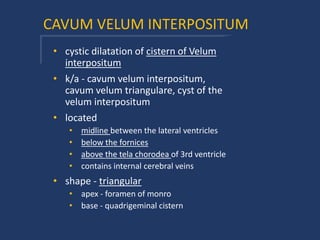
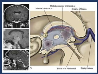


The third ventricle is a midline cavity located between the two thalami and hypothalami. It communicates with the lateral ventricles via the foramen of Monroe and with the fourth ventricle via the cerebral aqueduct. The third ventricle's roof is formed by the fornix and tela choroidea, while its floor extends from the optic chiasm to the posterior perforated substance. The third ventricle can be accessed surgically through various anterior or posterior approaches between brain structures such as the fornix.


