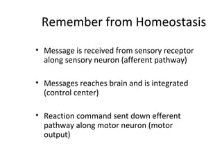
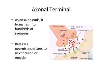
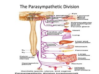
The nervous system has two main divisions: the central nervous system (CNS) and the peripheral nervous system (PNS). The autonomic nervous system (ANS) is a division of the PNS and controls involuntary body functions. It has two branches - the sympathetic and parasympathetic systems. The sympathetic system activates the fight or flight response while the parasympathetic system activates the rest and digest response. Both systems involve a two-neuron chain with a preganglionic neuron originating in the CNS and a postganglionic neuron that releases neurotransmitters like acetylcholine and norepinephrine.
Peripheral Nervous System, Audumbar MaliAudumbar Mali
Peripheral Nervous System,
Types of PNS,
Spinal nerves,
Types of neuron (3 basic types),
Plexus,
Cranial nerves,
Autonomic nervous system,
Structure of Neuron,
Human Anatomy and Physiology-I,
Syllabus As per PCI,
B. Pharm-I
You can watch the video on my you tube channel: https://youtu.be/I0FaX-iQfa0
Medulla oblongata or more simply medulla is part of brain stem which forms base of the brain stem. It contains pyramid, olive and above pyramidal structure, there is decussation of pyramids which explains why each part of brain controls opposite part of body. Adding to that medulla also has several nuclei which controls activity of cardiovascular system and respiratory system. Medulla also has nuclei for controlling reflexes of vomiting, swallowing, hiccuping, coughing and sneezing. It has also nuclei for test, hearing and balance. Medulla also contains nuclei of cranial nerve number VIII, IX, X, XI and XII.
cholenergic & adrenergic receptors
simplified physiology
دي عباره عن مجموعه من الفيديوهات بنشرح فيها فسيولوجي
بشكل مبسط وسهل تقدر تذاكر منها
تقدرو تتابعوا باقي الفيديوهات علي قناتنا
https://www.youtube.com/playlist?list=PLrkqYOkxk2KKAxHlRwZ8aWXkBq2KCvU_B
وكمان تقدروا تتابعو الجديد علي صفحتنا ع الفيس بوك
https://www.facebook.com/simplifiedphysiology
ولو في اي استفسار
https://www.facebook.com/medo.naeem99
http://ask.fm/medonaeem99
PHYSIOLOGY OF ANS(AUTONOMIC NERVOUS SYSTEM)
Sympathetic Responses
Parasympathetic Responses
Autonomic Interactions
Control of Autonomic Nervous System Function
This presentation was delivered over two days to second year pharmacy students enrolled in a course in pharmacology & toxicology. This lecture is designed to accompany Goodman & Gilman's (12e) chapter 11.
Individualized Webcam facilitated and e-Classroom USMLE Step 1 Tutorials with Dr. Cray. For questions or more information.. drcray@imhotepvirtualmedsch.com
vn nbsxjhx 59595959His temperature is 38.2°C (100.8°F), pulse is 110/min, respirations are 20/min, and blood pressure
is 80/60 mm Hg. The upper and lower extremities are cold and clammy. Pulmonary artery catheterization shows a
pulmonary capillary wedge pressure of 23 mm Hg (N=5-16). Which of the following is the most likely explanation for
these findings?
0 A) Inadequate volume replacement
0 B) Intra-abdominal hemorrhage
0 C) Myocardial infarction
0 D) Pulmonary embolism
a quick visual understanding of what actually nervous tissue is made up of at cellular level its functions nerve cell types chemical synapse detailed structure of neuron
NVBDCP.pptx Nation vector borne disease control programSapna Thakur
NVBDCP was launched in 2003-2004 . Vector-Borne Disease: Disease that results from an infection transmitted to humans and other animals by blood-feeding arthropods, such as mosquitoes, ticks, and fleas. Examples of vector-borne diseases include Dengue fever, West Nile Virus, Lyme disease, and malaria.
- Video recording of this lecture in English language: https://youtu.be/lK81BzxMqdo
- Video recording of this lecture in Arabic language: https://youtu.be/Ve4P0COk9OI
- Link to download the book free: https://nephrotube.blogspot.com/p/nephrotube-nephrology-books.html
- Link to NephroTube website: www.NephroTube.com
- Link to NephroTube social media accounts: https://nephrotube.blogspot.com/p/join-nephrotube-on-social-media.html
Title: Sense of Smell
Presenter: Dr. Faiza, Assistant Professor of Physiology
Qualifications:
MBBS (Best Graduate, AIMC Lahore)
FCPS Physiology
ICMT, CHPE, DHPE (STMU)
MPH (GC University, Faisalabad)
MBA (Virtual University of Pakistan)
Learning Objectives:
Describe the primary categories of smells and the concept of odor blindness.
Explain the structure and location of the olfactory membrane and mucosa, including the types and roles of cells involved in olfaction.
Describe the pathway and mechanisms of olfactory signal transmission from the olfactory receptors to the brain.
Illustrate the biochemical cascade triggered by odorant binding to olfactory receptors, including the role of G-proteins and second messengers in generating an action potential.
Identify different types of olfactory disorders such as anosmia, hyposmia, hyperosmia, and dysosmia, including their potential causes.
Key Topics:
Olfactory Genes:
3% of the human genome accounts for olfactory genes.
400 genes for odorant receptors.
Olfactory Membrane:
Located in the superior part of the nasal cavity.
Medially: Folds downward along the superior septum.
Laterally: Folds over the superior turbinate and upper surface of the middle turbinate.
Total surface area: 5-10 square centimeters.
Olfactory Mucosa:
Olfactory Cells: Bipolar nerve cells derived from the CNS (100 million), with 4-25 olfactory cilia per cell.
Sustentacular Cells: Produce mucus and maintain ionic and molecular environment.
Basal Cells: Replace worn-out olfactory cells with an average lifespan of 1-2 months.
Bowman’s Gland: Secretes mucus.
Stimulation of Olfactory Cells:
Odorant dissolves in mucus and attaches to receptors on olfactory cilia.
Involves a cascade effect through G-proteins and second messengers, leading to depolarization and action potential generation in the olfactory nerve.
Quality of a Good Odorant:
Small (3-20 Carbon atoms), volatile, water-soluble, and lipid-soluble.
Facilitated by odorant-binding proteins in mucus.
Membrane Potential and Action Potential:
Resting membrane potential: -55mV.
Action potential frequency in the olfactory nerve increases with odorant strength.
Adaptation Towards the Sense of Smell:
Rapid adaptation within the first second, with further slow adaptation.
Psychological adaptation greater than receptor adaptation, involving feedback inhibition from the central nervous system.
Primary Sensations of Smell:
Camphoraceous, Musky, Floral, Pepperminty, Ethereal, Pungent, Putrid.
Odor Detection Threshold:
Examples: Hydrogen sulfide (0.0005 ppm), Methyl-mercaptan (0.002 ppm).
Some toxic substances are odorless at lethal concentrations.
Characteristics of Smell:
Odor blindness for single substances due to lack of appropriate receptor protein.
Behavioral and emotional influences of smell.
Transmission of Olfactory Signals:
From olfactory cells to glomeruli in the olfactory bulb, involving lateral inhibition.
Primitive, less old, and new olfactory systems with different path
Couples presenting to the infertility clinic- Do they really have infertility...Sujoy Dasgupta
Dr Sujoy Dasgupta presented the study on "Couples presenting to the infertility clinic- Do they really have infertility? – The unexplored stories of non-consummation" in the 13th Congress of the Asia Pacific Initiative on Reproduction (ASPIRE 2024) at Manila on 24 May, 2024.
Knee anatomy and clinical tests 2024.pdfvimalpl1234
This includes all relevant anatomy and clinical tests compiled from standard textbooks, Campbell,netter etc..It is comprehensive and best suited for orthopaedicians and orthopaedic residents.
Recomendações da OMS sobre cuidados maternos e neonatais para uma experiência pós-natal positiva.
Em consonância com os ODS – Objetivos do Desenvolvimento Sustentável e a Estratégia Global para a Saúde das Mulheres, Crianças e Adolescentes, e aplicando uma abordagem baseada nos direitos humanos, os esforços de cuidados pós-natais devem expandir-se para além da cobertura e da simples sobrevivência, de modo a incluir cuidados de qualidade.
Estas diretrizes visam melhorar a qualidade dos cuidados pós-natais essenciais e de rotina prestados às mulheres e aos recém-nascidos, com o objetivo final de melhorar a saúde e o bem-estar materno e neonatal.
Uma “experiência pós-natal positiva” é um resultado importante para todas as mulheres que dão à luz e para os seus recém-nascidos, estabelecendo as bases para a melhoria da saúde e do bem-estar a curto e longo prazo. Uma experiência pós-natal positiva é definida como aquela em que as mulheres, pessoas que gestam, os recém-nascidos, os casais, os pais, os cuidadores e as famílias recebem informação consistente, garantia e apoio de profissionais de saúde motivados; e onde um sistema de saúde flexível e com recursos reconheça as necessidades das mulheres e dos bebês e respeite o seu contexto cultural.
Estas diretrizes consolidadas apresentam algumas recomendações novas e já bem fundamentadas sobre cuidados pós-natais de rotina para mulheres e neonatos que recebem cuidados no pós-parto em unidades de saúde ou na comunidade, independentemente dos recursos disponíveis.
É fornecido um conjunto abrangente de recomendações para cuidados durante o período puerperal, com ênfase nos cuidados essenciais que todas as mulheres e recém-nascidos devem receber, e com a devida atenção à qualidade dos cuidados; isto é, a entrega e a experiência do cuidado recebido. Estas diretrizes atualizam e ampliam as recomendações da OMS de 2014 sobre cuidados pós-natais da mãe e do recém-nascido e complementam as atuais diretrizes da OMS sobre a gestão de complicações pós-natais.
O estabelecimento da amamentação e o manejo das principais intercorrências é contemplada.
Recomendamos muito.
Vamos discutir essas recomendações no nosso curso de pós-graduação em Aleitamento no Instituto Ciclos.
Esta publicação só está disponível em inglês até o momento.
Prof. Marcus Renato de Carvalho
www.agostodourado.com
TEST BANK for Operations Management, 14th Edition by William J. Stevenson, Ve...kevinkariuki227
TEST BANK for Operations Management, 14th Edition by William J. Stevenson, Verified Chapters 1 - 19, Complete Newest Version.pdf
New Directions in Targeted Therapeutic Approaches for Older Adults With Mantl...i3 Health
i3 Health is pleased to make the speaker slides from this activity available for use as a non-accredited self-study or teaching resource.
This slide deck presented by Dr. Kami Maddocks, Professor-Clinical in the Division of Hematology and
Associate Division Director for Ambulatory Operations
The Ohio State University Comprehensive Cancer Center, will provide insight into new directions in targeted therapeutic approaches for older adults with mantle cell lymphoma.
STATEMENT OF NEED
Mantle cell lymphoma (MCL) is a rare, aggressive B-cell non-Hodgkin lymphoma (NHL) accounting for 5% to 7% of all lymphomas. Its prognosis ranges from indolent disease that does not require treatment for years to very aggressive disease, which is associated with poor survival (Silkenstedt et al, 2021). Typically, MCL is diagnosed at advanced stage and in older patients who cannot tolerate intensive therapy (NCCN, 2022). Although recent advances have slightly increased remission rates, recurrence and relapse remain very common, leading to a median overall survival between 3 and 6 years (LLS, 2021). Though there are several effective options, progress is still needed towards establishing an accepted frontline approach for MCL (Castellino et al, 2022). Treatment selection and management of MCL are complicated by the heterogeneity of prognosis, advanced age and comorbidities of patients, and lack of an established standard approach for treatment, making it vital that clinicians be familiar with the latest research and advances in this area. In this activity chaired by Michael Wang, MD, Professor in the Department of Lymphoma & Myeloma at MD Anderson Cancer Center, expert faculty will discuss prognostic factors informing treatment, the promising results of recent trials in new therapeutic approaches, and the implications of treatment resistance in therapeutic selection for MCL.
Target Audience
Hematology/oncology fellows, attending faculty, and other health care professionals involved in the treatment of patients with mantle cell lymphoma (MCL).
Learning Objectives
1.) Identify clinical and biological prognostic factors that can guide treatment decision making for older adults with MCL
2.) Evaluate emerging data on targeted therapeutic approaches for treatment-naive and relapsed/refractory MCL and their applicability to older adults
3.) Assess mechanisms of resistance to targeted therapies for MCL and their implications for treatment selection
Prix Galien International 2024 Forum ProgramLevi Shapiro
June 20, 2024, Prix Galien International and Jerusalem Ethics Forum in ROME. Detailed agenda including panels:
- ADVANCES IN CARDIOLOGY: A NEW PARADIGM IS COMING
- WOMEN’S HEALTH: FERTILITY PRESERVATION
- WHAT’S NEW IN THE TREATMENT OF INFECTIOUS,
ONCOLOGICAL AND INFLAMMATORY SKIN DISEASES?
- ARTIFICIAL INTELLIGENCE AND ETHICS
- GENE THERAPY
- BEYOND BORDERS: GLOBAL INITIATIVES FOR DEMOCRATIZING LIFE SCIENCE TECHNOLOGIES AND PROMOTING ACCESS TO HEALTHCARE
- ETHICAL CHALLENGES IN LIFE SCIENCES
- Prix Galien International Awards Ceremony
Pulmonary Thromboembolism - etilogy, types, medical- Surgical and nursing man...VarunMahajani
Disruption of blood supply to lung alveoli due to blockage of one or more pulmonary blood vessels is called as Pulmonary thromboembolism. In this presentation we will discuss its causes, types and its management in depth.
These lecture slides, by Dr Sidra Arshad, offer a quick overview of physiological basis of a normal electrocardiogram.
Learning objectives:
1. Define an electrocardiogram (ECG) and electrocardiography
2. Describe how dipoles generated by the heart produce the waveforms of the ECG
3. Describe the components of a normal electrocardiogram of a typical bipolar leads (limb II)
4. Differentiate between intervals and segments
5. Enlist some common indications for obtaining an ECG
Study Resources:
1. Chapter 11, Guyton and Hall Textbook of Medical Physiology, 14th edition
2. Chapter 9, Human Physiology - From Cells to Systems, Lauralee Sherwood, 9th edition
3. Chapter 29, Ganong’s Review of Medical Physiology, 26th edition
4. Electrocardiogram, StatPearls - https://www.ncbi.nlm.nih.gov/books/NBK549803/
5. ECG in Medical Practice by ABM Abdullah, 4th edition
6. ECG Basics, http://www.nataliescasebook.com/tag/e-c-g-basics
Flu Vaccine Alert in Bangalore Karnatakaaddon Scans
As flu season approaches, health officials in Bangalore, Karnataka, are urging residents to get their flu vaccinations. The seasonal flu, while common, can lead to severe health complications, particularly for vulnerable populations such as young children, the elderly, and those with underlying health conditions.
Dr. Vidisha Kumari, a leading epidemiologist in Bangalore, emphasizes the importance of getting vaccinated. "The flu vaccine is our best defense against the influenza virus. It not only protects individuals but also helps prevent the spread of the virus in our communities," he says.
This year, the flu season is expected to coincide with a potential increase in other respiratory illnesses. The Karnataka Health Department has launched an awareness campaign highlighting the significance of flu vaccinations. They have set up multiple vaccination centers across Bangalore, making it convenient for residents to receive their shots.
To encourage widespread vaccination, the government is also collaborating with local schools, workplaces, and community centers to facilitate vaccination drives. Special attention is being given to ensuring that the vaccine is accessible to all, including marginalized communities who may have limited access to healthcare.
Residents are reminded that the flu vaccine is safe and effective. Common side effects are mild and may include soreness at the injection site, mild fever, or muscle aches. These side effects are generally short-lived and far less severe than the flu itself.
Healthcare providers are also stressing the importance of continuing COVID-19 precautions. Wearing masks, practicing good hand hygiene, and maintaining social distancing are still crucial, especially in crowded places.
Protect yourself and your loved ones by getting vaccinated. Together, we can help keep Bangalore healthy and safe this flu season. For more information on vaccination centers and schedules, residents can visit the Karnataka Health Department’s official website or follow their social media pages.
Stay informed, stay safe, and get your flu shot today!
2. Composition of Nervous System
• Two main divisions
– 1) Central Nervous
system (CNS) brain and
spinal chord
– 2)Peripheral Nervous
System (PNS) nerves
3. Remember from Homeostasis
• Message is received from sensory receptor
along sensory neuron (afferent pathway)
• Messages reaches brain and is integrated
(control center)
• Reaction command sent down efferent
pathway along motor neuron (motor
output)
4. Types of Motor Actions
• Somatic
– Happens in skeletal
muscle
– voluntary
• Autonomic
– Happens in smooth and
cardiac muscle
– Involuntary
– 2 parts
• Sympathetic and
parasympathetic
5. Cells of the Nervous System
• Broken down into two
groups
– 1) supporting cells
– 2) neurons
6. Examples of Neuroglia (supporting cells)
• Astrocytes – anchor
neurons to capillaries
• Microglia – phagocytes
(digest debris and dead
cells)
• Ependymal cells- ciliated;
always on surface near
spinal fluid; circulates
fluid
7. One more Neuroglial cell (in CNS)
• Oligodendrocytes –
fatty; insulated nerve
fibers
• Produce myelin sheath
which surround and
insulate the nerve fiber
8. In PNS
• Instead of
oligodendrocytes, they
have Schwann cells,
which insulate the
nerve fiber
• Satellite cells – form
protective layer around
nerve cell body
10. Parts of a Neuron
• 1)Cell Body- contains
nucleus
• 2) fiber (process)-
carries message to next
neuron
– Toward cell body =
dendrites
– Away from cell body =
axon
11. Axonal Terminal
• As an axon ends, it
branches into
hundreds of
synapses.
• Releases
neurotransmitters to
next neuron or
muscle
12. Myelination of Neurons
• In CNS, the fatty
covering is
oligodendrocyte
• In PNS, fatty myelin
forms Schwann cells,
which increase
transmission rate.
• Gaps between
Schwann cells= Nodes
of Ranvier
13. INTRODUCTION
• Much of the action of the body in maintaining
such as cardiovascular, gastrointestinal and
thermal homeostasis occurs through the
autonomic nervous system (ANS).
• The ANS is our primary defense against
challenges, to maintain homeostasis. It
provides involuntary control and organization
of both maintenance and stress responses.
13
15. DIFFeReNCe
beTweeNSomatic Autonomic
Organ supplied Skeletal muscles All other organs
Distal most synapse Within CNS Outside the CNS(i.e. ganglia)
Nerve fibers Myelinated Preganglionic - myelinated
Postganglionic- nonmyelinated
Peripheral plexus
formation
Absent Present
Efferent transmitter ACH ACH, Nor-adrenaline
Effect of nerve section
on organ supplied
Paralysis and Atrophy Activity maintained, no
Atrophy 15
17. Comparison of Autonomic and
Somatic Motor Systems
• Autonomic nervous system
– Chain of two motor neurons
• Preganglionic neuron
• Postganglionic neuron
– Conduction is slower due to thinly or
unmyelinated axons
Pre-ganglionic
Ganglion
Post-ganglionic
18. Autonomic Nervous System
• Often work in
opposition
• Cooperate to fine-
tune homeostasis
• Regulated by the
brain;
hypothalamus, pons
and medulla
• Can also be
regulated by spinal
reflexes; no higher
order input
• Pathways both
consist of a two
neuron system
Preganglionic neuron autonomic ganglion postganglionic neuron target
from CNS outside CNS
19. Fig. 45.34(TE Art)Hypothalamus activates
sympathetic division of
nervous system
Heart rate, blood pressure,
and respiration increase
Blood flow to
skeletal muscles
increases
Stomach
contractions
are inhibited
Adrenal medulla
secretes
epinephrine and
norepinephrine
20.
21. Sympathetic
Fight or Flight, Dealing with stress,
thoracolumber,
intermediolateral column, T1 -L2
Parasympathetic
Rest and Digest, Vegging
Craniosacral S2-S4,
22. Sympathetic nerve endings also activate the release of NE and E from the adrenal
medulla
Enhances effects of NE from sympathetic nerve endings
24. Sympathetic
• Sometimes called the
“thoracolumbar” division
• Short preganglionic neurons;
long postganglionic neurons;
ganglia are called the chain
ganglia
• Preganglionic neurons secrete
Ach onto nicotinic receptors
• Postganglionic neurons
secrete NE on to α or β
receptors
• Target tissues are smooth
muscle, cardiac muscle,
endocrine glands, brown fat
25. Parasympathetic
•Sometimes called the
“cranio-sacral division
•Long preganglionic
neurons;
•short postganglionic
neurons (often in the
target organ)
•Preganglionic neurons
secrete Ach on to
nicotinic receptors
•Postganglionic neurons
secrete Ach on to
muscarinic receptors
•Target tissues are
smooth muscle,
cardiac muscle,
exocrine glands, brown
fat
28. Similarities between Sympathetic & ParasympatheticSimilarities between Sympathetic & Parasympathetic
• Both are efferent (motor) systems: “visceromotor”
• Both involve regulation of the “internal” environment
generally outside of our conscious control:
“autonomous”
• Both involve 2 neurons that synapse in a peripheral
ganglion and Innervate glands, smooth muscle,
cardiac muscle
CNS ganglion
preganglionic
neuron
postganglionic
neuron
glands
smooth
muscle
cardiac
muscle
29. Differences between Sympathetic & ParasympatheticDifferences between Sympathetic & Parasympathetic
Location of Preganglionic Cell Bodies
Thoracolumbar
T1 – L2/L3 levels
of the spinal cord
Craniosacral
Brain: CN III, VII, IX, X
Spinal cord: S2 – S4
Sympathetic Parasympathetic
30. Sympathetic
CNS ganglion
short preganglionic
neuron
long postganglionic
neuron
target
Parasympathetic
CNS ganglion
long preganglionic
neuron
target
short postganglionic
neuron
Differences between Sympathetic & ParasympatheticDifferences between Sympathetic & Parasympathetic
Relative Lengths of Neurons
31. Parasympathetic
Overview of the Autonomic Nervous SystemOverview of the Autonomic Nervous System
Differences between Sympathetic & ParasympatheticDifferences between Sympathetic & Parasympathetic
Neurotransmitters
ACh, +
NE (ACh at sweat glands),
+ / -, α & ß receptors
ACh, + / -
muscarinic receptors
• All preganglionics release acetylcholine (ACh) & are excitatory (+)
• Symp. postgangl. — norepinephrine (NE) & are excitatory (+) or inhibitory (-)
• Parasymp. postgangl. — ACh & are excitatory (+) or inhibitory (-)
Sympathetic
• Excitation or inhibition is a receptor-dependent & receptor-mediated response
ACh, +
32. Overview of the Autonomic Nervous SystemOverview of the Autonomic Nervous System
Differences between Sympathetic & ParasympatheticDifferences between Sympathetic & Parasympathetic
Target Tissues
ParasympatheticSympathetic
• Organs of head, neck,
trunk, & external genitalia
• Organs of head, neck,
trunk, & external genitalia
• Adrenal medulla
• Sweat glands in skin
• Arrector muscles of hair
• ALL vascular smooth muscle
» Sympathetic system is distributed to essentially all
tissues (because of vascular smooth muscle)
» Parasympathetic system never reaches limbs or
body wall (except for external genitalia)
33. Overview of ANSOverview of ANS
Functional Differences
Sympathetic
• “Fight or flight”
• Catabolic (expend energy)
Parasympathetic
• “Feed & breed”, “rest &
digest”
• Homeostasis
» Dual innervation of many
organs — having a brake
and an accelerator provides
more control
34.
35. The reflex arc
The autonomic reflex
arc
The somatic reflex
arc
Origin Lateral horn cells Anterior horn cells
Efferent Relay in autonomic
ganglia outside the
CNS.
Supply the effector
organ directly.
Inter
neuron
------------------------ present
Effector
organs
Smooth , cardiac
muscles
skeletal
44. Sympathetic System: Postganglionic Cell BodiesSympathetic System: Postganglionic Cell Bodies
Paravertebral
ganglia
Prevertebral
ganglia
• celiac ganglion
• sup. mesent. g.
• inf. mesent. g.
aorta
sympathetic
trunk (chain)
1. Paravertebral ganglia
• Located along sides of vertebrae
• United by preganglionics into Sympathetic Trunk
• Preganglionic neurons are thoracolumbar (T1–L2/L3)
but postganglionic neurons are cervical to coccyx
• Some preganglionics ascend or descend in trunk
synapse at
same level
ascend to
synapse at
higher level
descend to
synapse at
lower level
45. Sympathetic System: Postganglionic Cell BodiesSympathetic System: Postganglionic Cell Bodies
Paravertebral
ganglia
Prevertebral
ganglia
• celiac ganglion
• sup. mesent. g.
• inf. mesent. g.
aorta
sympathetic
trunk (chain)
2. Prevertebral (preaortic) ganglia
• Located anterior to abdominal aorta, in plexuses
surrounding its major branches
• Preganglionics reach prevertebral ganglia via
abdominopelvic splanchnic nerves
abdominopelvic
splanchnic
nerve
47. Sympathetic System: SummarySympathetic System: Summary
T1
L2
4- somatic
tissues
(body wall, limbs)
visceral tissues
(organs)
postganglionics
via 31 spinal
nerves
to somatic tissues
of neck, body wall,
and limbs
sympathetic
trunk
prevertebral
ganglia
2- Cardiopulmonary
Splanchnics: postganglionic
fibers to thoracic viscera
3- Abdominopelvic
Splanchnics: preganglionic
fibers to prevertebral ganglia,
postganglionic fibers to
abdominopelvic viscera
1- Cervical division
48. 1- Cervical division
Origin: T1-2
Course: preganglionic fibres reach the sympathetic
chain and then ascend upwards to relay
in the superior cervical ganglion.
Postganglionic neuron: pass from ganglion
to the following organs:-
• EYE: pupil dilatation, widening of palpebral fissure, exophthalmos,
Vasoconstriction of eye b.v. and Relaxation of ciliary muscle.
• Salivary gland : trophic secretion, Vasoconstriction of its blood vessels and
Squeezing of salivary secretion.
• Lacrimal gland: Trophic secretion and Vasoconstriction.
• Face skin blood vessel: Vasoconstriction of (Pale color).
• Sweet secretion: copious secretion.
• Hair: erection due to contraction of erector pilae muscles..
• Cerebral vessels: Weak vasoconstriction
50. (2) Cardiopulmonary division
Origin: Lateral horn cells of upper 4-5 thoracic segments.
Course: Preganglionic neurons reach the sympathetic chain to relay in
the three cervical ganglion and upper four thoracic ganglion.
The postganglionic arise from these ganglia supply the following
structures:-
• Heart: Increase all properties of cardiac muscle (contraction,
rhythmicity, excitability, conductivity.
• Coronary vessels, its sympathetic supply. At first it causes
vasoconstriction, and then it causes vasodilatation due to
accumulation of metabolites.
• Bronchi: Broncho dilation, decrease bronchial secretions and
vasoconstriction of pulmonary blood vessels.
52. 3- Splanchnic division
Origin: lateral horn cells of the lower six thoracic and upper four lumber segments.
Course: Preganglionic neurons originate from these segments reach the sympathetic chain
where they pass without relay, and then they divided into two branches:
(1) Greater splanchnic nerve
(2) Lesser splanchnic nerve.
Greater splanchnic nerve:
• Origin: Preganglionic nerves fibers emerge from lateral horn cells of lower six thoracic
segments and then relay in the collateral ganglion in the abdomen.
• Course: Postganglionic nerve fibers arise from these ganglia (celiac, superior mesenteric and
inferior mesenteric ganglia) and supply the abdominal organs causing the following effects:
• Vasoconstriction: of most arteries of stomach, small intestine, proximal part of large
intestine, kidney, pancreas and liver.
• Relaxation of the musculature of: stomach, small intestine and proximal part of large
intestine.
• Contraction of sphincters: of the stomach and intestine leading to (food retention).
• Contraction of the capsule: of the spleen leading to evacuation of about 200 ml of blood.
• Breakdown of the glucose in the liver: (glycogenolysis) leading to increase of blood glucose
level.
• Stimulation of adrenal medulla: Secrete adrenaline and noradrenalin.
56. Lesser splanchnic nerve
Origin: Preganglionic nerve fibers originate from the lateral horn cells of
the 12 thoracic and upper two lumber segments.
Course: 2 nerves from both sides unite together forming the presacral
nerve, which proceeds to pelvis and divided into two branches
(hypogastric nerves), then relay in the inferior mesenteric ganglion.
Postganglionic nerve fiber supplies the following pelvic viscera:
Urinary bladder: Relaxation of its wall.
– Contraction of internal urethral sphincter.
– Leading to urine retention.
Rectum:
– Relaxation of the distal part of large intestine.
– Relaxation of the rectum wall.
– Contraction of the internal anal sphincter.
– Leading to feces retention.
57. Genital organs:
- Vasoconstriction of its blood vessels.
–Leading to shrinkage of penis and clitoris.
Vas deferens:
- Contraction of its wall, and wall of
seminal vesicles, ejaculatory ducts and
prostate
- Leading to ejaculation.
59. (4) Somatic division
Origin: Preganglionic nerve fibers arise from all lateral horn
cells of all sympathetic segments, and then relay in the
cervical and sympathetic chain ganglia.
Course: Postganglionic nerve fibers emerge from these
ganglia proceeds outside the central nervous system to
return back to spinal cord to join the spinal nerve when it
comes out from the anterior horn cells, and supply the
following structures:
Skin:
• Vasoconstriction giving the pale color of the skin.
• Stimulation of the sweet glands, the eccrine glands give copious secretion,
while the apocrine glands give thick odoriferous secretion.
• Hair erection.
Skeletal muscle:
• Its blood vessels show vasodilatation (V.D.) due to cholinergic effect or
vasoconstriction (V.C.) due to a adrenergic effect.
• The type of stimulation depends upon the nature of stimulation.
• Muscles: its stimulation causing delayed fatigue and early recovery.
60. 4- somatic tissues
(body wall, limbs)
postganglionics
via 31 spinal nerves
to somatic tissues of neck,
body wall, and limbs
sympathetic
trunk
62. The Role of the Adrenal Medulla in the
Sympathetic Division
• Major organ of the sympathetic nervous
system
• Secretes great quantities epinephrine (a little
norepinephrine)
• Stimulated to secrete by preganglionic
sympathetic fibers
64. ParasympatheticParasympathetic
PathwaysPathways
Cranial outflow
• CN III, VII, IX, X
• Four ganglia in head
• Vagus nerve (CN X) is major
preganglionic parasymp.
supply to thorax & abdomen
• Synapse in ganglia within
wall of the target organs (e.g.,
enteric plexus of GI tract)
Sacral outflow
• S2–S4 via pelvic splanchnics
• Hindgut, pelvic viscera, and
external genitalia
Clinical Relevance
» Surgery for colorectal cancer
puts pelvic splanchnics at risk
» Damage causes bladder &
sexual dysfunction
65. The Parasympathetic Division
• Cranial outflow
– Comes from the brain
– Innervates organs of the head, neck, thorax, and
abdomen
• Sacral outflow
– Supplies remaining abdominal and pelvic organs
67. Cranial Nerves
• Attach to the brain and pass through foramina
of the skull
• Numbered from I–XII
• Cranial nerves I and II attach to the forebrain
– All others attach to the brain stem
• Primarily serve head and neck structures
– The vagus nerve (X) extends into the abdomen
77. CN IX: Glossopharyngeal Nerve
• Sensory and motor innervation of structures of
the tongue and pharynx
• Taste
78. CN X: Vagus Nerve
• A mixed sensory and motor nerve
• Main parasympathetic nerve
– “Wanders” into thorax and abdomen
79. CN XI: Accessory Nerve
• An accessory part of the vagus nerve
• Somatic motor function of pharynx, larynx, neck
muscles
80. CN XII: Hypoglossal Nerve
• Runs inferior to the tongue
– Innervates the tongue muscles
81. Cranial Outflow
• Preganglionic fibers run via:
– Oculomotor nerve (III)
– Facial nerve (VII)
– Glossopharyngeal nerve (IX)
– Vagus nerve (X)
• Cell bodies located in cranial nerve nuclei in
the brain stem
82. CN III: Oculomotor Nerve
Origin: Edinger-Westphal nucleus at
midbrain.
Course:
preganglionic from E-W nucleus to rely
in the ciliary ganglion.
Postganglionic supply:
1- pupillconstrictor muscle
2- ciliary muscle.
3- four of the extrinsic eye muscles.
Its stimulation leads to miosis,
accommodation to neat vision and
movements of the eye ball.
84. CN VII: Facial Nerve
Origin: The superior salivary nucleus which is a part of facial
nucleus in the lower part of pons.
Course: Preganglionic nerve fibers run in the chorda tympani
nerve which is a part of facial nerve and relay in:-
- Submaxillary ganglion
- Sphenopalatine ganglion.
• Postganglionic nerve arises from Submaxillary ganglion
supply submandibular and sublingual salivary glands and
anterior 2/3 of the tongue.
• Postganglionic nerve arises from Sphenopalatine ganglion
supply the mucosa of the soft palate and nasopharynx and
Lacrimal glands.
• Its stimulation causes vasodilatation and secretion at their
effector organs.
85. CN VII: Facial Nerve
• Innervates muscles of facial expression
• Sensory innervation of face
• Taste
86. CN IX: Glossopharyngeal Nerve
Origin: Glossopharyngeal nerve nucleus in the
upper part of the medulla oblongata called
inferior salivary nucleus, and then relay in the
otic ganglion.
Course: Postganglionic nerve fibers arise from
otic ganglion supply the parotid salivary gland
and posterior 1/3 of the tongue
Its stimulation causes vasodilatation and
secretion at their effector organs
87. CN IX: Glossopharyngeal Nerve
• Sensory and motor innervation of structures of
the tongue and pharynx
• Taste
88. CN X: Vagus Nerve
Origin: Dorsal vagus nucleus in medulla oblongata
Course: Postganglionic nerve fibers from the terminal ganglia
which supplied from dorsal vagus nucleus and supply the
following structures:
• HEART: The vagus nerve supplies the both auricles and
don't supply the ventricles (and this called vagus escape
phenomena).
• Its stimulation produces inhibition of all cardiac properties
(decrease heart rate, decrease contractility and decrease
conductivity).
• Its stimulation causes vasoconstriction of coronary vessels and
reduction of O2 consumption by cardiac muscle.
• These responses lead to bradycardia.
89. • Lungs: Vagus stimulation causes:
• Bronchoconstriction.
• Increased bronchial secretion.
• Vasodilatation of pulmonary blood vessels.
• These responses lead to precipitation of asthma.
Gastrointestinal tract: Vagus stimulation causes:
• Contraction of walls of esophagus, stomach, small intestine and proximal
part of large intestine.
• Relaxation of their corresponding sphincter.
• These responses promote deglutition, increased secretion of GIT and
evacuation of foods.
• Gall bladder: Vagus stimulation causes:
• Contraction of the gall bladder wall.
• Relaxation of its sphincter.
• These responses lead to evacuation of the gall bladder.
91. Sacral Outflow
Origin: Preganglionic nerve fibers arise from the lateral
horn cells of the 2nd, 3rd and 4th sacral segments.
Course: These preganglionic passes without relay, then the
right and left branches unit together to form the pelvic
nerve, the pelvic nerve relay in the terminal ganglia,
where the postganglionic nerve fibers emerge and
supply the following structures:-
Urinary bladder: parasympathetic stimulation causes:
- Contraction of the bladder wall
- Relaxation of its sphincter.
- These responses lead to micturition.
92. Rectum and descending colon:
parasympathetic stimulation causes:
- Contraction of its wall.
- Relaxation of internal anal sphincter.
- These responses lead to defecation.
Seminal vesicles and prostate:
parasympathetic stimulation -causes:
- Secretion of these glands.
Erectile tissue: parasympathetic stimulation causes:
- Vasodilatation which lead to erection.
93. Chemical transmission
The traveling of signal in the nervous system between
different neurons is mediated by the effect of a
chemical substance released at the nerve terminal
called chemical transmitter.
In the sympathetic nervous system the chemical
transmitter is adrenaline, noradrenaline or
sometimes acetylcholine.
When the chemical transmitter is adrenaline the nerve
fiber is called adrenergic, but when the chemical
transmitter is acetylcholine, the nerve fiber is called
cholinergic.
94. Nerves Contact Other Cells at Synapses
• The synapse is the relay point where information is
conveyed from neuron to neuron by chemical transmitters.
• At a synapse the axon usually enlarges to from a button '
which is the information delivering part of the junction.
• The terminal button contains tiny spherical structures
called synaptic vesicles, each of which can hold several
thousand molecules of chemical transmitter.
• On the arrival of a nerve impulse at the terminal button,
some the vesicles discharge their contents into the narrow
cleft that separates the membrane of another cell's
dendrite, which is designated to receive the chemical
message.
95. • Chemical transmitters carry the signal across
synapses
• Chemical transmitters are made and stored in
the presynaptic terminal
• The transmitter diffuses across the synaptic
gap and binds to a receptor in the
postsynaptic membrane.
• Binding of the Transmitter Produces an
excitatory postsynaptic potential EPSP or
inhibitory postsynaptic potential IPSP
96. The Transmitter is Broken down and Recycled
• Once the signal has been delivered the
transmitter must be removed so that new
signals may be received
• In some cases the transmitter is broken down
by an enzyme in the synapse
• In other cases the transmitter is recycled- it is
transported back into the presynaptic nerve
• In still other cases these 2 methods are
combined
97. Acetylcholine
• Important neurotransmitter in central and
peripheral nervous systems.
• Acetylcholine is synthesized in the nerve
terminal.
1- Acetyl-coenzyme A (AcCoA) is
manufacured in mitochondria.
2- Choline is accumulated in the teminals by
active uptake from interstitial fluid.
3- AcCoA + choline = acetylcholine.
98. Acetylcholine storage
• Acetylcholine is stored in vesciles in the verve terminal after
its synthesis, each vesicle contains approximatly 104
Ach
molecules, which are released as a single packet.
Acetylcholine release
The arrival of the action potential to the nerve terminal, it leads
to increase in the permeability of the terminal to Ca++ influx.
• Ca++ recat with synapsin that bind the vesciles, which on its
unbinding the vesciles sweeps to attach to the presynaptic
membrane.
• The vesciles rupture and the acetylcholine released to the
synaptic cleft.
• Acetylcholine act on its specific receptors on the postsynaptic
membrane.
99. Acetylcholine release sites
1-Preganglionic nerve fibres of both
sympathetic and parasympathetic divisions
of the autonomic nervous system.
2-Postganglionic nerves of the
parasympathetic division.
3- The sympathetic innervation of sweet
glands.
4- Neuromuscular junction.
5- Autonomic ganglion to the adrenal gland.
101. Acetylcholine inactivation
In synaptic cleft, Acetylcholinesterase
breaks it down into acetate and choline.
50% of choline then re up taken into
presynaptic neuron.
102. Acetylcholine receptors
Acetylcholine effects on the tissue are the result of its
action on the receptor present in the membrane of the
effector cells.
Several types of Ach receptors have been characterized by
their sensetivity to agonists (which mimic the action of
Ach) or antagonists (which specifically block the action of
Ach).
• Two types of cholinergic receptors are well known:
• Nicotinic receptors which are easily activated by agonist
molocule such as nicotine and
• Muscarinic receptors: which are sensitive to muscarine.
103. Cholinergic receptors
Nicotinic receptors
(Central)
Muscarinic receptors
(peripheral )
Types Two types:-
Ganglionic
Neruomuscular
M1, M2 (cardiac), M3
(glandular&smooth
muscle) M4
(brain).M5,M6 and M7.
Stimulated
by
Nicotine in small
doses, Ach,
metacholine
Muscarine, Ach,
carbarcholine
Blocked by Nicoitin in large doses-
decameyhonium
d-tubourarine-
Atropine
scopolamine
site Autonomic ganglia
M.E.P
Adrenal medulla
Preganglionic neuron.
Parasympathetic
(pre-postganglionic)
Sympathetic
postganglionic nerve
endings (sweat glands
& skeletal muscle).
104. Nicotinic Receptors
• Located in the ganglia of both the
PSNS and SNS
• Named “nicotinic” because can be stimulated
by the alkaloid nicotine
105. Muscarinic Receptors
• Located postsynaptically:
– Smooth muscle
– Cardiac muscle
– Glands of parasympathetic fibers
– Effector organs of cholinergic sympathetic fibers
• Named “muscarinic” because can be
stimulated by the alkaloid muscarine
109. Drugs Affecting the
Autonomic Nervous System
Parasympathomimetic drugs:
These are drugs which exert an action similar to
acetylcholine and there are two types:-
- Drugs directly stimulate cholinergic receptors -
Drugs inhibit cholinesterase enzyme.
Parasympatholytic Drugs:
These drugs antagonize the action of
acetylcholine.
110. Cholinergic Agents
• Drugs that stimulate the parasympathetic
nervous system (PSNS).
• Drugs that mimic the effects of the PSNS
neurotransmitter
• Acetylcholine (ACh)
111. Parasympathomimetic drugs
These are drugs which exert an action similar to the action of
acetylcholine and it is divided into two groups:
(A) Drugs that directly stimulate the cholinergic receptors: These
include Ach derivatives that not hydrolyzed rapidly by
cholinesterase e.g. metacholine, carbachol, poiolocarpine and
muscarine.
(B) Drugs that inhibit the cholinesterase enzyme: These drugs preserve
the action of Ach by preventing the action of cholinesterase enzyme
and they are two types:-
(1) Drugs which has a reversible effect i.e. their action is temporary e.g. eserine
(phyostigmine) and prostigmine (neostigmine).
• - Eserine: is a generalized drugs which causes generalized blocking allover the
body, thus we use it locally as an eye drops in treatment of glaucoma otherwise it
will cause generalized parasympathetic effect.
• - Neostigmine:It was used in treatment of myasthenia gravis due to its direct
action on the motor end plate.
(2) Drugs which have irreversible effect i.e. their action are
prolonged e.g. parathion (an insecticide) and D.F.P.
(Diisopropyflurophosphate), which is a toxic nerve gas.
112. Parasympatholytic Drugs
• These drugs which antagonize the action of
Ach by one of the following mechanisms:-
• Competitive inhibition: These drugs occupy
the Ach receptors and present its action.
• Persistent depolarization: These drugs cause
prolonged depolarization of Ach receptor thus
they prevent the excitation of the receptor by
the released Ach.
113. Parasympatholytic drugs
Muscarinic like action
blockers
Ganglion blockers Neuromuscular blocker
These drugs block the
action of Ach at
cholinergic receptors by
blocking the action of
Ach at muscarinic
receptors
These drugs block the
action of Ach at nicotinic
recpotors
These drugs block the
nicotinic like action of Ach
at neuromuscular junction.
e.g.-
AtropineHomatropine
Hyoscine
e.g.
-Nicotine in large doses.
- Arfonad
- Hexamethonium
e.g.
- curare
Mechanism of action-
competitive inhibition
Competitive inhibition.
-Persistent depolarization
Competitive inhibition.
Clinical use:
Atropine used for:--
dilation of pupil- relive
spasm- prevent
bronchial secretion
- Ganglion blocker used
for blocking conduction in
sympathetic ganglion of
hypertension.
- Curare is used as a
muscle relaxant
115. DHBR
NADP+
NADPH
from phe, diet, or protein
breakdown
Tyrosine L-Dopa
H2OO2
Tyrosine hydroxylase
(rate-determining step)
BH2BH4
1
Dopa
decarboxylase
CO2
Dopamine
pyridoxal
phosphate
2
Dopamine hydroxylase
ascorbate
H2O
Norepinephrine
O2
3
PNMT
SAM SAH
Epinephrine
4
Biosynthesis of catecholamines. BH2/BH4, dihydro/tetrahydrobiopterin; DHBR,
dihydrobiopterin reductase; PNMT, phenylethanolamine N-CH3 transferase; SAH, S-
adenosylhomocysteine; SAM, S-adenosylmethionine
Parkinson’s disease: local
deficiency of dopamine
synthesis; L-dopa boosts
productionPNMT specific to
adrenal medulla
SAM from
metabolism of
Met
DPN OHase in neuro-
scretory granules
116. ........
acetylcholine
Adrenal Medulla
Chromaffin Cell
Neuron
Acute
regulation
Tyrosine
L-Dopa DPN
DPN
↓
NE
granule
induction
Chronic
regulation
Stress
Hypothalamus
ACTH
Cortisol
from adrenal
cortex via intra-
adrenal portal
system
Epinephrine
PNMT
NE
neuro-
secretory
granules
E E E
NE E
Regulation of the release of
catecholamines and synthesis of
epinephrine in the adrenal
medulla chromaffin cell.
promotes
exocytosis
⊕
................
E
EE
ENE
E
E E
NE
E
Ca2+
117. Norepinephrine
Epinephrine COMT + MAO
Vanillylmandelic acid
Degradation of epinephrine, norepinephrine and dopamine via
monoamine oxidase (MAO) and catechol O methyl-‑ ‑
transferase (COMT)
Neuronal re-uptake and degradation of catecholamines quickly
terminates hormonal or neurotransmitter activity.
Cocaine binds to dopamine receptor to block re-uptake of dopamine
Dopamine continues to stimulate receptors of the postsynaptic nerve.
Dopamine Homovanillic acid
COMT + MAO
118. Table 1. Classification of Adrenergic Hormone Receptors
Receptor Agonists
Second
Messenger
G protein
alpha1
(α1
) E>NE IP3
/Ca2+
; DAG Gq
alpha2
(α2
) NE>E ↓ cyclic AMP Gi
beta1
(β1
) E=NE ↑ cyclic AMP Gs
beta2
(β2
) E>>NE ↑ cyclic AMP Gs
E = epinephrine; NE = norepinephrine
Synthetic agonists:
isoproterenol binds to beta receptors
phenylephrine binds to alpha receptors (nose spray action)
Synthetic antagonists:
propranolol binds to beta receptors
phentolamine binds to alpha receptors
120. Table 2. Metabolic and muscle contraction responses to catecholamine binding to
various adrenergic receptors. Responses in italics indicate decreases of the indicated
process (i.e., decreased flux through a pathway or muscle relaxation)
Process
α1
-receptor
(IP3
, DAG)
α2
-
receptor
(↓ cAMP)
β1
-
receptor
(↑ cAMP)
β2
-receptor
(↑ cAMP)
Carbohydrat
e
metabolism
↑ liver
glycogenolysis
No effect No effect
↑liver/muscle
glycogenolysis;
↑ liver gluconeogenesis;
↓ glycogenesis
Fat
metabolism
No effect ↓ lipolysis ↑ lipolysis No effect
Hormone
secretion
No effect
↓ insulin
secretion
No effect
↑ insulin and glucagon
secretion
Muscle
contraction
Smooth
muscle - blood
vessels,
genitourinary
tract
Smooth
muscle -
some
vascular;
GI tract
relaxation
Myocardial
-↑ rate,
force
Smooth muscle
relaxation - bronchi,
blood vessels,
GI tract, genitourinary
tract
121. ⊕
β1 or β2
receptor
ATP cyclic AMP
Gs
β
γ
αs
β
γ
GTP
inactive
adenylyl
cyclase
γ
β
GTP
ACTIVE
adenylyl
cyclase
inactive
adenylyl
cyclase
α2 receptor
Figure 5. Mechanisms of β1, β2, and α2 agonist effects on adenylyl cyclase activity
Gi
β
γ
αi
GTP
αs
GTP
αi
X
122. "FIGHT OR FLIGHT" RESPONSE
epinephrine/ norepinephrine major elements in the "fight or flight" response
acute, integrated adjustment of many complex processes in organs vital to the
response (e.g., brain, muscles, cardiopulmonary system, liver)
occurs at the expense of other organs less immediately involved (e.g., skin, GI).
epinephrine:
rapidly mobilizes fatty acids as the primary fuel for muscle action
increases muscle glycogenolysis
mobilizes glucose for the brain by ↑ hepatic glycogenolysis/
gluconeogenesis
preserves glucose for CNS by ↓ insulin release leading to reduced glucose
uptake by muscle/ adipose
increases cardiac output
norepinephrine elicits responses of the CV system - ↑ blood flow and ↓ insulin
secretion.
123. OH OP
[2]
degradation
to VMA
insulin activation of protein
phosphatase to dephosphorylate
enzymes[7]
α
[5]
γ
β
GTPase
αGDP
epinephrine
phosphorylation
of β-receptor by
β-ARK decreases
activity even with
bound hormone
OH OH
[3]
OP OP
[4]
OPOP
binding of β-arrestin
further inactivates
receptor despite
bound hormone
AC
cAMPATP
activated PKA
phosphorylates
enzymes
[6]
AMP
phosphodiesterase
GTP
[1]
dissociation
Figure 6. Mechanisms for terminating the signal generated by epinephrine
binding to a β-adrenergic receptor
124. Β1 found on heart muscle and in certain cells of the kidney
B2 found in certain blood vessels, smooth muscle of airways; found where sympathetic
neurons ARE NOT
Α1 receptors are found most commonly in sympathetic target tissues
A2 receptors are found in the GI tract and pancreas (relaxation)