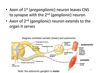
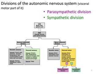
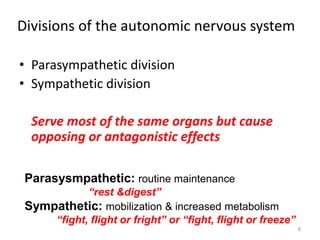
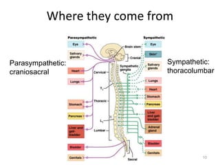
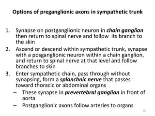
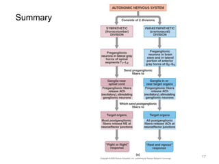
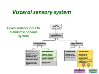
The Autonomic Nervous System
The Autonomic
Nervous System
Visceral sensory
Visceral motor
&
Autonomic nervous system
• The autonomic nervous system is the
subdivision of the peripheral nervous system
that regulates body activities that are
generally not under conscious control
• Visceral motor innervates non-skeletal (non-
somatic) muscles
• Visceral sensory will be covered later
3
• ANS is the subdivision of the peripheral nervous
system that regulates body activities that are
generally not under conscious control
• Visceral motor innervates non-skeletal (non-somatic)
muscles
• Composed of a special group of neurons serving:
– Cardiac muscle (the heart)
– Smooth muscle (walls of viscera and blood vessels)
– Internal organs
– Skin
4
To repeat…
Basic anatomical difference between the motor
pathways of the voluntary somatic nervous system
(to skeletal muscles) and those of the autonomic
nervous system
• Somatic division:
– Cell bodies of motor neurons reside in CNS (brain or spinal
cord)
– Their axons (sheathed in spinal nerves) extend all the way
to their skeletal muscles
• Autonomic system: chains of two motor neurons
– 1st = preganglionic neuron (in brain or cord)
– 2nd = gangionic neuron (cell body in ganglion outside CNS)
– Slower because lightly or unmyelinated
(see next diagram)
5
• Axon of 1st (preganglionic) neuron leaves CNS
to synapse with the 2nd (ganglionic) neuron
• Axon of 2nd (ganglionic) neuron extends to the
organ it serves
6
Diagram contrasts somatic (lower) and autonomic:
autonomic
somatic
Note: the autonomic ganglion is motor
this dorsal
root ganglion
is sensory
Divisions of the autonomic nervous system (visceral
motor part of it)
• Parasympathetic division
• Sympathetic division
7
Divisions of the autonomic nervous system
• Parasympathetic division
• Sympathetic division
Serve most of the same organs but cause
opposing or antagonistic effects
8
Parasysmpathetic: routine maintenance
“rest &digest”
Sympathetic: mobilization & increased metabolism
“fight, flight or fright” or “fight, flight or freeze”
9
Where they come from
10
Parasympathetic:
craniosacral
Sympathetic:
thoracolumbar
Parasympathetic nervous system
“rest & digest”
• Also called the craniosacral system because
all its preganglionic neurons are in the brain
stem or sacral levels of the spinal cord
– Cranial nerves III,VII, IX and X
– In lateral horn of gray matter from S2-S4
• Only innervate internal organs (not skin)
• Acetylcholine is neurotransmitter at end
organ as well as at preganglionic synapse:
“cholinergic”
11
Parasympathetic continued
• Cranial outflow
– III - pupils constrict
– VII - tears, nasal mucus, saliva
– IX – parotid salivary gland
– X (Vagus n) – visceral organs of thorax & abdomen:
• Stimulates digestive glands
• Increases motility of smooth muscle of digestive tract
• Decreases heart rate
• Causes bronchial constriction
• Sacral outflow (S2-4): form pelvic splanchnic nerves
– Supply 2nd half of large intestine
– Supply all the pelvic (genitourinary) organs
12
Parasympathetic
(only look at this if
it helps you)
13
Sympathetic nervous system
“fight, flight or fright”
• Also called thoracolumbar system: all its neurons are in
lateral horn of gray matter from T1-L2
• Lead to every part of the body (unlike parasymp.)
– Easy to remember that when nervous, you sweat; when afraid,
hair stands on end; when excited blood pressure rises
(vasoconstriction): these sympathetic only
– Also causes: dry mouth, pupils to dilate, increased heart &
respiratory rates to increase O2 to skeletal muscles, and liver
to release glucose
• Norepinephrine (aka noradrenaline) is neurotransmitter
released by most postganglionic fibers (acetylcholine in
preganglionic): “adrenergic”
14
Sympathetic nervous system continued
• Regardless of target, all begin
same
• Preganglionic axons exit
spinal cord through ventral
root and enter spinal nerve
• Exit spinal nerve via
communicating ramus
• Enter sympathetic
trunk/chain where
postganglionic neurons are
• Has three options…
15
Options of preganglionic axons in sympathetic trunk
1. Synapse on postganglionic neuron in chain ganglion
then return to spinal nerve and follow its branch to
the skin
2. Ascend or descend within sympathetic trunk, synapse
with a posganglionic neuron within a chain ganglion,
and return to spinal nerve at that level and follow
branches to skin
3. Enter sympathetic chain, pass through without
synapsing, form a splanchnic nerve that passes
toward thoracic or abdominal organs
– These synapse in prevertebral ganglion in front of
aorta
– Postganglionic axons follow arteries to organs
16
17
Summary
Visceral sensory system
18
Gives sensory input to
autonomic nervous
system
Visceral sensory neurons
• Monitor temperature, pain, irritation, chemical changes and
stretch in the visceral organs
– Brain interprets as hunger, fullness, pain, nausea, well-being
• Receptors widely scattered – localization poor (e.g. which part
is giving you the gas pain?)
• Visceral sensory fibers run within autonomic nerves,
especially vagus and sympathetic nerves
– Sympathetic nerves carry most pain fibers from visceral organs of body
trunk
• Simplified pathway: sensory neurons to spinothalamic tract to
thalamus to cerebral cortex
• Visceral pain is induced by stretching, infection and cramping
of internal organs but seldom by cutting .
19

Autonomic Nervous System (1).ppt

  • 1.
  • 2.
    The Autonomic Nervous System Visceralsensory Visceral motor &
  • 3.
    Autonomic nervous system •The autonomic nervous system is the subdivision of the peripheral nervous system that regulates body activities that are generally not under conscious control • Visceral motor innervates non-skeletal (non- somatic) muscles • Visceral sensory will be covered later 3
  • 4.
    • ANS isthe subdivision of the peripheral nervous system that regulates body activities that are generally not under conscious control • Visceral motor innervates non-skeletal (non-somatic) muscles • Composed of a special group of neurons serving: – Cardiac muscle (the heart) – Smooth muscle (walls of viscera and blood vessels) – Internal organs – Skin 4 To repeat…
  • 5.
    Basic anatomical differencebetween the motor pathways of the voluntary somatic nervous system (to skeletal muscles) and those of the autonomic nervous system • Somatic division: – Cell bodies of motor neurons reside in CNS (brain or spinal cord) – Their axons (sheathed in spinal nerves) extend all the way to their skeletal muscles • Autonomic system: chains of two motor neurons – 1st = preganglionic neuron (in brain or cord) – 2nd = gangionic neuron (cell body in ganglion outside CNS) – Slower because lightly or unmyelinated (see next diagram) 5
  • 6.
    • Axon of1st (preganglionic) neuron leaves CNS to synapse with the 2nd (ganglionic) neuron • Axon of 2nd (ganglionic) neuron extends to the organ it serves 6 Diagram contrasts somatic (lower) and autonomic: autonomic somatic Note: the autonomic ganglion is motor this dorsal root ganglion is sensory
  • 7.
    Divisions of theautonomic nervous system (visceral motor part of it) • Parasympathetic division • Sympathetic division 7
  • 8.
    Divisions of theautonomic nervous system • Parasympathetic division • Sympathetic division Serve most of the same organs but cause opposing or antagonistic effects 8 Parasysmpathetic: routine maintenance “rest &digest” Sympathetic: mobilization & increased metabolism “fight, flight or fright” or “fight, flight or freeze”
  • 9.
  • 10.
    Where they comefrom 10 Parasympathetic: craniosacral Sympathetic: thoracolumbar
  • 11.
    Parasympathetic nervous system “rest& digest” • Also called the craniosacral system because all its preganglionic neurons are in the brain stem or sacral levels of the spinal cord – Cranial nerves III,VII, IX and X – In lateral horn of gray matter from S2-S4 • Only innervate internal organs (not skin) • Acetylcholine is neurotransmitter at end organ as well as at preganglionic synapse: “cholinergic” 11
  • 12.
    Parasympathetic continued • Cranialoutflow – III - pupils constrict – VII - tears, nasal mucus, saliva – IX – parotid salivary gland – X (Vagus n) – visceral organs of thorax & abdomen: • Stimulates digestive glands • Increases motility of smooth muscle of digestive tract • Decreases heart rate • Causes bronchial constriction • Sacral outflow (S2-4): form pelvic splanchnic nerves – Supply 2nd half of large intestine – Supply all the pelvic (genitourinary) organs 12
  • 13.
    Parasympathetic (only look atthis if it helps you) 13
  • 14.
    Sympathetic nervous system “fight,flight or fright” • Also called thoracolumbar system: all its neurons are in lateral horn of gray matter from T1-L2 • Lead to every part of the body (unlike parasymp.) – Easy to remember that when nervous, you sweat; when afraid, hair stands on end; when excited blood pressure rises (vasoconstriction): these sympathetic only – Also causes: dry mouth, pupils to dilate, increased heart & respiratory rates to increase O2 to skeletal muscles, and liver to release glucose • Norepinephrine (aka noradrenaline) is neurotransmitter released by most postganglionic fibers (acetylcholine in preganglionic): “adrenergic” 14
  • 15.
    Sympathetic nervous systemcontinued • Regardless of target, all begin same • Preganglionic axons exit spinal cord through ventral root and enter spinal nerve • Exit spinal nerve via communicating ramus • Enter sympathetic trunk/chain where postganglionic neurons are • Has three options… 15
  • 16.
    Options of preganglionicaxons in sympathetic trunk 1. Synapse on postganglionic neuron in chain ganglion then return to spinal nerve and follow its branch to the skin 2. Ascend or descend within sympathetic trunk, synapse with a posganglionic neuron within a chain ganglion, and return to spinal nerve at that level and follow branches to skin 3. Enter sympathetic chain, pass through without synapsing, form a splanchnic nerve that passes toward thoracic or abdominal organs – These synapse in prevertebral ganglion in front of aorta – Postganglionic axons follow arteries to organs 16
  • 17.
  • 18.
    Visceral sensory system 18 Givessensory input to autonomic nervous system
  • 19.
    Visceral sensory neurons •Monitor temperature, pain, irritation, chemical changes and stretch in the visceral organs – Brain interprets as hunger, fullness, pain, nausea, well-being • Receptors widely scattered – localization poor (e.g. which part is giving you the gas pain?) • Visceral sensory fibers run within autonomic nerves, especially vagus and sympathetic nerves – Sympathetic nerves carry most pain fibers from visceral organs of body trunk • Simplified pathway: sensory neurons to spinothalamic tract to thalamus to cerebral cortex • Visceral pain is induced by stretching, infection and cramping of internal organs but seldom by cutting . 19