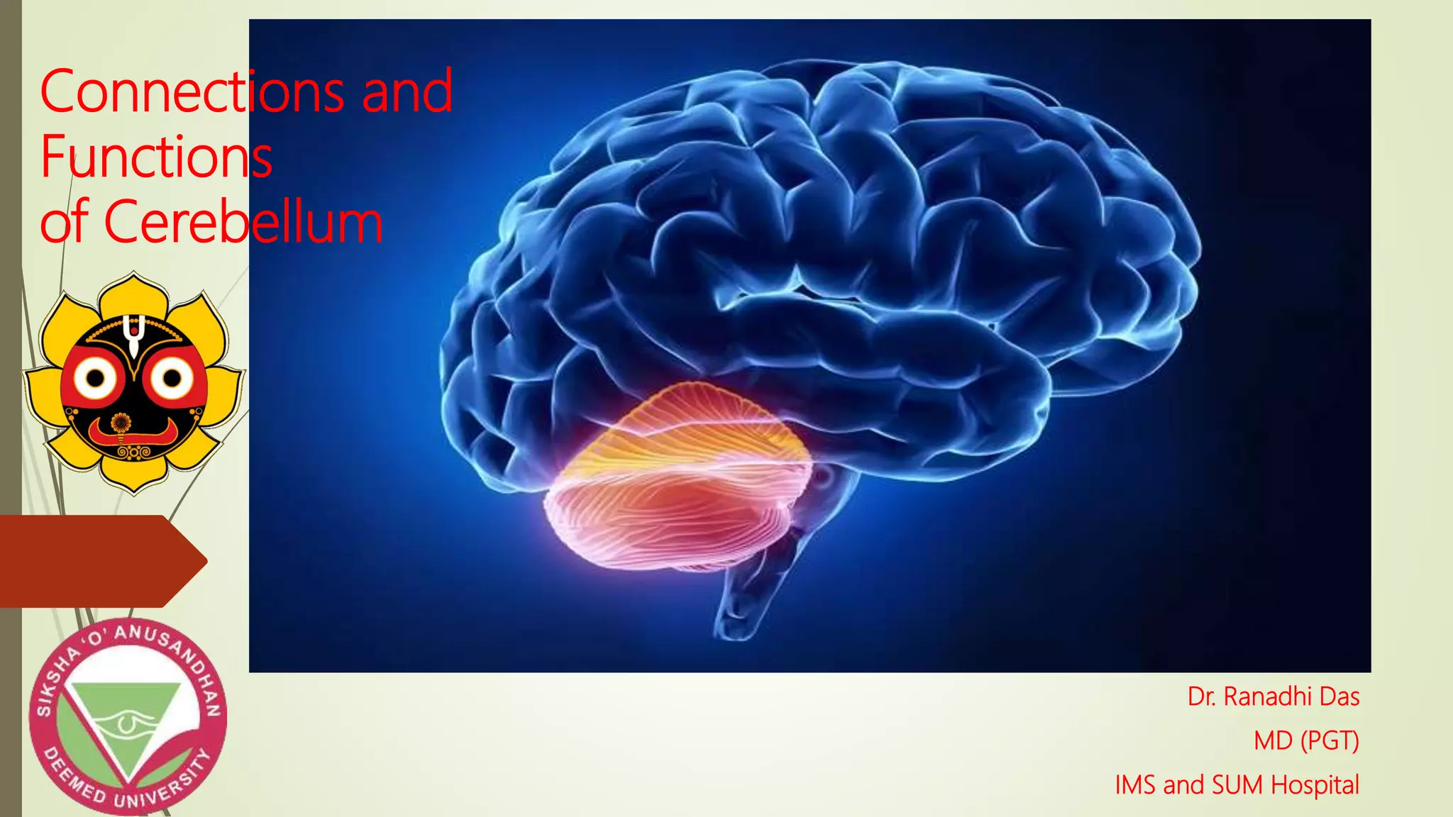
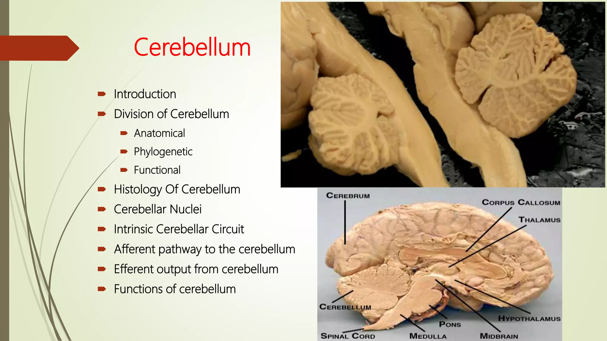
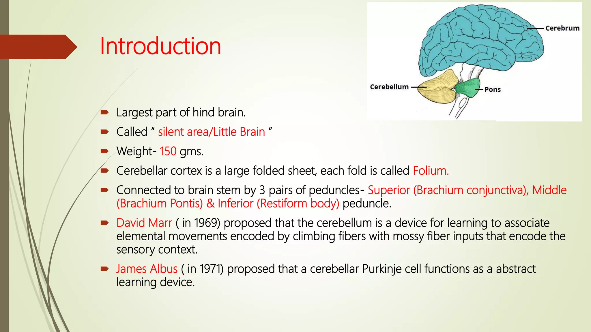
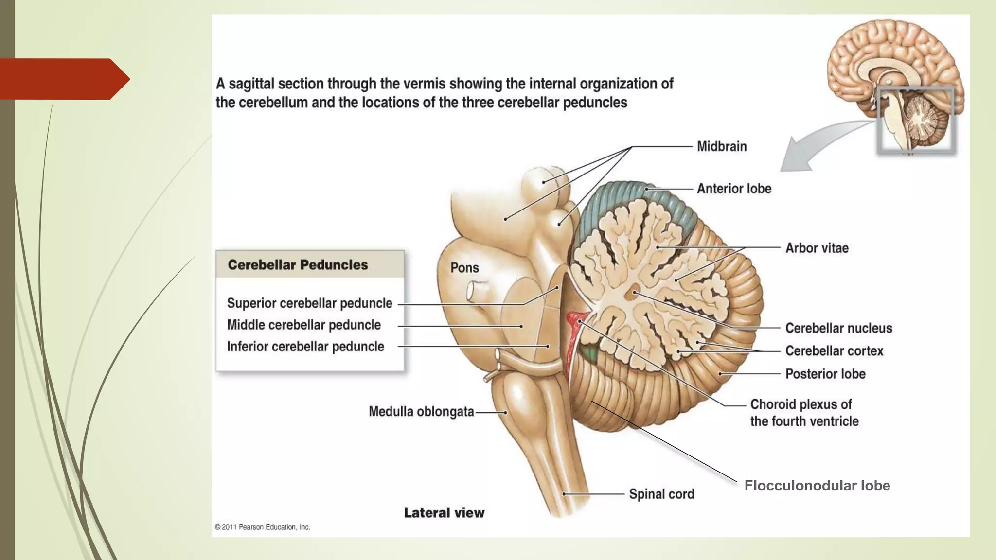
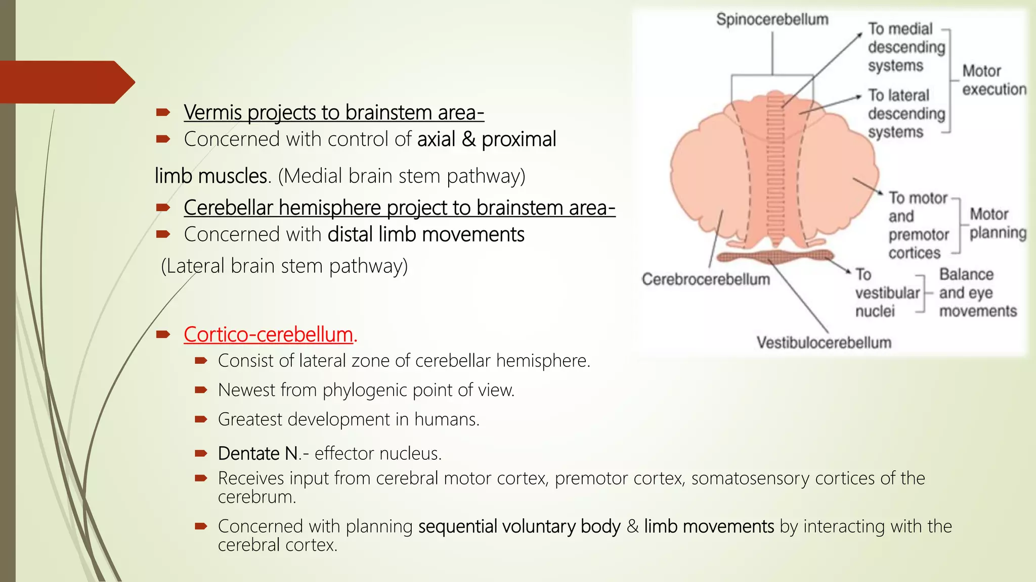
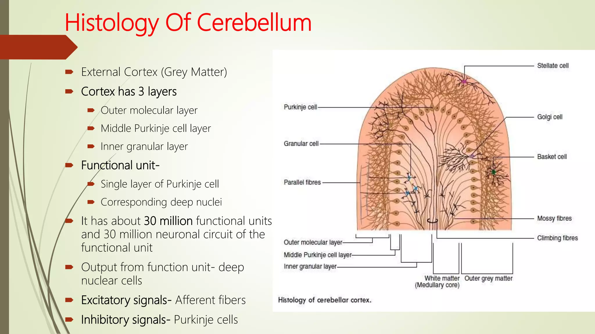
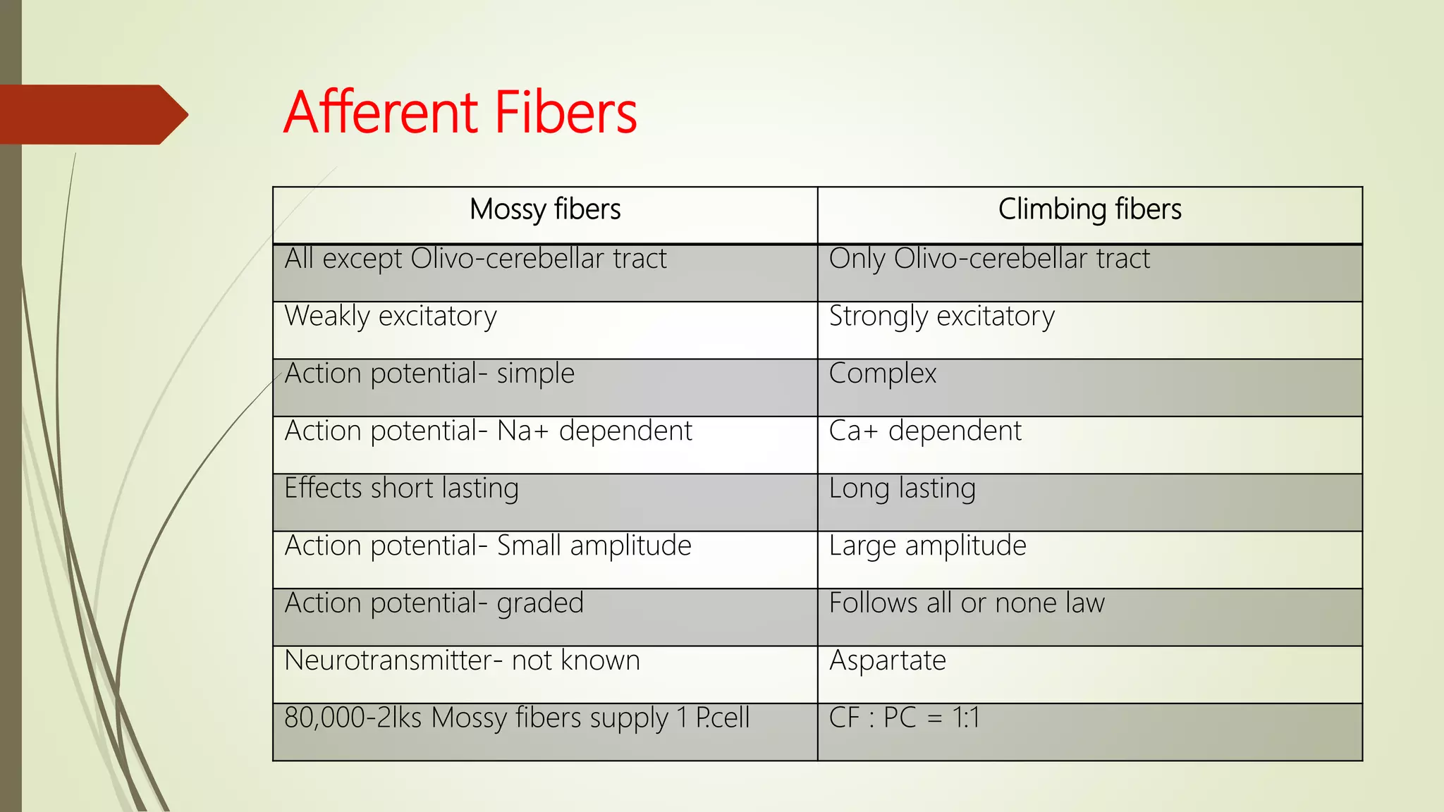
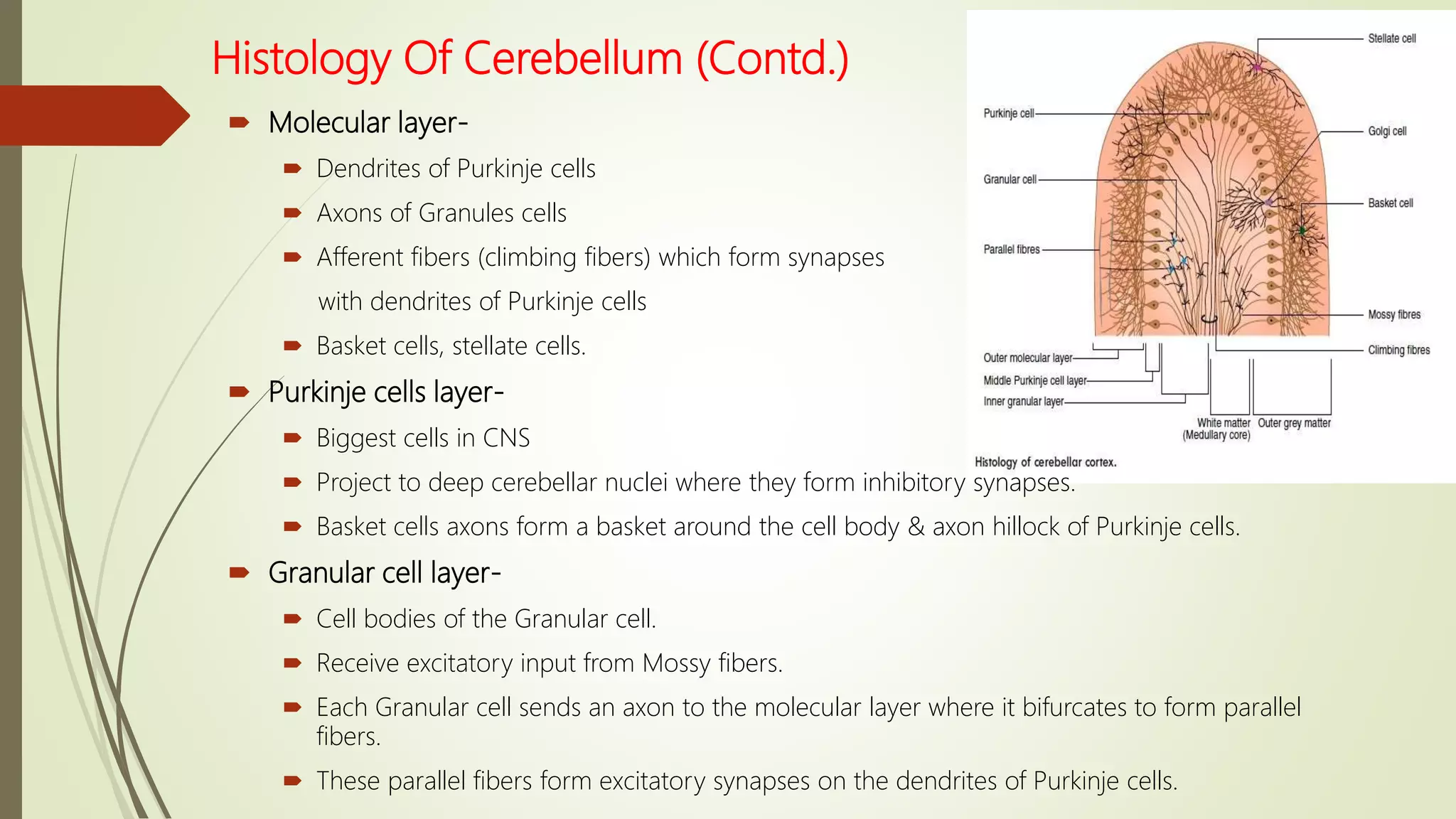
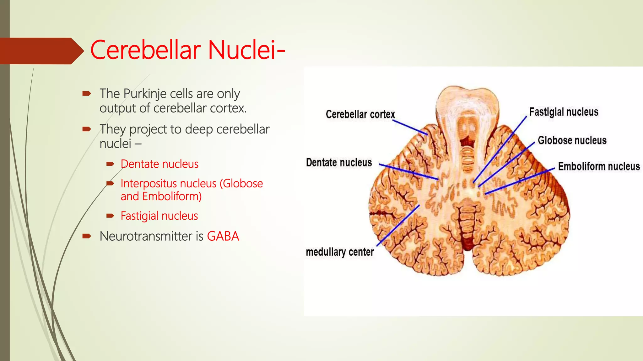
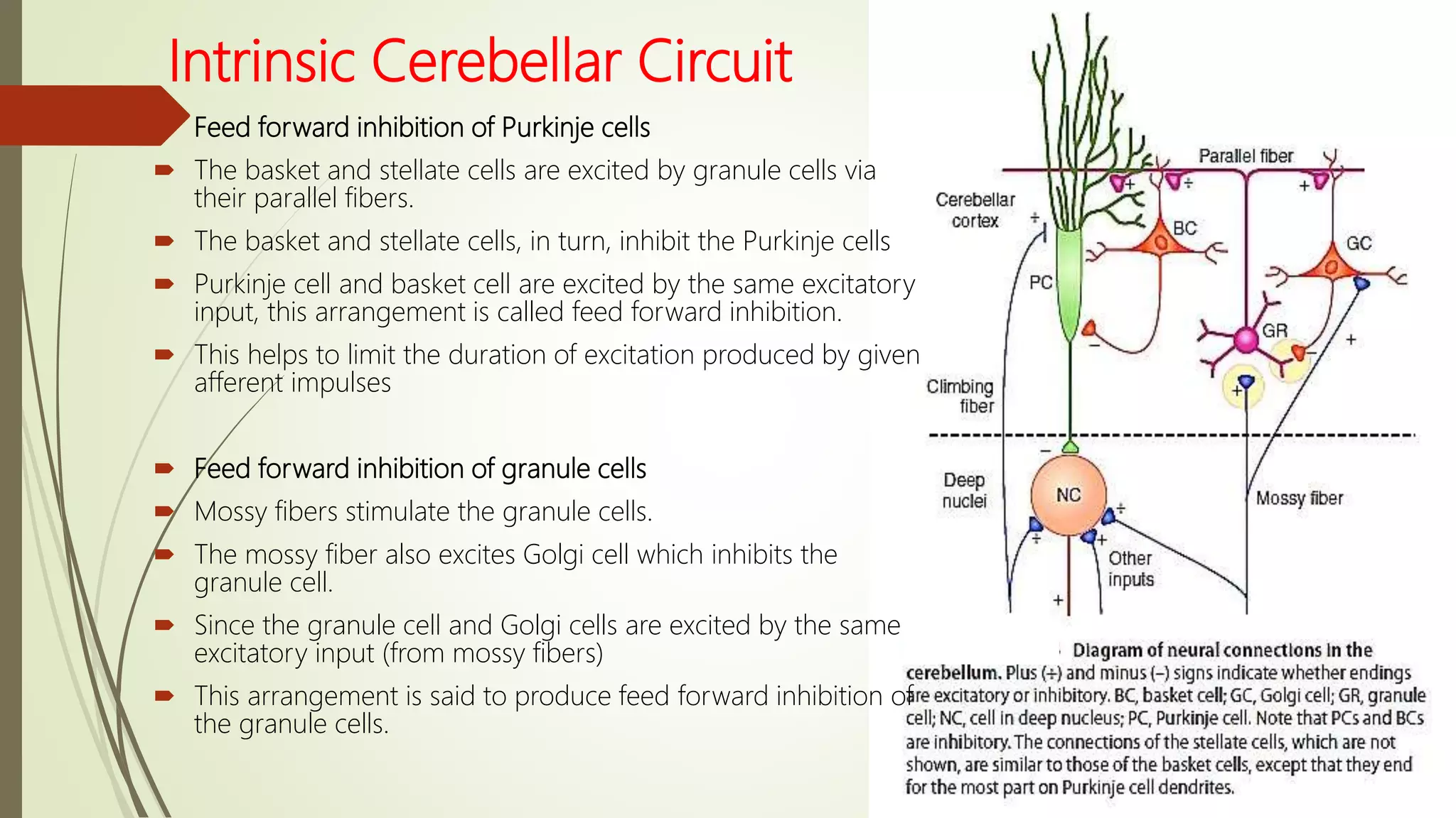
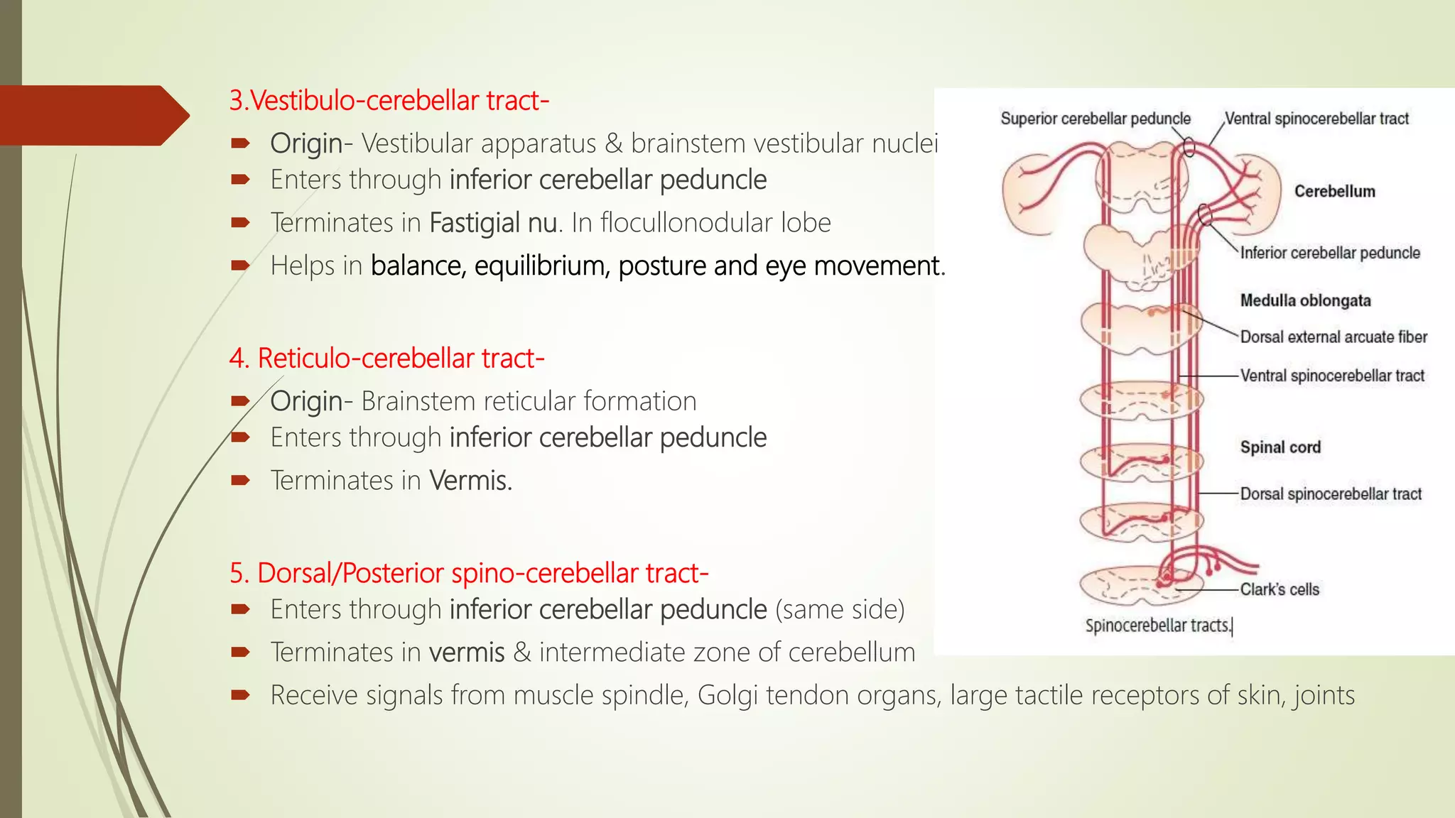
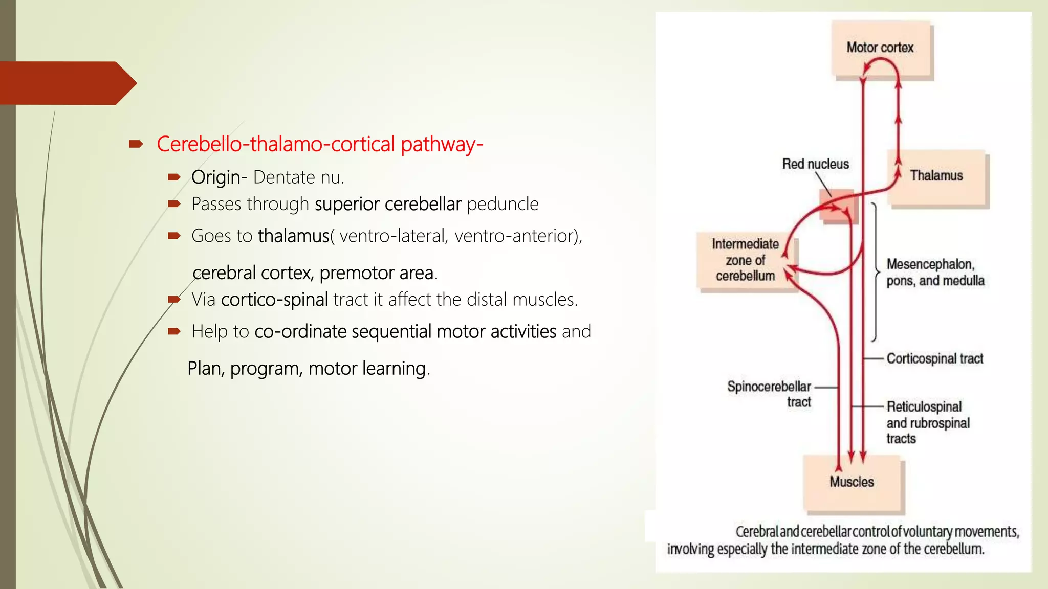
Dr. Ranadhi Das
MD (PGT)
IMS and SUM Hospital
Connections and
Functions
of Cerebellum
Cerebellum
 Introduction
 Division of Cerebellum
 Anatomical
 Phylogenetic
 Functional
 Histology Of Cerebellum
 Cerebellar Nuclei
 Intrinsic Cerebellar Circuit
 Afferent pathway to the cerebellum
 Efferent output from cerebellum
 Functions of cerebellum
Introduction
 Largest part of hind brain.
 Called “ silent area/Little Brain ”
 Weight- 150 gms.
 Cerebellar cortex is a large folded sheet, each fold is called Folium.
 Connected to brain stem by 3 pairs of peduncles- Superior (Brachium conjunctiva), Middle
(Brachium Pontis) & Inferior (Restiform body) peduncle.
 David Marr ( in 1969) proposed that the cerebellum is a device for learning to associate
elemental movements encoded by climbing fibers with mossy fiber inputs that encode the
sensory context.
 James Albus ( in 1971) proposed that a cerebellar Purkinje cell functions as a abstract
learning device.
Flocculonodular lobe
Anatomical Divisions
1.Anterior lobe
 Lingula
 Centralis
 Culmen
2.Posterior lobe
 Simplex
 Declive
 Folium
 Tuber
 Ansiform lobe
 Paramedian lobe
3.Flocculonodular lobe-
 Pyramis
 Uvula
 Nodule
 Flocullus.
 A narrow band down the center of the cerebellum called Vermis.
 It controls most of the cerebellar functions.
 The axial portion of the body is topographically represented in the vermis.
Phylogenetic Divisions
 Archicerebellum
 Flocculonodular lobe
 Palaeocerebellum
 Anterior lobe- Lingula, Centralis, Culmen, Simplex
 Posterior lobe – pyramis, uvula & parafllocculus.
 Neocerebellum
 Whole posterior lobe- Declive, Folium, Tuber, Ansiform lobe, Paramedian lobe.
Functional Divisions
 Vestibulo-cerebellum.
 Consists of Flocculonodular lobe,
adjacent part of vermis, Lingula
 Oldest part
 Has Vestibular connections
 N. fastigial – effector N.
 Control body posture, equilibrium & eye movements
 Control axial and proximal limbs.
 Spino-cerebellum.
 Consists of most of the vermis and adjacent intermediate zone on both side of vermis.
 N. Interpositus ( N.Globose & N. Emboliformis) – effector N.
 Receives proprioceptive impulses from the body & prepare a efference copy of motor plan from
the cerebral cortex.
 Vermis projects to brainstem area-
 Concerned with control of axial & proximal
limb muscles. (Medial brain stem pathway)
 Cerebellar hemisphere project to brainstem area-
 Concerned with distal limb movements
(Lateral brain stem pathway)
 Cortico-cerebellum.
 Consist of lateral zone of cerebellar hemisphere.
 Newest from phylogenic point of view.
 Greatest development in humans.
 Dentate N.- effector nucleus.
 Receives input from cerebral motor cortex, premotor cortex, somatosensory cortices of the
cerebrum.
 Concerned with planning sequential voluntary body & limb movements by interacting with the
cerebral cortex.
Histology Of Cerebellum
 External Cortex (Grey Matter)
 Cortex has 3 layers
 Outer molecular layer
 Middle Purkinje cell layer
 Inner granular layer
 Functional unit-
 Single layer of Purkinje cell
 Corresponding deep nuclei
 It has about 30 million functional units
and 30 million neuronal circuit of the
functional unit
 Output from function unit- deep
nuclear cells
 Excitatory signals- Afferent fibers
 Inhibitory signals- Purkinje cells
Afferent Fibers
Mossy fibers Climbing fibers
All except Olivo-cerebellar tract Only Olivo-cerebellar tract
Weakly excitatory Strongly excitatory
Action potential- simple Complex
Action potential- Na+ dependent Ca+ dependent
Effects short lasting Long lasting
Action potential- Small amplitude Large amplitude
Action potential- graded Follows all or none law
Neurotransmitter- not known Aspartate
80,000-2lks Mossy fibers supply 1 P.cell CF : PC = 1:1
Histology Of Cerebellum (Contd.)
 Molecular layer-
 Dendrites of Purkinje cells
 Axons of Granules cells
 Afferent fibers (climbing fibers) which form synapses
with dendrites of Purkinje cells
 Basket cells, stellate cells.
 Purkinje cells layer-
 Biggest cells in CNS
 Project to deep cerebellar nuclei where they form inhibitory synapses.
 Basket cells axons form a basket around the cell body & axon hillock of Purkinje cells.
 Granular cell layer-
 Cell bodies of the Granular cell.
 Receive excitatory input from Mossy fibers.
 Each Granular cell sends an axon to the molecular layer where it bifurcates to form parallel
fibers.
 These parallel fibers form excitatory synapses on the dendrites of Purkinje cells.
Cerebellar Nuclei-
 The Purkinje cells are only
output of cerebellar cortex.
 They project to deep cerebellar
nuclei –
 Dentate nucleus
 Interpositus nucleus (Globose
and Emboliform)
 Fastigial nucleus
 Neurotransmitter is GABA
Intrinsic Cerebellar Circuit
 Feed forward inhibition of Purkinje cells
 The basket and stellate cells are excited by granule cells via
their parallel fibers.
 The basket and stellate cells, in turn, inhibit the Purkinje cells
 Purkinje cell and basket cell are excited by the same excitatory
input, this arrangement is called feed forward inhibition.
 This helps to limit the duration of excitation produced by given
afferent impulses
 Feed forward inhibition of granule cells
 Mossy fibers stimulate the granule cells.
 The mossy fiber also excites Golgi cell which inhibits the
granule cell.
 Since the granule cell and Golgi cells are excited by the same
excitatory input (from mossy fibers)
 This arrangement is said to produce feed forward inhibition of
the granule cells.
 Feedback inhibition of granule cells
 the granule cell is excited by the mossy fibers.
 The axon of granule cell excites the Golgi cell
dendrites, whose axon inhibits the granule cell.
 Excitation of the granule cell is rapidly stopped by a
negative feedback loop.
 Called feedback inhibition of granule cells.
 The Reverberating circuit
 The granule cells and Purkinje cells form a
reverberating (echoing) circuit.
 Function is to revive and strengthen the non-
synapses, when two neurons discharge by repeatedly
and synchronously.
 This principle was developed by Hebb
Afferent Pathway To The Cerebellum
1.Cortico-ponto-cerebellar pathway-
 Origin- Cerebral motor cortex, premotor cortex, somatosensory cortex
 Passes by Pontine nu.
 Ponto-cerebellar tract enter through Middle cerebellar peduncle (Opposite side)
 Distributed mainly to lateral zone of cerebellar hemisphere.
2.Olivo-cerebellar tract-
 Passes from inferior Olivary complex in medulla.
 Enters through Inferior cerebellar peduncle(Opposite side)
 Relay all over the cerebellum.
 Receive proprioceptive input from whole body.
 The inferior Olivary complex is stimulated by-
 Cerebral motor cortex
 Basal ganglia
 Reticular formation
 Spinal cord
3.Vestibulo-cerebellar tract-
 Origin- Vestibular apparatus & brainstem vestibular nuclei
 Enters through inferior cerebellar peduncle
 Terminates in Fastigial nu. In flocullonodular lobe
 Helps in balance, equilibrium, posture and eye movement.
4. Reticulo-cerebellar tract-
 Origin- Brainstem reticular formation
 Enters through inferior cerebellar peduncle
 Terminates in Vermis.
5. Dorsal/Posterior spino-cerebellar tract-
 Enters through inferior cerebellar peduncle (same side)
 Terminates in vermis & intermediate zone of cerebellum
 Receive signals from muscle spindle, Golgi tendon organs, large tactile receptors of skin, joints
6. Ventral/Anterior spino-cerebellar tract-
 Enters through superior cerebellar peduncle (same side)
 Terminates on both side of the cerebellum
 Receive signals from anterior horn cell of spinal cord
 Receive proprioceptive and exteroceptive (touch, pressure) signals.
 Convey information about length and tension of muscle fibers (unconscious proprioceptive
sensation)
 Most rapid conduction pathway.
7. Cuneocerebellar tract-
 Carries proprioceptive impulse from head and neck.
8. Tecto-cerebellar tract-
 Carries auditory and visual impulse via inferior and superior colliculi.
 Controlling the movement of the head and neck in response to auditory and visual stimuli.
Efferent Output From Cerebellum
 Cerebello-vestibular pathway-
 Origin- Fastigial nu. At flocullonodular lobe of cerebellum.
 Passes through Inferior cerebellar peduncle.
 Signal goes to brainstem reticular formation and vestibular nu. by reticulo-spinal tract and
vestibulo-spinal tract
 To axial and proximal muscles.
 Maintain the posture, tone, equilibrium, balance and eye movements.
 Cerebello-rubro-thalamo-cortical pathway-
 Origin- Interpositus nu. Of Intermediate zone of cerebellar hemisphere
 Passes through superior cerebellar peduncle
 Signals goes to thalamus, cerebral cortex, basal ganglia, red nucleus, reticular formation.
 Helps to co-ordinate reciprocal contraction of agonist & antagonist muscles in the distal muscles,
compare them.
 Helps in ongoing movement.
 Cerebello-thalamo-cortical pathway-
 Origin- Dentate nu.
 Passes through superior cerebellar peduncle
 Goes to thalamus( ventro-lateral, ventro-anterior),
cerebral cortex, premotor area.
 Via cortico-spinal tract it affect the distal muscles.
 Help to co-ordinate sequential motor activities and
Plan, program, motor learning.
Functions of Cerebellum
 It helps to control the tone of the muscles on the same side of the body.
 Control the rate, range, force & direction of movement.(Synergia)
 Control rapid muscular movements, like running, typing, talking. Cerebellum helps in co-
ordination of these activities, sequencing them & correcting them.
 It received continuously update information on the desired programme of muscular
contractions.
 It has a comparator function.
 It helps in planning the next sequential movement in advance while the present
movement is going on.
 It helps in timing of the movement, controls ballistic and saccadic movements.
 Helps in predicting from a changing visual scene, how fast he is approaching an object.
 Helps in applying brakes where necessary.
 Helps in smooth coordinated movements by virtue of efference copy.
 Helps in speech.
 Helps in posture and equilibrium.
Cerebellum-Connections and Functions

Cerebellum-Connections and Functions

  • 1.
    Dr. Ranadhi Das MD(PGT) IMS and SUM Hospital Connections and Functions of Cerebellum
  • 2.
    Cerebellum  Introduction  Divisionof Cerebellum  Anatomical  Phylogenetic  Functional  Histology Of Cerebellum  Cerebellar Nuclei  Intrinsic Cerebellar Circuit  Afferent pathway to the cerebellum  Efferent output from cerebellum  Functions of cerebellum
  • 3.
    Introduction  Largest partof hind brain.  Called “ silent area/Little Brain ”  Weight- 150 gms.  Cerebellar cortex is a large folded sheet, each fold is called Folium.  Connected to brain stem by 3 pairs of peduncles- Superior (Brachium conjunctiva), Middle (Brachium Pontis) & Inferior (Restiform body) peduncle.  David Marr ( in 1969) proposed that the cerebellum is a device for learning to associate elemental movements encoded by climbing fibers with mossy fiber inputs that encode the sensory context.  James Albus ( in 1971) proposed that a cerebellar Purkinje cell functions as a abstract learning device.
  • 4.
  • 5.
    Anatomical Divisions 1.Anterior lobe Lingula  Centralis  Culmen 2.Posterior lobe  Simplex  Declive  Folium  Tuber  Ansiform lobe  Paramedian lobe 3.Flocculonodular lobe-  Pyramis  Uvula  Nodule  Flocullus.
  • 6.
     A narrowband down the center of the cerebellum called Vermis.  It controls most of the cerebellar functions.  The axial portion of the body is topographically represented in the vermis.
  • 7.
    Phylogenetic Divisions  Archicerebellum Flocculonodular lobe  Palaeocerebellum  Anterior lobe- Lingula, Centralis, Culmen, Simplex  Posterior lobe – pyramis, uvula & parafllocculus.  Neocerebellum  Whole posterior lobe- Declive, Folium, Tuber, Ansiform lobe, Paramedian lobe.
  • 8.
    Functional Divisions  Vestibulo-cerebellum. Consists of Flocculonodular lobe, adjacent part of vermis, Lingula  Oldest part  Has Vestibular connections  N. fastigial – effector N.  Control body posture, equilibrium & eye movements  Control axial and proximal limbs.  Spino-cerebellum.  Consists of most of the vermis and adjacent intermediate zone on both side of vermis.  N. Interpositus ( N.Globose & N. Emboliformis) – effector N.  Receives proprioceptive impulses from the body & prepare a efference copy of motor plan from the cerebral cortex.
  • 9.
     Vermis projectsto brainstem area-  Concerned with control of axial & proximal limb muscles. (Medial brain stem pathway)  Cerebellar hemisphere project to brainstem area-  Concerned with distal limb movements (Lateral brain stem pathway)  Cortico-cerebellum.  Consist of lateral zone of cerebellar hemisphere.  Newest from phylogenic point of view.  Greatest development in humans.  Dentate N.- effector nucleus.  Receives input from cerebral motor cortex, premotor cortex, somatosensory cortices of the cerebrum.  Concerned with planning sequential voluntary body & limb movements by interacting with the cerebral cortex.
  • 10.
    Histology Of Cerebellum External Cortex (Grey Matter)  Cortex has 3 layers  Outer molecular layer  Middle Purkinje cell layer  Inner granular layer  Functional unit-  Single layer of Purkinje cell  Corresponding deep nuclei  It has about 30 million functional units and 30 million neuronal circuit of the functional unit  Output from function unit- deep nuclear cells  Excitatory signals- Afferent fibers  Inhibitory signals- Purkinje cells
  • 11.
    Afferent Fibers Mossy fibersClimbing fibers All except Olivo-cerebellar tract Only Olivo-cerebellar tract Weakly excitatory Strongly excitatory Action potential- simple Complex Action potential- Na+ dependent Ca+ dependent Effects short lasting Long lasting Action potential- Small amplitude Large amplitude Action potential- graded Follows all or none law Neurotransmitter- not known Aspartate 80,000-2lks Mossy fibers supply 1 P.cell CF : PC = 1:1
  • 12.
    Histology Of Cerebellum(Contd.)  Molecular layer-  Dendrites of Purkinje cells  Axons of Granules cells  Afferent fibers (climbing fibers) which form synapses with dendrites of Purkinje cells  Basket cells, stellate cells.  Purkinje cells layer-  Biggest cells in CNS  Project to deep cerebellar nuclei where they form inhibitory synapses.  Basket cells axons form a basket around the cell body & axon hillock of Purkinje cells.  Granular cell layer-  Cell bodies of the Granular cell.  Receive excitatory input from Mossy fibers.  Each Granular cell sends an axon to the molecular layer where it bifurcates to form parallel fibers.  These parallel fibers form excitatory synapses on the dendrites of Purkinje cells.
  • 13.
    Cerebellar Nuclei-  ThePurkinje cells are only output of cerebellar cortex.  They project to deep cerebellar nuclei –  Dentate nucleus  Interpositus nucleus (Globose and Emboliform)  Fastigial nucleus  Neurotransmitter is GABA
  • 14.
    Intrinsic Cerebellar Circuit Feed forward inhibition of Purkinje cells  The basket and stellate cells are excited by granule cells via their parallel fibers.  The basket and stellate cells, in turn, inhibit the Purkinje cells  Purkinje cell and basket cell are excited by the same excitatory input, this arrangement is called feed forward inhibition.  This helps to limit the duration of excitation produced by given afferent impulses  Feed forward inhibition of granule cells  Mossy fibers stimulate the granule cells.  The mossy fiber also excites Golgi cell which inhibits the granule cell.  Since the granule cell and Golgi cells are excited by the same excitatory input (from mossy fibers)  This arrangement is said to produce feed forward inhibition of the granule cells.
  • 15.
     Feedback inhibitionof granule cells  the granule cell is excited by the mossy fibers.  The axon of granule cell excites the Golgi cell dendrites, whose axon inhibits the granule cell.  Excitation of the granule cell is rapidly stopped by a negative feedback loop.  Called feedback inhibition of granule cells.  The Reverberating circuit  The granule cells and Purkinje cells form a reverberating (echoing) circuit.  Function is to revive and strengthen the non- synapses, when two neurons discharge by repeatedly and synchronously.  This principle was developed by Hebb
  • 16.
    Afferent Pathway ToThe Cerebellum 1.Cortico-ponto-cerebellar pathway-  Origin- Cerebral motor cortex, premotor cortex, somatosensory cortex  Passes by Pontine nu.  Ponto-cerebellar tract enter through Middle cerebellar peduncle (Opposite side)  Distributed mainly to lateral zone of cerebellar hemisphere. 2.Olivo-cerebellar tract-  Passes from inferior Olivary complex in medulla.  Enters through Inferior cerebellar peduncle(Opposite side)  Relay all over the cerebellum.  Receive proprioceptive input from whole body.  The inferior Olivary complex is stimulated by-  Cerebral motor cortex  Basal ganglia  Reticular formation  Spinal cord
  • 17.
    3.Vestibulo-cerebellar tract-  Origin-Vestibular apparatus & brainstem vestibular nuclei  Enters through inferior cerebellar peduncle  Terminates in Fastigial nu. In flocullonodular lobe  Helps in balance, equilibrium, posture and eye movement. 4. Reticulo-cerebellar tract-  Origin- Brainstem reticular formation  Enters through inferior cerebellar peduncle  Terminates in Vermis. 5. Dorsal/Posterior spino-cerebellar tract-  Enters through inferior cerebellar peduncle (same side)  Terminates in vermis & intermediate zone of cerebellum  Receive signals from muscle spindle, Golgi tendon organs, large tactile receptors of skin, joints
  • 18.
    6. Ventral/Anterior spino-cerebellartract-  Enters through superior cerebellar peduncle (same side)  Terminates on both side of the cerebellum  Receive signals from anterior horn cell of spinal cord  Receive proprioceptive and exteroceptive (touch, pressure) signals.  Convey information about length and tension of muscle fibers (unconscious proprioceptive sensation)  Most rapid conduction pathway. 7. Cuneocerebellar tract-  Carries proprioceptive impulse from head and neck. 8. Tecto-cerebellar tract-  Carries auditory and visual impulse via inferior and superior colliculi.  Controlling the movement of the head and neck in response to auditory and visual stimuli.
  • 19.
    Efferent Output FromCerebellum  Cerebello-vestibular pathway-  Origin- Fastigial nu. At flocullonodular lobe of cerebellum.  Passes through Inferior cerebellar peduncle.  Signal goes to brainstem reticular formation and vestibular nu. by reticulo-spinal tract and vestibulo-spinal tract  To axial and proximal muscles.  Maintain the posture, tone, equilibrium, balance and eye movements.  Cerebello-rubro-thalamo-cortical pathway-  Origin- Interpositus nu. Of Intermediate zone of cerebellar hemisphere  Passes through superior cerebellar peduncle  Signals goes to thalamus, cerebral cortex, basal ganglia, red nucleus, reticular formation.  Helps to co-ordinate reciprocal contraction of agonist & antagonist muscles in the distal muscles, compare them.  Helps in ongoing movement.
  • 20.
     Cerebello-thalamo-cortical pathway- Origin- Dentate nu.  Passes through superior cerebellar peduncle  Goes to thalamus( ventro-lateral, ventro-anterior), cerebral cortex, premotor area.  Via cortico-spinal tract it affect the distal muscles.  Help to co-ordinate sequential motor activities and Plan, program, motor learning.
  • 21.
    Functions of Cerebellum It helps to control the tone of the muscles on the same side of the body.  Control the rate, range, force & direction of movement.(Synergia)  Control rapid muscular movements, like running, typing, talking. Cerebellum helps in co- ordination of these activities, sequencing them & correcting them.  It received continuously update information on the desired programme of muscular contractions.  It has a comparator function.  It helps in planning the next sequential movement in advance while the present movement is going on.  It helps in timing of the movement, controls ballistic and saccadic movements.  Helps in predicting from a changing visual scene, how fast he is approaching an object.  Helps in applying brakes where necessary.  Helps in smooth coordinated movements by virtue of efference copy.  Helps in speech.  Helps in posture and equilibrium.