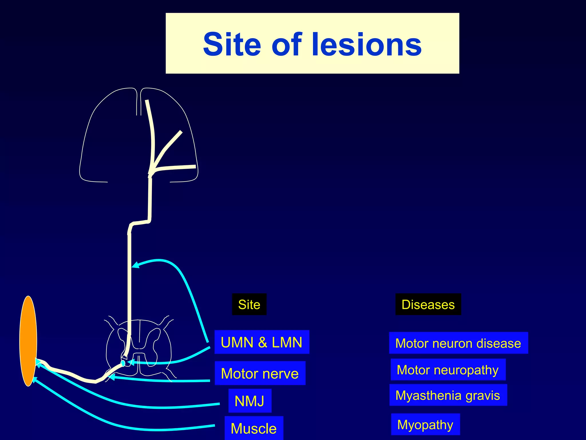
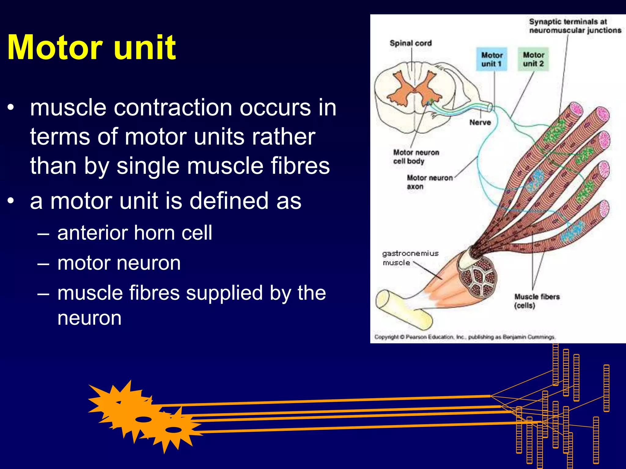
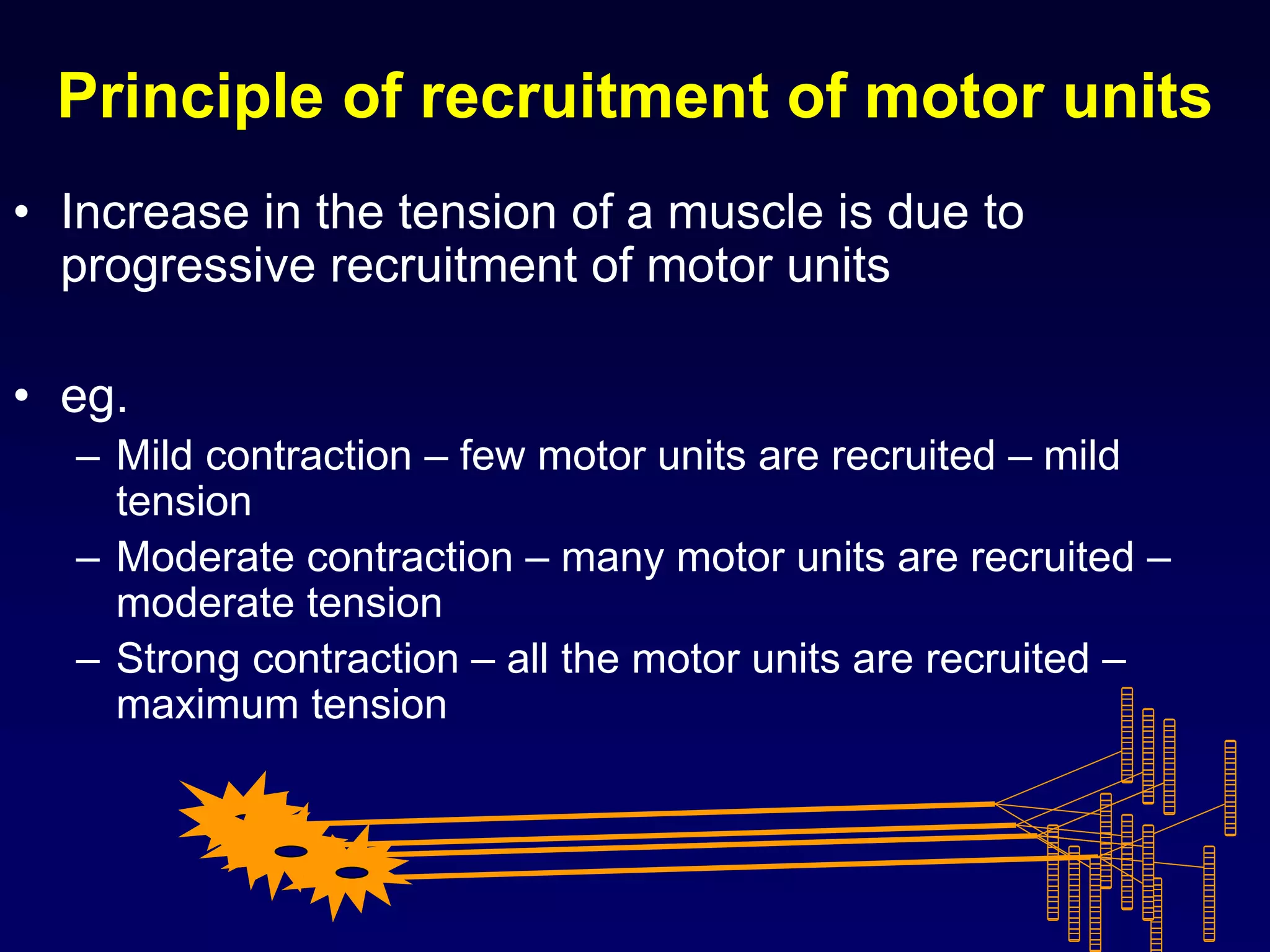
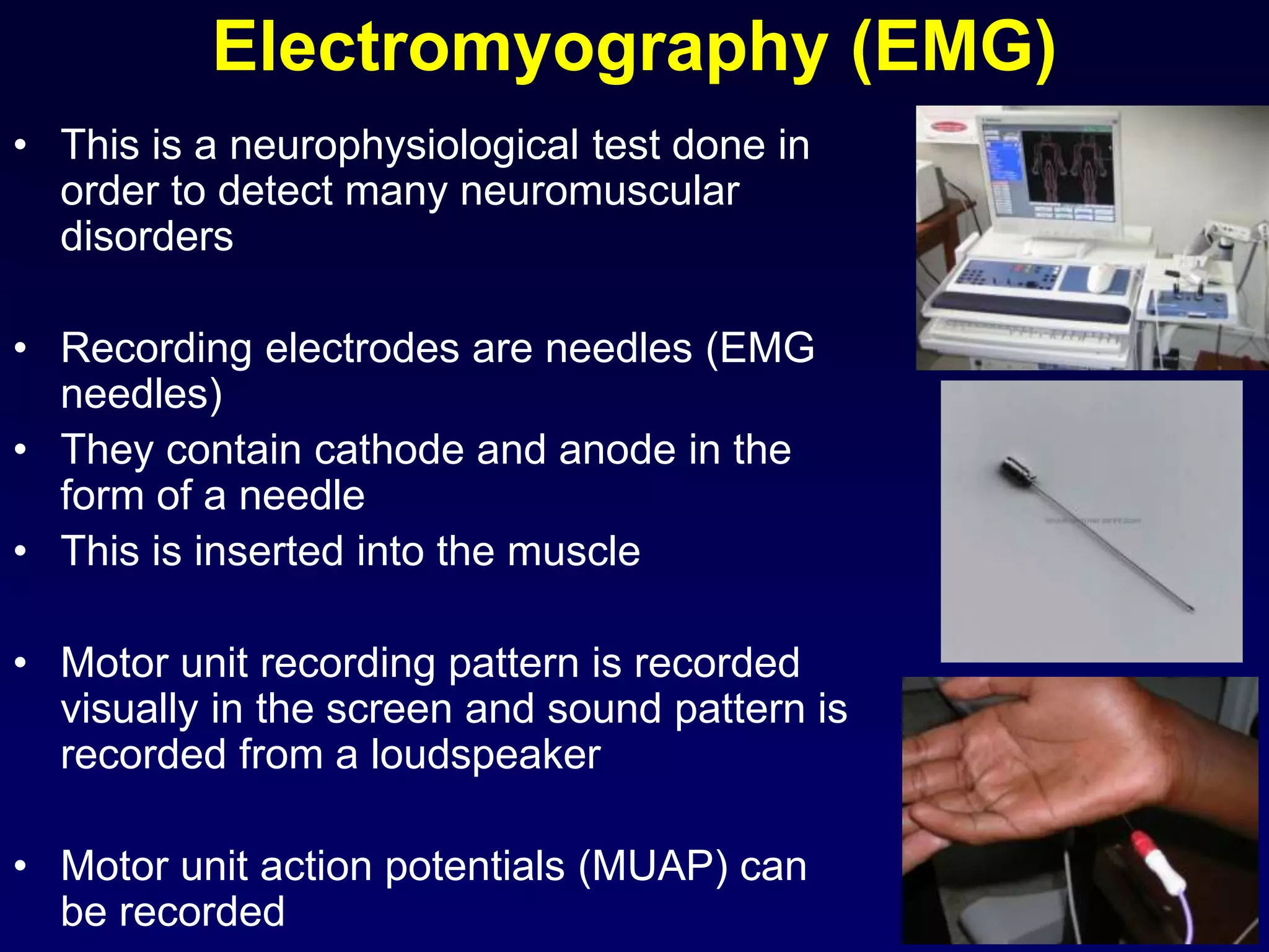
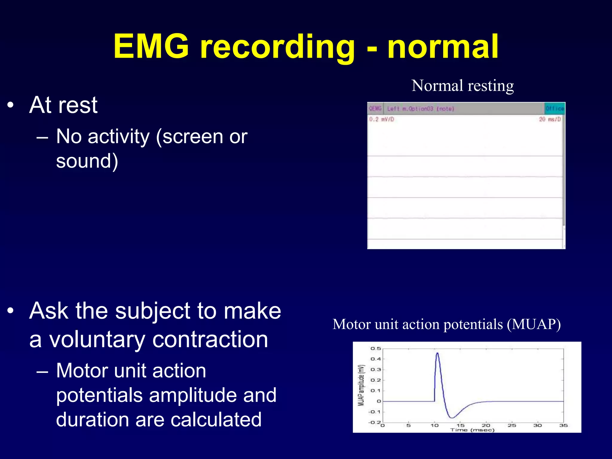
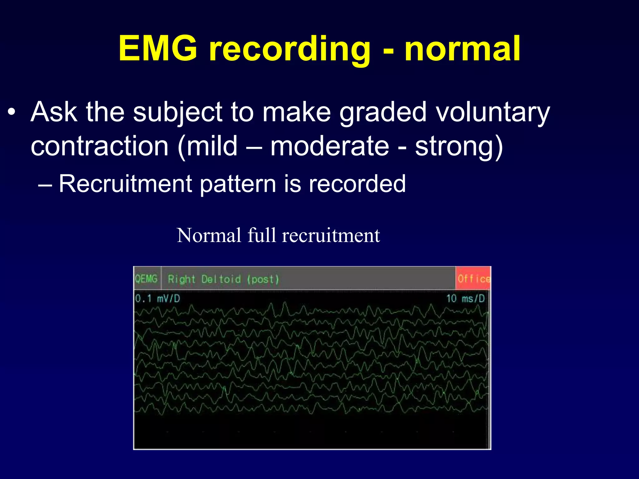
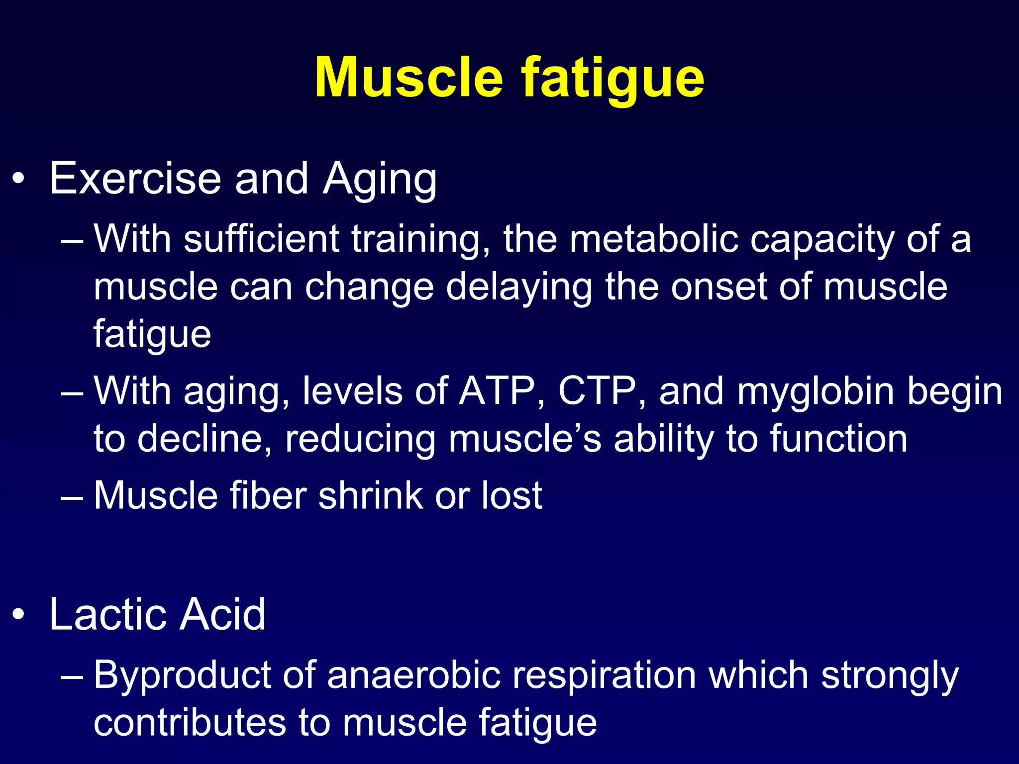
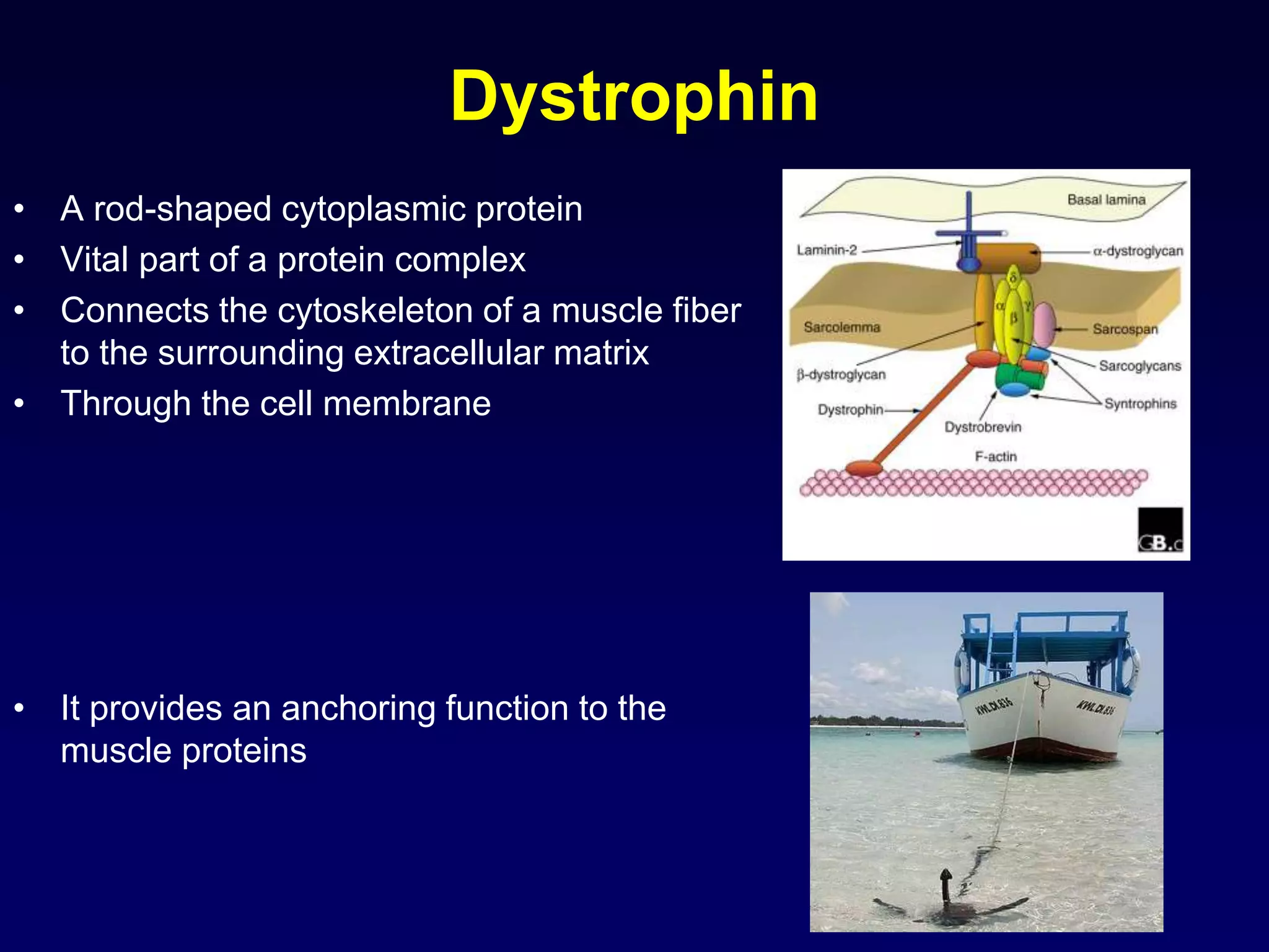
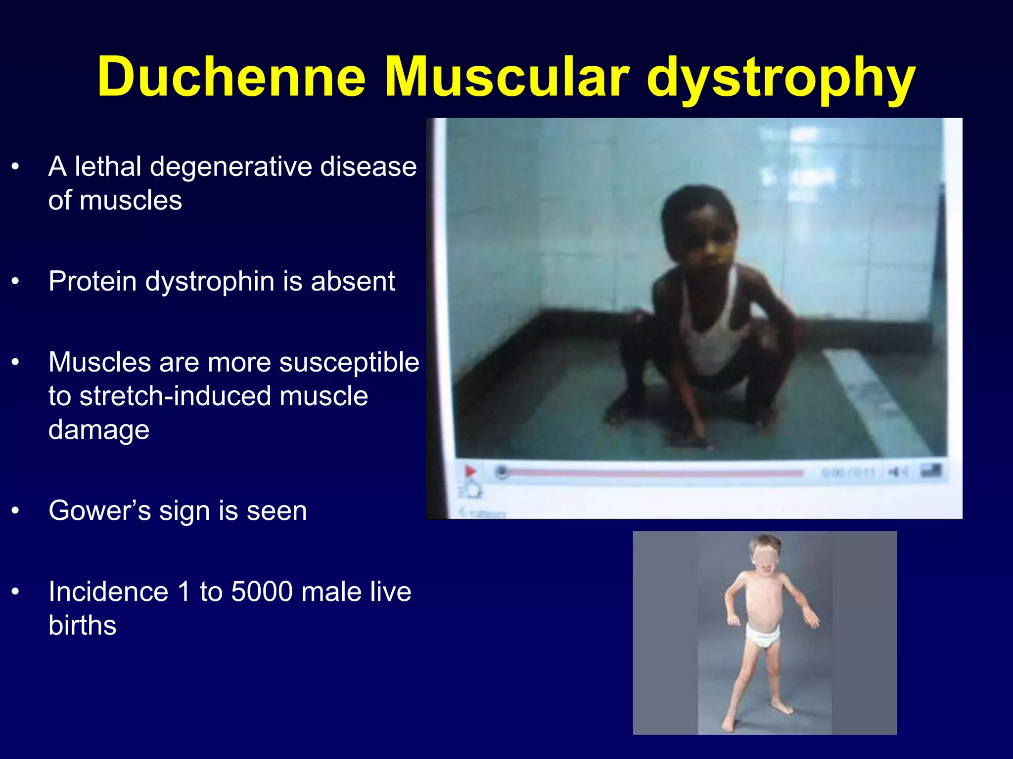
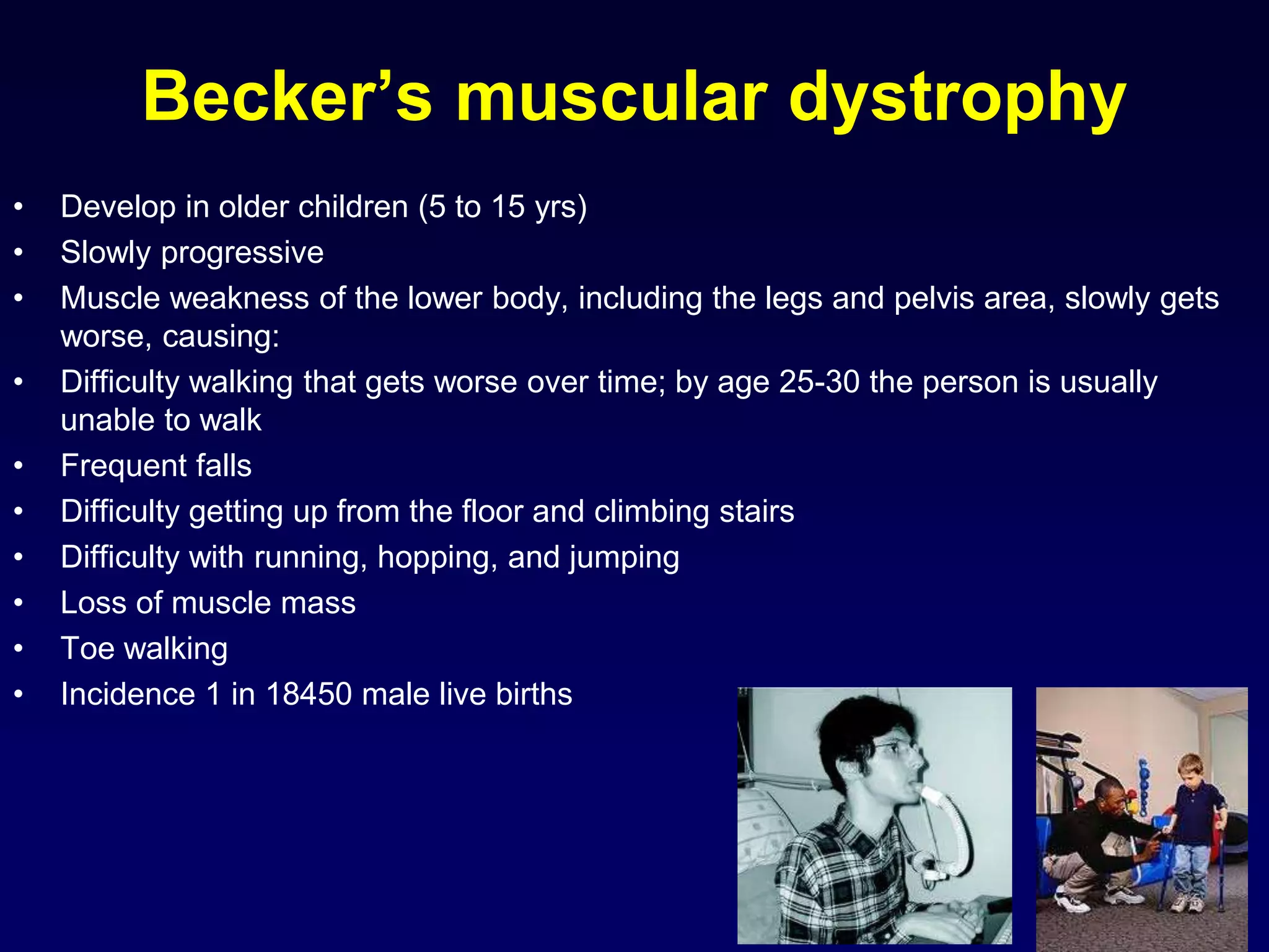
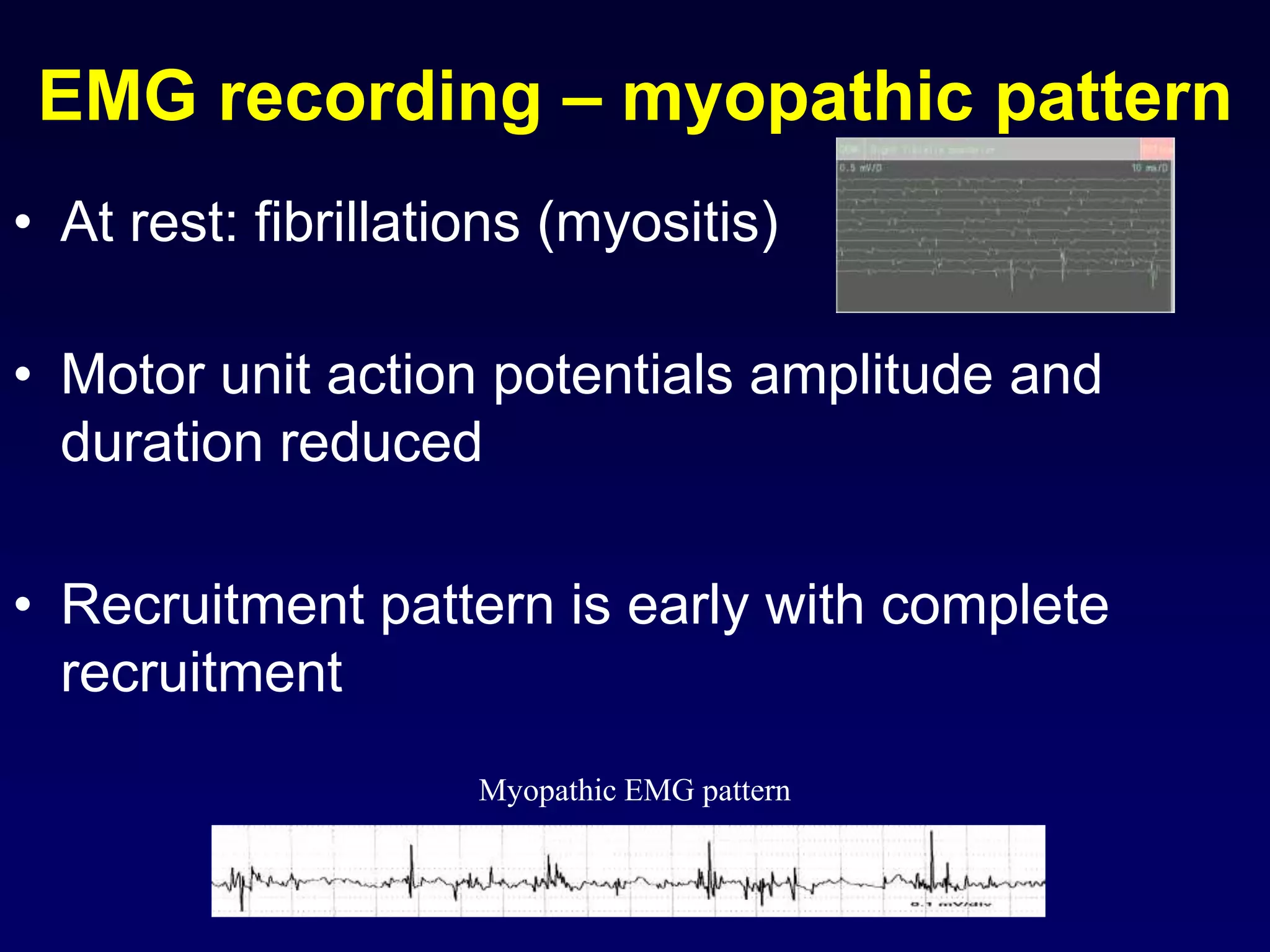
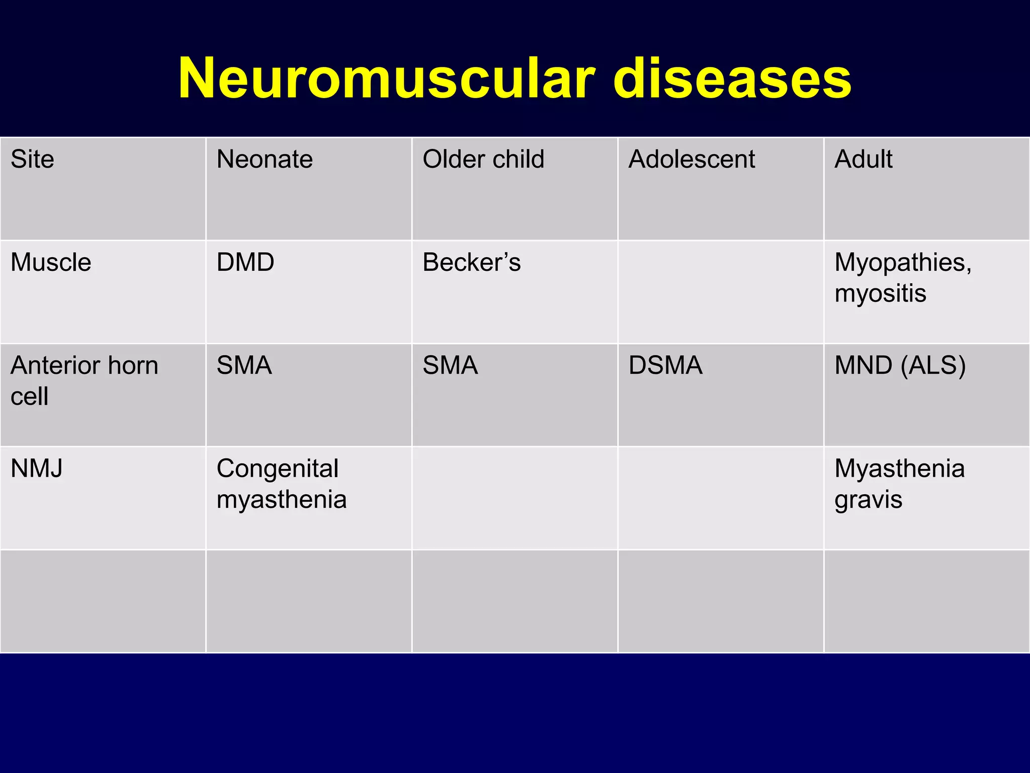
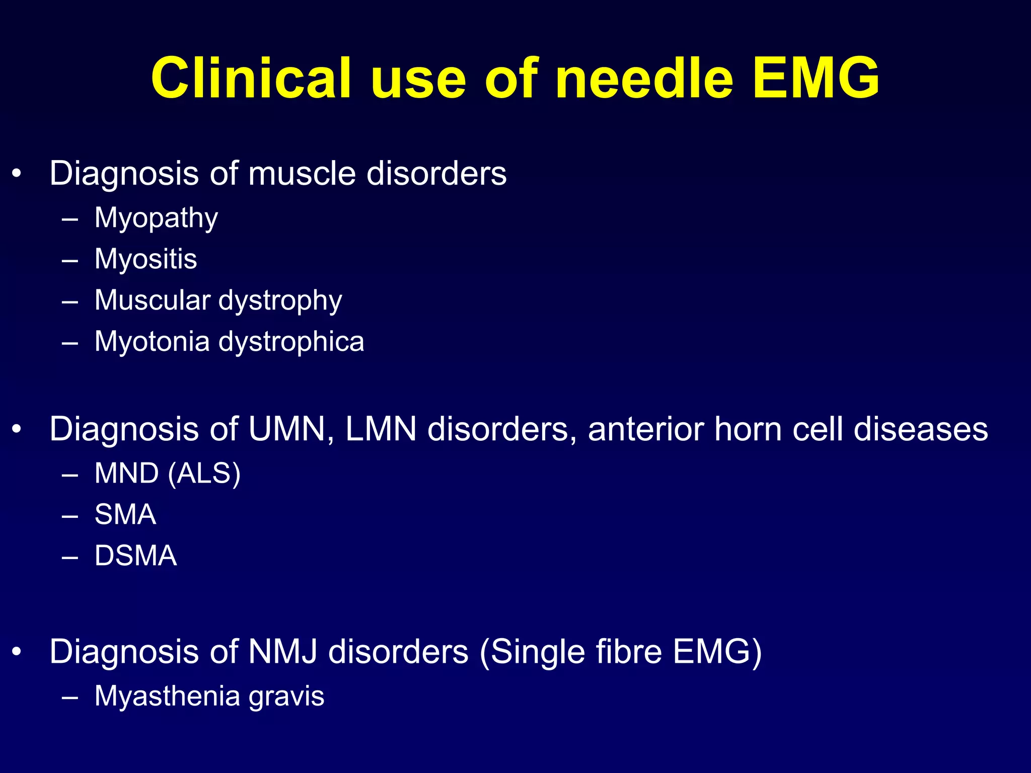
1. The document discusses various mechanisms of muscle dysfunction including disorders at the motor neuron, neuromuscular junction, and muscle levels.
2. Electromyography is described as a key test to detect neuromuscular disorders by examining motor unit recruitment patterns and action potentials.
3. Specific muscle and nerve diseases are outlined like myopathies, motor neuropathies, myasthenia gravis, muscular dystrophies, motor neuron disease and spinal muscular atrophy.
4. The roles, features, causes and electromyography patterns of these diseases are explained to characterize different types of muscle dysfunction.
The presentation focuses on one of the important aspects of Neurophysiology-- The sesnsorimotor integration for planning and execution of movement.
It highlights on the brain regions associated with motor functions, the crosstalk between association areas, hierarchical levels of movement execution and the diseases related to it.
skeletal, cardiac & smooth Muscles by Thiru Murugan.pptxthiru murugan
Unit III – The Muscular System - Anatomy
Types and structure of muscles
Muscle groups
Alterations in disease
Applications and implications in nursing
Muscle:
Muscle is a soft tissue and it is one of the 4 basic tissues, along with nervous tissue, epithelium, and connective tissue.
Muscles helps in movement, support and protection of internal organs.
Muscles can perform variety of functions
Muscles tissue is made up of cells called “MYOCYTES” or muscle fibers.
There are more than 600 muscles in the human body. A kind of elastic tissue makes up each muscle, which consists of thousands, or tens of thousands, of small muscle fibers.
Types of Muscles: There are 3 main types of muscles
Skeletal muscle
Cardiac muscle
Smooth muscle
Skeletal muscle:
These are having close relationship to the bone or skeleton, so called Skeletal muscles
It present in limbs and related body parts & It form about 40% of body weight.
Under microscope the skeletal muscles fibers shows prominent striations, so called “Striated Muscles” & It is also known as “Voluntary Muscles” (movements are under our control)
Structure of Skeletal muscle:
Muscle fibers shows transverse striations under light microscope so it is called “striated muscles”
The nucleus is located peripherally.
Each skeletal muscle is an organ that consists of numerous cells called muscle fibers.
Each muscle fibers surrounded by “ Endomysium”
Inside each skeletal muscle, muscle fibers are organized into bundles, called fascicles, each fascicle surrounded by perimysium.
The whole muscle is covered by “epimysium”
Each skeletal muscle has three layers: endomysium, perimysium and epimysium
Muscle fibers:
Muscle is composed of many long cylindrical-shaped elongated fibres called muscle fibers
Length varies according to the size and shape of the muscles.
The actual arrangement of the fibres depending on the function of the muscle.
Each muscle fibers covered by a membrane is called the sarcolemma.
The cytoplasm of a muscle fiber is called Sarcoplasm
In sarcoplasm there are many mitochondria and bundles of fine longitudinal thread like part is called “myofibrils”
Microscopic structure of myofibrils:
A myofibril (also known as a muscle fibril or sarcostyle) is a basic rod-like part of a muscle cell.
Muscles are composed of tubular cells called myocytes, known as muscle fibres in striated muscle, and these cells in turn contain many chains of myofibrils.
They are created during embryonic development in a process known as myogenesis.
Under light microscope each myofibril consist of 2 bands:
Light band or “I” Band and Dark band or “A” Band
The alternating pattern of these bands results in the striated appearance of skeletal muscle.
Light band or “I” Band:
The I-bands (isotropic in polarized light) appear light in color.
I band divided into 2 portions by a narrow dark line called “Z” line or “Z” Disc.
This “Z” line is formed by protein which does not permit the light.
The part in between 2 “Z” lines called “sarc
NVBDCP.pptx Nation vector borne disease control programSapna Thakur
NVBDCP was launched in 2003-2004 . Vector-Borne Disease: Disease that results from an infection transmitted to humans and other animals by blood-feeding arthropods, such as mosquitoes, ticks, and fleas. Examples of vector-borne diseases include Dengue fever, West Nile Virus, Lyme disease, and malaria.
Muktapishti is a traditional Ayurvedic preparation made from Shoditha Mukta (Purified Pearl), is believed to help regulate thyroid function and reduce symptoms of hyperthyroidism due to its cooling and balancing properties. Clinical evidence on its efficacy remains limited, necessitating further research to validate its therapeutic benefits.
Title: Sense of Taste
Presenter: Dr. Faiza, Assistant Professor of Physiology
Qualifications:
MBBS (Best Graduate, AIMC Lahore)
FCPS Physiology
ICMT, CHPE, DHPE (STMU)
MPH (GC University, Faisalabad)
MBA (Virtual University of Pakistan)
Learning Objectives:
Describe the structure and function of taste buds.
Describe the relationship between the taste threshold and taste index of common substances.
Explain the chemical basis and signal transduction of taste perception for each type of primary taste sensation.
Recognize different abnormalities of taste perception and their causes.
Key Topics:
Significance of Taste Sensation:
Differentiation between pleasant and harmful food
Influence on behavior
Selection of food based on metabolic needs
Receptors of Taste:
Taste buds on the tongue
Influence of sense of smell, texture of food, and pain stimulation (e.g., by pepper)
Primary and Secondary Taste Sensations:
Primary taste sensations: Sweet, Sour, Salty, Bitter, Umami
Chemical basis and signal transduction mechanisms for each taste
Taste Threshold and Index:
Taste threshold values for Sweet (sucrose), Salty (NaCl), Sour (HCl), and Bitter (Quinine)
Taste index relationship: Inversely proportional to taste threshold
Taste Blindness:
Inability to taste certain substances, particularly thiourea compounds
Example: Phenylthiocarbamide
Structure and Function of Taste Buds:
Composition: Epithelial cells, Sustentacular/Supporting cells, Taste cells, Basal cells
Features: Taste pores, Taste hairs/microvilli, and Taste nerve fibers
Location of Taste Buds:
Found in papillae of the tongue (Fungiform, Circumvallate, Foliate)
Also present on the palate, tonsillar pillars, epiglottis, and proximal esophagus
Mechanism of Taste Stimulation:
Interaction of taste substances with receptors on microvilli
Signal transduction pathways for Umami, Sweet, Bitter, Sour, and Salty tastes
Taste Sensitivity and Adaptation:
Decrease in sensitivity with age
Rapid adaptation of taste sensation
Role of Saliva in Taste:
Dissolution of tastants to reach receptors
Washing away the stimulus
Taste Preferences and Aversions:
Mechanisms behind taste preference and aversion
Influence of receptors and neural pathways
Impact of Sensory Nerve Damage:
Degeneration of taste buds if the sensory nerve fiber is cut
Abnormalities of Taste Detection:
Conditions: Ageusia, Hypogeusia, Dysgeusia (parageusia)
Causes: Nerve damage, neurological disorders, infections, poor oral hygiene, adverse drug effects, deficiencies, aging, tobacco use, altered neurotransmitter levels
Neurotransmitters and Taste Threshold:
Effects of serotonin (5-HT) and norepinephrine (NE) on taste sensitivity
Supertasters:
25% of the population with heightened sensitivity to taste, especially bitterness
Increased number of fungiform papillae
Integrating Ayurveda into Parkinson’s Management: A Holistic ApproachAyurveda ForAll
Explore the benefits of combining Ayurveda with conventional Parkinson's treatments. Learn how a holistic approach can manage symptoms, enhance well-being, and balance body energies. Discover the steps to safely integrate Ayurvedic practices into your Parkinson’s care plan, including expert guidance on diet, herbal remedies, and lifestyle modifications.
These lecture slides, by Dr Sidra Arshad, offer a quick overview of the physiological basis of a normal electrocardiogram.
Learning objectives:
1. Define an electrocardiogram (ECG) and electrocardiography
2. Describe how dipoles generated by the heart produce the waveforms of the ECG
3. Describe the components of a normal electrocardiogram of a typical bipolar lead (limb II)
4. Differentiate between intervals and segments
5. Enlist some common indications for obtaining an ECG
6. Describe the flow of current around the heart during the cardiac cycle
7. Discuss the placement and polarity of the leads of electrocardiograph
8. Describe the normal electrocardiograms recorded from the limb leads and explain the physiological basis of the different records that are obtained
9. Define mean electrical vector (axis) of the heart and give the normal range
10. Define the mean QRS vector
11. Describe the axes of leads (hexagonal reference system)
12. Comprehend the vectorial analysis of the normal ECG
13. Determine the mean electrical axis of the ventricular QRS and appreciate the mean axis deviation
14. Explain the concepts of current of injury, J point, and their significance
Study Resources:
1. Chapter 11, Guyton and Hall Textbook of Medical Physiology, 14th edition
2. Chapter 9, Human Physiology - From Cells to Systems, Lauralee Sherwood, 9th edition
3. Chapter 29, Ganong’s Review of Medical Physiology, 26th edition
4. Electrocardiogram, StatPearls - https://www.ncbi.nlm.nih.gov/books/NBK549803/
5. ECG in Medical Practice by ABM Abdullah, 4th edition
6. Chapter 3, Cardiology Explained, https://www.ncbi.nlm.nih.gov/books/NBK2214/
7. ECG Basics, http://www.nataliescasebook.com/tag/e-c-g-basics
Tom Selleck Health: A Comprehensive Look at the Iconic Actor’s Wellness Journeygreendigital
Tom Selleck, an enduring figure in Hollywood. has captivated audiences for decades with his rugged charm, iconic moustache. and memorable roles in television and film. From his breakout role as Thomas Magnum in Magnum P.I. to his current portrayal of Frank Reagan in Blue Bloods. Selleck's career has spanned over 50 years. But beyond his professional achievements. fans have often been curious about Tom Selleck Health. especially as he has aged in the public eye.
Follow us on: Pinterest
Introduction
Many have been interested in Tom Selleck health. not only because of his enduring presence on screen but also because of the challenges. and lifestyle choices he has faced and made over the years. This article delves into the various aspects of Tom Selleck health. exploring his fitness regimen, diet, mental health. and the challenges he has encountered as he ages. We'll look at how he maintains his well-being. the health issues he has faced, and his approach to ageing .
Early Life and Career
Childhood and Athletic Beginnings
Tom Selleck was born on January 29, 1945, in Detroit, Michigan, and grew up in Sherman Oaks, California. From an early age, he was involved in sports, particularly basketball. which played a significant role in his physical development. His athletic pursuits continued into college. where he attended the University of Southern California (USC) on a basketball scholarship. This early involvement in sports laid a strong foundation for his physical health and disciplined lifestyle.
Transition to Acting
Selleck's transition from an athlete to an actor came with its physical demands. His first significant role in "Magnum P.I." required him to perform various stunts and maintain a fit appearance. This role, which he played from 1980 to 1988. necessitated a rigorous fitness routine to meet the show's demands. setting the stage for his long-term commitment to health and wellness.
Fitness Regimen
Workout Routine
Tom Selleck health and fitness regimen has evolved. adapting to his changing roles and age. During his "Magnum, P.I." days. Selleck's workouts were intense and focused on building and maintaining muscle mass. His routine included weightlifting, cardiovascular exercises. and specific training for the stunts he performed on the show.
Selleck adjusted his fitness routine as he aged to suit his body's needs. Today, his workouts focus on maintaining flexibility, strength, and cardiovascular health. He incorporates low-impact exercises such as swimming, walking, and light weightlifting. This balanced approach helps him stay fit without putting undue strain on his joints and muscles.
Importance of Flexibility and Mobility
In recent years, Selleck has emphasized the importance of flexibility and mobility in his fitness regimen. Understanding the natural decline in muscle mass and joint flexibility with age. he includes stretching and yoga in his routine. These practices help prevent injuries, improve posture, and maintain mobilit
- Video recording of this lecture in English language: https://youtu.be/kqbnxVAZs-0
- Video recording of this lecture in Arabic language: https://youtu.be/SINlygW1Mpc
- Link to download the book free: https://nephrotube.blogspot.com/p/nephrotube-nephrology-books.html
- Link to NephroTube website: www.NephroTube.com
- Link to NephroTube social media accounts: https://nephrotube.blogspot.com/p/join-nephrotube-on-social-media.html
Rasamanikya is a excellent preparation in the field of Rasashastra, it is used in various Kushtha Roga, Shwasa, Vicharchika, Bhagandara, Vatarakta, and Phiranga Roga. In this article Preparation& Comparative analytical profile for both Formulationon i.e Rasamanikya prepared by Kushmanda swarasa & Churnodhaka Shodita Haratala. The study aims to provide insights into the comparative efficacy and analytical aspects of these formulations for enhanced therapeutic outcomes.
1. Mechanisms of
dysfunction of
muscles
Prof. Vajira Weerasinghe
Professor of Physiology, Faculty of Medicine
and Consultant Neurophysiologist, Teaching Hospital, Peradeniya
Lecture is available at www.slideshare.net/vajira54
2. Objectives
1. Recall the physiology of the motor unit and its
neural control
2. Outline how disorders at different levels in the
control mechanisms affect muscle function
3. Site of lesions
UMN & LMN
Motor nerve
NMJ
Muscle
Motor neuron disease
Diseases
Motor neuropathy
Myasthenia gravis
Myopathy
Site
4. Motor unit
• muscle contraction occurs in
terms of motor units rather
than by single muscle fibres
• a motor unit is defined as
– anterior horn cell
– motor neuron
– muscle fibres supplied by the
neuron
5. Principle of recruitment of motor units
• Increase in the tension of a muscle is due to
progressive recruitment of motor units
• eg.
– Mild contraction – few motor units are recruited – mild
tension
– Moderate contraction – many motor units are recruited –
moderate tension
– Strong contraction – all the motor units are recruited –
maximum tension
6. Electromyography (EMG)
• This is a neurophysiological test done in
order to detect many neuromuscular
disorders
• Recording electrodes are needles (EMG
needles)
• They contain cathode and anode in the
form of a needle
• This is inserted into the muscle
• Motor unit recording pattern is recorded
visually in the screen and sound pattern is
recorded from a loudspeaker
• Motor unit action potentials (MUAP) can
be recorded
7. EMG recording - normal
• At rest
– No activity (screen or
sound)
• Ask the subject to make
a voluntary contraction
– Motor unit action
potentials amplitude and
duration are calculated
Normal resting
Motor unit action potentials (MUAP)
8. EMG recording - normal
• Ask the subject to make graded voluntary
contraction (mild – moderate - strong)
– Recruitment pattern is recorded
Normal full recruitment
10. Muscle fatigue
• Decline in muscle force generated over
sustained periods of activity or due to
pathological issues
• Causes
– Ion imbalance within muscle
– Nervous Fatigue and Loss of Desire
– Metabolic Fatigue
– Exercise and Aging
– Lactic Acid Accumulation
11. Muscle fatigue
Ion Imbalance
– Contraction of a muscle requires Ca ions to interact
with troponin, exposing the actin binding site to
myosin head
– Osmotically active molecules outside the muscles
are lost through sweating
– Difficult for the required Ca ions to be delivered to
muscle fiber
12. Muscle fatigue
• Nervous Fatigue and Loss Of Desire
– Nerves are responsible for controlling the
contraction and force of muscles
• Metabolic Fatigue
– Accumulation of metabolites from Mg ions, induce
fatigue by interfering with release of Ca ions
– Reduction in sensitivity of troponin to Ca ions
13. Muscle fatigue
• Exercise and Aging
– With sufficient training, the metabolic capacity of a
muscle can change delaying the onset of muscle
fatigue
– With aging, levels of ATP, CTP, and myglobin begin
to decline, reducing muscle’s ability to function
– Muscle fiber shrink or lost
• Lactic Acid
– Byproduct of anaerobic respiration which strongly
contributes to muscle fatigue
14. Muscle cramps
• At one time or another most athletes will experience a muscle cramp
• Muscle cramps are involuntary
• Usually a painful contraction of skeletal muscle
• Muscle cramps will usually occur without warning
• Muscle cramps are usually a warning to an athlete as the muscle cramp is
often a symptom of a more serious physical issue that requires attention
15. Muscle cramps
• Some common causes of muscle cramps include
– fatigue
– strenuous exercise
– overuse of particular muscle groups
– a failure to stretch or properly warm up prior to activity
– dehydration and the related problem of sodium deficiency
– low blood sugar (glucose) levels
– magnesium deficiency
– calcium deficiency
– the presence of the hydrogen ion that is a byproduct of lactic acid formation in working
muscles
– thyroid gland irregularity
– kidney dysfunction
– side effects of certain medications
– neuromuscular disorder
• In order for an athlete to prevent a muscle cramp occurring the causes
above should be monitored continuously.
16.
17. Muscle strain
• A muscle strain is damage caused by
overstretching the muscle
23. Dystrophin
• A rod-shaped cytoplasmic protein
• Vital part of a protein complex
• Connects the cytoskeleton of a muscle fiber
to the surrounding extracellular matrix
• Through the cell membrane
• It provides an anchoring function to the
muscle proteins
24. Duchenne Muscular dystrophy
• A lethal degenerative disease
of muscles
• Protein dystrophin is absent
• Muscles are more susceptible
to stretch-induced muscle
damage
• Gower’s sign is seen
• Incidence 1 to 5000 male live
births
25. Becker’s muscular dystrophy
• Develop in older children (5 to 15 yrs)
• Slowly progressive
• Muscle weakness of the lower body, including the legs and pelvis area, slowly gets
worse, causing:
• Difficulty walking that gets worse over time; by age 25-30 the person is usually
unable to walk
• Frequent falls
• Difficulty getting up from the floor and climbing stairs
• Difficulty with running, hopping, and jumping
• Loss of muscle mass
• Toe walking
• Incidence 1 in 18450 male live births
26. EMG recording – myopathic pattern
• At rest: fibrillations (myositis)
• Motor unit action potentials amplitude and
duration reduced
• Recruitment pattern is early with complete
recruitment
Myopathic EMG pattern
27. Myotonia
• Two types
– Myotonia dystrophica
– Myotonia congenita
• Contraction is normal, relaxation is slow
– Depolarisation is normal
– Repolarisation will not take place normally
• Due to derangement of electrical activity in the muscle
membrane
– K+ Cl- channel derangements
• Channelopathy
28.
29. EMG recording – myotonia
• Rest:
– “Dive bomber sound”
– Prolonged continuous activity
– Triggered by needle position or percussion
31. MND (Motor neuron disease)
or ALS (amyotrophic lateral sclerosis)
• Affect adults (after 40 years)
• Features include weakness and wasting of limb muscles, tongue fasciculations,
dysarthria, dysphagia
• Slowly progressive and poor prognosis
• Both upper motor neuron and lower motor neuron are affected
• No cure or treatment
• Incidence 2-6 per 100,000
32. EMG recording – MND pattern
• At rest: fibrillations and fasciculations
• Motor unit action potentials amplitude
and duration increased, giant motor
units
• Recruitment pattern is reduced
Giant
motor units
33. SMA (Spinal muscular atrophy)
• Anterior horn cell disease
• Affect infants and older children
• Poor prognosis
• Muscle weakness and wasting
• Caused by an abnormal or missing gene responsible for the production of a
protein essential to motor neurons
• Incidence 1 in 10,000 live births
• Several types are present SMA type I, II and III
• Type I (Werdnig-Hoffman disease or infantile-onset SMA)
– Evident at birth or within the first few months
– Symptoms include floppy limbs and trunk, feeble movements of the arms and legs,
swallowing and feeding difficulties, and impaired breathing
• Type II (the intermediate form)
– Usually begins 6 and 18 months of age. Legs tend to be more impaired than arms
– Children with Type II may able to sit and some may be able to stand or walk with help
• Type III (Kugelberg-Welander disease)
– Appear between 2 and 17 years of age and include difficulty running, climbing steps, or rising
from a chair
34. DSMA (Distal spinal muscular atrophy)
• Anterior horn cell disease
• Affect adolesecents
• Main feature is a wasting of small muscles of the hand
• Non-progressive and benign
DSMA (distal spinal muscular atrophy)