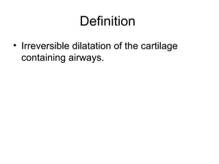
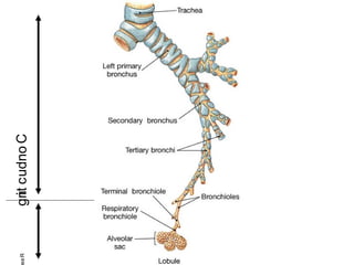
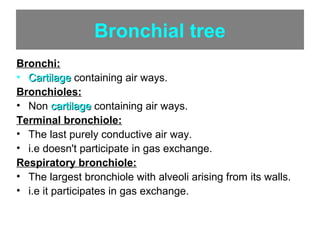
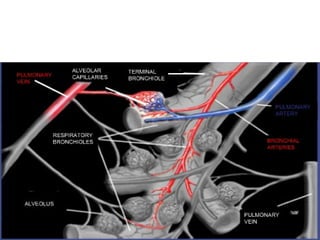
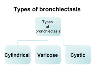
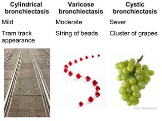
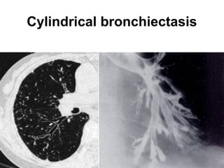
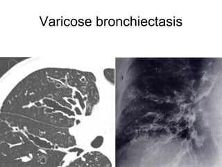
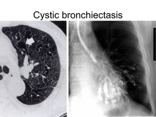
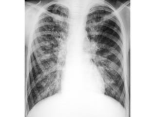
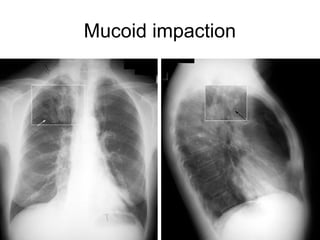
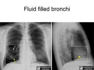
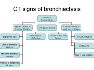
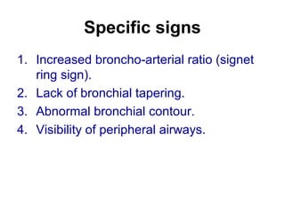
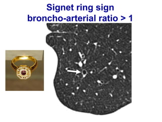
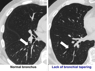
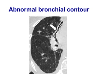
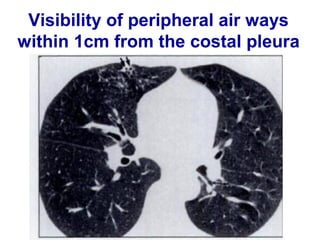
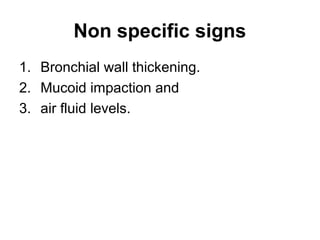
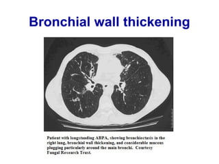
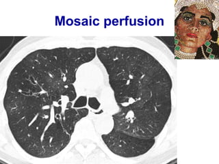
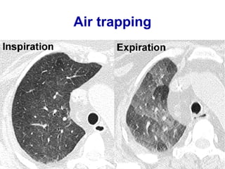
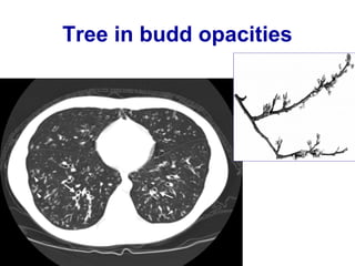
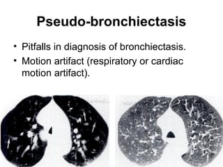
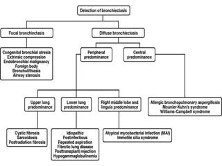
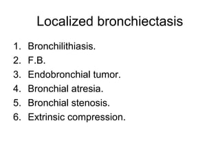
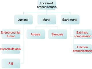
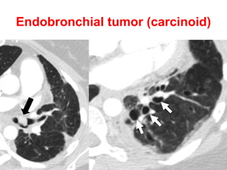
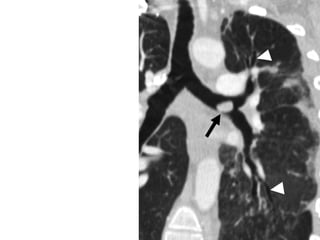
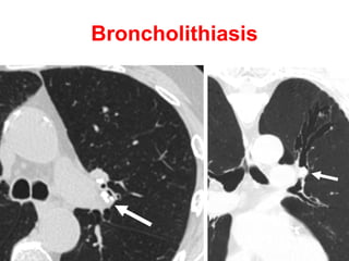
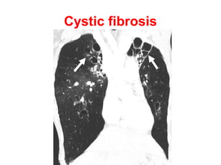
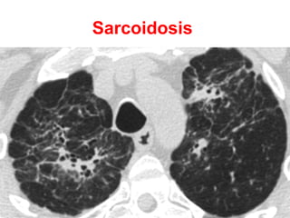
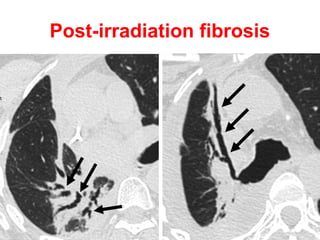
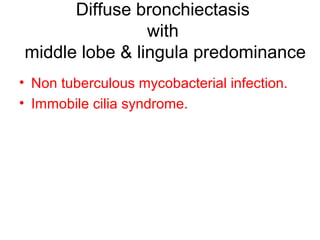
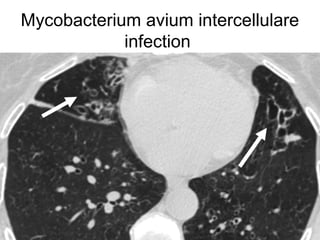
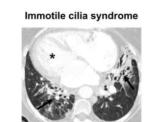
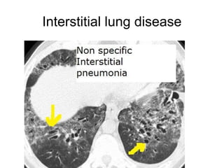
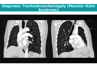
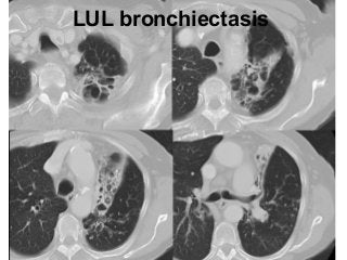
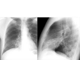
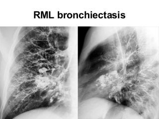
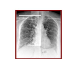
Bronchiectasis
Dr / Hytham Nafady
Definition
• Irreversible dilatation of the cartilage
containing airways.
Bronchial tree
Types of bronchiectasis
Cylindrical
bronchiectasis
Varicose
bronchiectasis
Cystic
bronchiectasis
Mild Moderate Sever
Tram track
appearance
String of beads Cluster of grapes
Cylindrical bronchiectasis
Varicose bronchiectasis
Cystic bronchiectasis
Plain radiographic signs of
bronchiectasis
Mucoid impaction
Fluid filled bronchi
CTsigns of bronchiectasis
•Signet ring sign (broncho-arterial ratio > 1).
•Lack of bronchial tapering.
•Abnormal bronchial contour.
•Visibility of peripheral air ways.
Signet ring sign
broncho-arterial ratio > 1
Normal bronchus Lack of bronchial tapering
Abnormal bronchial contour
Visibility of peripheral air ways
within 1cm from the costal pleura
• Peribronchial cuffing (thickened hazy bronchial
wall).
• Finger in glove opacities (mucus filled bronchi).
• Multiple air fluid levels (fluid filled bronchi).
Bronchial wall thickening
Mucoid impaction
Multiple air fluid levels
• Mosaic perfusion.
• Air trapping.
• Tree in bud opacities.
Mosaic perfusion
Air trapping
Tree in budd opacities
Pseudo-bronchiectasis
• Pitfalls in diagnosis of bronchiectasis.
• Motion artifact (respiratory or cardiac
motion artifact).
Endobronchial tumor
(carcinoid)
Broncholithiasis
Bronchial atresia
Bronchial stenosis
Post-irradiation fibrosis
(traction bronchiectasis)
Diffuse bronchiectasis
with central predominance
• ABPA.
• Mounier Kuhn syndrome.
• William Campbell syndrome.
ABPA
Mounier Kuhn syndrome
tracheobronchomegaly
William Campbell syndrome
Congenital cystic bronchiectasis that results from a
deficiency of cartilage in the 4th
to 6th
order bronchi
Diffuse bronchiectasis
with upper lobe predominance
• Cystic fibrosis.
• Sarcoidosis.
Cystic fibrosis
Sarcoidosis
Diffuse bronchiectasis
with middle lobe & lingula predominance
• Non tuberculous mycobacterial infection.
• Immobile cilia syndrome.
Mycobacterium avium
intercellulare infection
Immotile cilia syndrome
Bronchiectasis
with lower lobe predominance
1. Idiopathic (40% of cases).
2. Recurrent childhood infection.
3. Repeated aspiration.
4. Interstitial lung disease.
Interstitial lung disease
Case
• 65-year-old man with a chronic productive
cough and progressive dyspnea on
exertion.
Atlas of bronchiectasis
Bronchietasis
Lack of bronchial
tapering
+
Tree in bud
opacities
LUL bronchiectasis
RML bronchiectasis
Bronchiectasis
Bronchiectasis
Bronchiectasis

Bronchiectasis