Downloaded 139 times

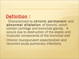
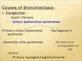

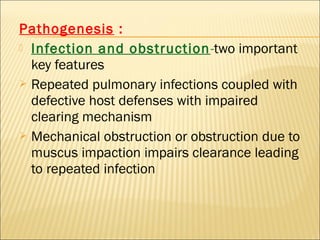

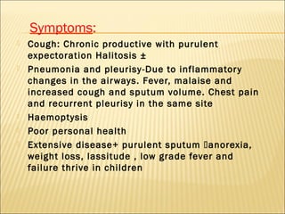
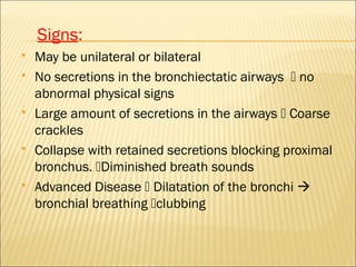
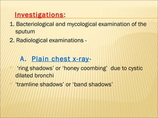


Bronchiectasis is a chronic lung condition characterized by abnormal dilatation of the bronchi. It occurs due to destruction of the elastic and muscular components of the bronchial wall from repeated pulmonary infections. Common causes include cystic fibrosis, pneumonia, tuberculosis, and allergic bronchopulmonary aspergillosis. Symptoms include chronic cough with purulent sputum, pneumonia, hemoptysis, and poor health. Diagnosis involves sputum culture, chest x-ray, and high-resolution CT scan of the chest. Management includes chest physiotherapy, antibiotics, and sometimes surgery for uncontrolled infections or hemorrhage. Complications can include recurrent lung infections, abscesses, and respiratory failure.


![PERI-PROSTHETIC FRACTURE NAIL-PLATE CONSTRUCT [NPC].pptx](https://cdn.slidesharecdn.com/ss_thumbnails/drarunkumardrmohamedashrafperiprostheticfrasturenail-plateconstructnpc-260209164459-7e9d15a1-thumbnail.jpg?width=640&height=640&fit=bounds)


