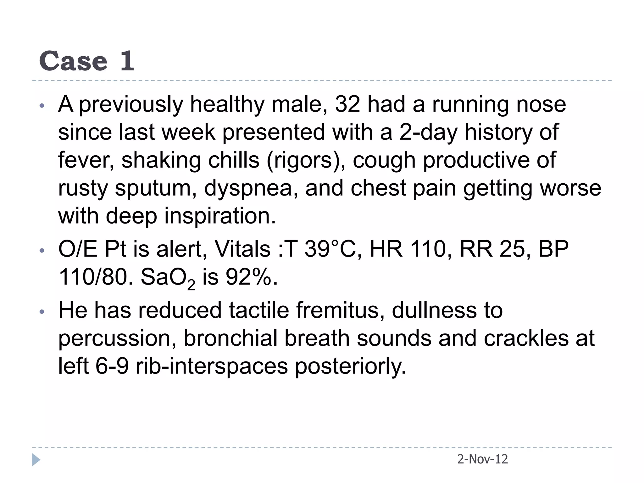
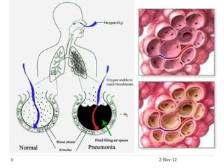
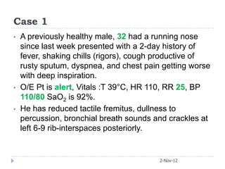
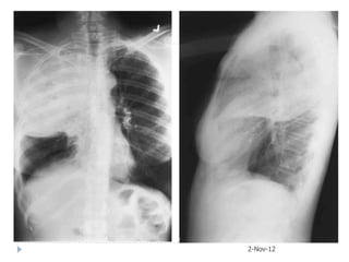
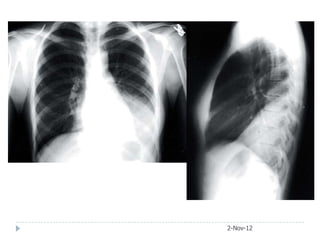
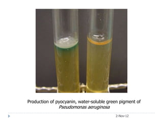
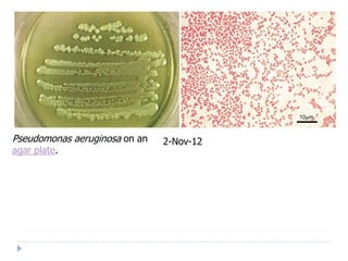
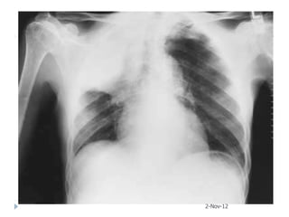
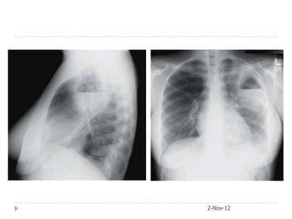
Case 1
•   A previously healthy male, 32 had a running nose
    since last week presented with a 2-day history of
    fever, shaking chills (rigors), cough productive of
    rusty sputum, dyspnea, and chest pain getting worse
    with deep inspiration.
•   O/E Pt is alert, Vitals :T 39°C, HR 110, RR 25, BP
    110/80. SaO2 is 92%.
•   He has reduced tactile fremitus, dullness to
    percussion, bronchial breath sounds and crackles at
    left 6-9 rib-interspaces posteriorly.


                                         2-Nov-12
2-Nov-12
What is your diagnosis?


                          2-Nov-12
Definition

 “ACUTE RESPIRATORY ILLNESS ASSOCIATED
   WITH PYREXIA AND COUGH, AND
   RECENTLY DEVELOPED RADIOGRAPHIC
   SIGNS OF CONSOLIDATION OF A PART OR
   PARTS OF ONE OR BOTH LUNGS”


 •   The commonest infectious cause of death
 •   Most of mortality at extremes of ages
 •   Most cases are treatable if diagnosed and
     treated with appropriate antibiotics
                                    2-Nov-12
2-Nov-12
How severe is our patient’s pneumonia?


                           2-Nov-12
Applying CURB-65 Rule


                        2-Nov-12
Case 1
•   A previously healthy male, 32 had a running nose
    since last week presented with a 2-day history of
    fever, shaking chills (rigors), cough productive of
    rusty sputum, dyspnea, and chest pain getting worse
    with deep inspiration.
•   O/E Pt is alert, Vitals :T 39°C, HR 110, RR 25, BP
    110/80 SaO2 is 92%.
•   He has reduced tactile fremitus, dullness to
    percussion, bronchial breath sounds and crackles at
    left 6-9 rib-interspaces posteriorly.


                                         2-Nov-12
Classification of pneumonias

• BRONCHO-pneumonia
• LOBAR pneumonia
 • SEGMENTAL pneumonia
 • SUBSEGMENTAL pneumonia
And
 ‘Double’ pneumonia


                            2-Nov-12
2-Nov-12
Distribution of lung involvement in
lobar pneumonia and bronchopneumonia
                              2-Nov-12
2-Nov-12
Classification of pneumonias
•   COMMUNITY ACQUIRED (CAP)
•   HOSPITAL ACQUIRED (HAP) (Nosocomial)
•   VENTILATOR ASSOCIATED (VAP)
•   Healthcare-associated (HCAP)
•   ASPIRATION (Alcoholics/epileptics/comatosed)
•   IMMUNOCOMPROMISED PATIENT (PICP) OR
    (HIV – associated)


                                      2-Nov-12
Classification of pneumonias
•   Hospital-acquired (or nosocomial) pneumonia (HAP) is
    pneumonia that occurs 48 hours or more after admission and
    did not appear to be incubating at the time of admission.
•   Ventilator-associated pneumonia (VAP) is a type of HAP
    that develops more than 48 to 72 hours after endotracheal
    intubation.

•   Healthcare-associated pneumonia (HCAP) is defined as
    pneumonia that occurs in a non-hospitalized patient with
    extensive healthcare contact, as defined by one or more of the
    following:
    o Hospitalization in an acute care hospital for two or more days
        within the prior 90 days
    o   Intravenous therapy, wound care, or intravenous chemotherapy
        within the prior 30 days
    o   Attendance at a hospital or hemodialysis clinic within the prior 30
        days
    o   Residence in a nursing home or other long-term 2-Nov-12
                                                         care facility
CAP - Microbiology
Typical CAP                Atypical CAP
 Strept. Pneumoniae        Influinza + other viruses
 Hemophilus influinzae     Mycoplasma
 Legionella pneumophila     pneumoniae
 Staphylococcus aureus     Legionella pneumophila

 Gram negative bacilli     Chlamydia Pneumoniae

 Moraxella catarrhalis     Chlamydia psittaci
                            Coxiella burneti


                                       2-Nov-12
CAP - Presentation
Typical CAP                     Atypical CAP
 an abrupt onset,               a progressive onset,
 high fever, chills,            fever without chills,
 productive cough,              dry cough,
 thoracic pain,                 headache, myalgia,
 focal clinical signs,          diffuse crackles,
 lobar or segmental             interstitial infiltrates on chest
  radiographic findings,          radiograph,
 leukocytosis, and              modest leukocytosis,
 sputum Gram stain that is      sputum Gram stain (and
  positive for                    possibly culture) that is
  bacteria, frequently of a       negative for bacteria
  single predominant type.       Mostly due to intracellular
 Mostly due to extracellular     bacteria or to viruses.
  bacteria such as S.
  pneumoniae, Staph
  aureus, and H. influenzae.                    2-Nov-12
Other Pneumonias - Microbiology
What are the differences?
HAP, VAP, and HCAP may be caused by
• Specific pathogens and can be polymicrobial.
• Common pathogens include
 •   Aerobic gram-negative bacilli (eg, Escherichia coli,
     Klebsiella pneumoniae, Pseudomonas aeruginosa,
     Enterobacter spp, Acinetobacter spp)
 •   Gram-positive cocci (eg, Staphylococcus aureus,
     including MRSA, Streptococcus spp).
 •   Viruses or fungi are significantly less common
• Organisms may be multi-drug resistant
                                            2-Nov-12
Case 2
•   A heavy smoker bank accountant of 46 years
    presented with high grade fever, worsening cough,
    little rusty sputum production and Rt pleuritic chest
    pain after a visit to Skardu two days ago (in the
    month of January).
•   O/E confused, RR 36/min, T 102 OF, P 110/min, BP
    80/60, Hb 16.8 g/dl, TLC 18000 (88% N), Urea 32
    mg/dl (5.3 mmol/L) Sputum Smear showed Gram
    Positive diplococci


                                            2-Nov-12
`


    2-Nov-12
Streptococcus pneumoniae are Gram-positive, lancet-shaped cocci in
couples
                                 Streptococcus pneumoniae
                                 A mucoid strain on blood agar
                                 showing alpha hemolysis (green
                                 zone surrounding colonies). Note
                                 the zone of inhibition around a
                                 filter paper disc impregnated with
                                 optochin. Viridans streptococci
                                 are not inhibited by optochin.
                                                  2-Nov-12
Streptococcus pneumoniae
Quellung (capsular swelling)
reaction can be used to
demonstrate the presence of
a specific capsular type of the
bacterium.


                                  2-Nov-12
Case 3
•   An 18 years old previously healthy medical student
    living in college hostel developed worsening dry
    cough with high grade pyrexia for three days. Two
    days ago he developed severe pain in Rt ear.
•   O/E Alert, Mildly Jaundiced, P: 120/min RR:32/min, T
    101OF, BP: 110/80
•   Labs: Hb 13.4, TLC 8800, Normal Dif. S Bil 2.5
    mg/dl ALT 34 iu/L, Urea 4 mmol/L


                                          2-Nov-12
2-Nov-12
2-Nov-12
2-Nov-12
2-Nov-12
Mycoplasma Pneumonia
Extrapulmonary manifestations
   CAHA (immune                   Arthralgia
    hemolytic anemia)              Cervical
   Meningitis,                     lymphadenopathy,
    meningoencephalitis            Bullous myringitis
   Myalgia                        Diarrhea
   Myocarditis, Pericarditis      Nausea and vomiting
   Skin eruptions                 Hepatitis
    (EM/SJS)


                                             2-Nov-12
Case 4
•   Male, 60, admitted to hospital with 2-wk H/O
    myalgia, headache, dyspnoea and cough without
    sputum.
•   O/E severely ill, cyanosed and delirious with
    T:39°C;      P:110/min,      RR:40/min       and
    BP:110/60mmHg.                    PO2:43mmHg,
    PCO2:37mmHg, TLC 4600/cumm and urea 4
    mmol/L.
•   He had received amoxicillin for 6 days before
    admission without improvement. CXR shows
    extensive bilateral multilobar consolidation. He
    kept birds as a hobby and one of his budgerigars
                                       2-Nov-12
The clinical
       diagnosis of
    psittacosis was
      subsequently
      confirmed by
serology tests. He
   was treated with
        intravenous
fluids, oxygen and
   tetracycline and
    recovered fully


    2-Nov-12
CT scan of the chest demonstrates patchy multifocal ground glass
attenuation opacities (arrows).
                                                2-Nov-12
Case 5
   65-year-old female presented with acute respiratory
    failure. She had been sick for two weeks with fever,
    confusion, diarrhea, cough, and purulent sputum
    production. Her medical checkup two months ago
    was unremarkable.
   Urea 11 mmol/L, Creatinine 3.2
   CXR and later CT chest obtained


                                           2-Nov-12
2-Nov-12
After3 weeks course of macrolide


                             2-Nov-12
Investigations in CAP
ROUTINE INVESTIGATIONS IN CAP
• Chest X-ray +/- CT chest
• Blood: CBC with reticulocytes
• Tests for microbiological identification:


                                       2-Nov-12
Differential diagnosis
PULMONARY INFARCTION
PULMONARY/PLEURAL TUBERCULOSIS
PULMONARY EDEMA
NEOPLASTIC DISEASE
SUBDIPHRAGMATIC INFLAMMATION
 Subphrenic abscess, Amebic liver abscess,
  cholycystitis, pancreatitis
RARE CONDITIONS
 Pulmonary eosinophilia, connective tissue
  diseases, Wegener’s granuloma

                                   2-Nov-12
Approach to a patient with CAP

  History               Examination           Chest x-ray

                        PNEUMONIA
                         diagnosed

                       CLUES TO LIKELY
                         PATHOGEN
•Environmental clues              •Bird or animal contact
•Season                           •Immunosuppression?
•Recent hotel stay                •Co-morbidity
•Current epidemic                 •Age


                                            2-Nov-12
Case 6
   A 34-year-old woman is admitted with a history of
    fever, chills, and reddish sputum like red currant
    jelly for 10 days. She is on pulse steroid therapy
    for lupus nephritis.
   On physical examination, pulse 113 bpm;
    temperature 101°F; respirations 35/min; blood
    pressure 110/78 mm Hg. She looks ill and has
    crackles in the right upper lung field.
   Lab data: Hb 12 g/dL; WBCs 25.0/μL; N 92%
    BUN 8 mmol/L; creatinine 1.7 mg/dL

                                        2-Nov-12
2-Nov-12
2-Nov-12
Klebsiella pneumoniae on a
MacConkey agar plate.


                             2-Nov-12
Klebsiella pneumonia. Downward bulging of the minor fissure (arrow)
due to massive enlargement of the right upper lobe with inflammatory
exudate.
                                                   2-Nov-12
Case 7
•   Male, 65, known diabetic with h/o 30 cpy
    presents with a 4-day history of productive cough
    with greenish sputum and shortness of breath.
    He has left-sided chest pain that is worse with
    deep inspiration and complained of fever and
    chills on the day of admission.
•   On physical exam, he has a temperature of
    103°F; pulse 120 bpm; respirations 32/min; BP
    100/68.
•   Lungs: increased tactile vocal fremitus with
    bronchial breath sounds on the left side
    posteriorly.                          2-Nov-12
2-Nov-12
Production of pyocyanin, water-soluble green pigment of
               Pseudomonas aeruginosa
                                          2-Nov-12
Pseudomonas aeruginosa on an   2-Nov-12
agar plate.
Case 8
A 53-year-old man with a bone marrow transplant
  presented with one month hitory of dry cough and
  low grade pyrexia. CXR and CT scan show bilateral
  dense airspace and ground-glass opacities
  associated with airway dilatation. The distribution is
  predominantly central and upper lung.


                                         2-Nov-12
2-Nov-12
PCP pneumonia in a young HIV-      CT scan of the chest demonstrates
positive patient.                  cystic air spaces of varying sizes that
CXR demonstrates                   are consistent with pneumatoceles.
predominantly central airspace
disease with peripheral sparing.


                                                       2-Nov-12
Pneumonia in the
immunocompromised host
Mechanism       Cause                    Organisms
                 Marrow Aplasia         Staph. aureus
Neutropenia      AMM                    Gram negative bacteria
                 Marrow infiltration    Candida/ Aspergillus
                   AIDS                 Strept. pneumoniae
T cell defect      CLL                  H. influinzae
                   Lymphoma             Staph. aureus
                   Immunosuppressants   Gram negative bacteria
                   BMT                  Pneumocystis carinii
                   Splenectomy          Myco. tuberculosis
                   CLL                  Strept. pneumoniae
 Antibody          Myeloma              H. influinzae
production


                                               2-Nov-12
Case 9
   A 56-year-old male non-smoker is admitted with
    shortness of breath, right sided chest pain, and
    productive cough. He has a history of seizure
    disorder and is on anticonvulsants. Phenytoin
    level is within therapeutic range.
   On examination, there is dullness to percussion
    in the right upper chest with decreased breath
    sounds. Sputum for AFB and fungi are negative
    on initial smear and cultures are pending.


                                        2-Nov-12
2-Nov-12
Case 10
   A 29-year-old man is admitted with cough, rusty
    sputum production, fever, chills, and decreased
    O2 saturation.
   His chest x-ray shows a right upper lobe
    nonhomogeneous opacity. He is treated with IV
    antibiotics but does not improve. On the fifth
    hospital day, CXR is repeated


                                       2-Nov-12
1: 15.09.2008 2: 20.09.2008
2-Nov-12
3: 12.10.2008
2-Nov-12
                4: 22.10.2008
Complications of pneumonia
   Respiratory failure
   Septicemia/ hypotension: ARF
   Atrial fibrillation
   Pleural effusion/ empyema
   Lung abscess
   Jaundice
   Pericarditis/myocarditis/endocarditis


                                            2-Nov-12
2-Nov-12

Pneumonia

  • 1.
    Case 1 • A previously healthy male, 32 had a running nose since last week presented with a 2-day history of fever, shaking chills (rigors), cough productive of rusty sputum, dyspnea, and chest pain getting worse with deep inspiration. • O/E Pt is alert, Vitals :T 39°C, HR 110, RR 25, BP 110/80. SaO2 is 92%. • He has reduced tactile fremitus, dullness to percussion, bronchial breath sounds and crackles at left 6-9 rib-interspaces posteriorly. 2-Nov-12
  • 2.
  • 3.
    What is yourdiagnosis? 2-Nov-12
  • 4.
    Definition “ACUTE RESPIRATORYILLNESS ASSOCIATED WITH PYREXIA AND COUGH, AND RECENTLY DEVELOPED RADIOGRAPHIC SIGNS OF CONSOLIDATION OF A PART OR PARTS OF ONE OR BOTH LUNGS” • The commonest infectious cause of death • Most of mortality at extremes of ages • Most cases are treatable if diagnosed and treated with appropriate antibiotics 2-Nov-12
  • 5.
  • 6.
    How severe isour patient’s pneumonia? 2-Nov-12
  • 7.
  • 8.
    Case 1 • A previously healthy male, 32 had a running nose since last week presented with a 2-day history of fever, shaking chills (rigors), cough productive of rusty sputum, dyspnea, and chest pain getting worse with deep inspiration. • O/E Pt is alert, Vitals :T 39°C, HR 110, RR 25, BP 110/80 SaO2 is 92%. • He has reduced tactile fremitus, dullness to percussion, bronchial breath sounds and crackles at left 6-9 rib-interspaces posteriorly. 2-Nov-12
  • 9.
    Classification of pneumonias •BRONCHO-pneumonia • LOBAR pneumonia • SEGMENTAL pneumonia • SUBSEGMENTAL pneumonia And  ‘Double’ pneumonia 2-Nov-12
  • 10.
  • 11.
    Distribution of lunginvolvement in lobar pneumonia and bronchopneumonia 2-Nov-12
  • 12.
  • 13.
    Classification of pneumonias • COMMUNITY ACQUIRED (CAP) • HOSPITAL ACQUIRED (HAP) (Nosocomial) • VENTILATOR ASSOCIATED (VAP) • Healthcare-associated (HCAP) • ASPIRATION (Alcoholics/epileptics/comatosed) • IMMUNOCOMPROMISED PATIENT (PICP) OR (HIV – associated) 2-Nov-12
  • 14.
    Classification of pneumonias • Hospital-acquired (or nosocomial) pneumonia (HAP) is pneumonia that occurs 48 hours or more after admission and did not appear to be incubating at the time of admission. • Ventilator-associated pneumonia (VAP) is a type of HAP that develops more than 48 to 72 hours after endotracheal intubation. • Healthcare-associated pneumonia (HCAP) is defined as pneumonia that occurs in a non-hospitalized patient with extensive healthcare contact, as defined by one or more of the following: o Hospitalization in an acute care hospital for two or more days within the prior 90 days o Intravenous therapy, wound care, or intravenous chemotherapy within the prior 30 days o Attendance at a hospital or hemodialysis clinic within the prior 30 days o Residence in a nursing home or other long-term 2-Nov-12 care facility
  • 15.
    CAP - Microbiology TypicalCAP Atypical CAP  Strept. Pneumoniae  Influinza + other viruses  Hemophilus influinzae  Mycoplasma  Legionella pneumophila pneumoniae  Staphylococcus aureus  Legionella pneumophila  Gram negative bacilli  Chlamydia Pneumoniae  Moraxella catarrhalis  Chlamydia psittaci  Coxiella burneti 2-Nov-12
  • 16.
    CAP - Presentation TypicalCAP Atypical CAP  an abrupt onset,  a progressive onset,  high fever, chills,  fever without chills,  productive cough,  dry cough,  thoracic pain,  headache, myalgia,  focal clinical signs,  diffuse crackles,  lobar or segmental  interstitial infiltrates on chest radiographic findings, radiograph,  leukocytosis, and  modest leukocytosis,  sputum Gram stain that is  sputum Gram stain (and positive for possibly culture) that is bacteria, frequently of a negative for bacteria single predominant type.  Mostly due to intracellular  Mostly due to extracellular bacteria or to viruses. bacteria such as S. pneumoniae, Staph aureus, and H. influenzae. 2-Nov-12
  • 17.
    Other Pneumonias -Microbiology What are the differences? HAP, VAP, and HCAP may be caused by • Specific pathogens and can be polymicrobial. • Common pathogens include • Aerobic gram-negative bacilli (eg, Escherichia coli, Klebsiella pneumoniae, Pseudomonas aeruginosa, Enterobacter spp, Acinetobacter spp) • Gram-positive cocci (eg, Staphylococcus aureus, including MRSA, Streptococcus spp). • Viruses or fungi are significantly less common • Organisms may be multi-drug resistant 2-Nov-12
  • 18.
    Case 2 • A heavy smoker bank accountant of 46 years presented with high grade fever, worsening cough, little rusty sputum production and Rt pleuritic chest pain after a visit to Skardu two days ago (in the month of January). • O/E confused, RR 36/min, T 102 OF, P 110/min, BP 80/60, Hb 16.8 g/dl, TLC 18000 (88% N), Urea 32 mg/dl (5.3 mmol/L) Sputum Smear showed Gram Positive diplococci 2-Nov-12
  • 19.
    ` 2-Nov-12
  • 20.
    Streptococcus pneumoniae areGram-positive, lancet-shaped cocci in couples Streptococcus pneumoniae A mucoid strain on blood agar showing alpha hemolysis (green zone surrounding colonies). Note the zone of inhibition around a filter paper disc impregnated with optochin. Viridans streptococci are not inhibited by optochin. 2-Nov-12
  • 21.
    Streptococcus pneumoniae Quellung (capsularswelling) reaction can be used to demonstrate the presence of a specific capsular type of the bacterium. 2-Nov-12
  • 22.
    Case 3 • An 18 years old previously healthy medical student living in college hostel developed worsening dry cough with high grade pyrexia for three days. Two days ago he developed severe pain in Rt ear. • O/E Alert, Mildly Jaundiced, P: 120/min RR:32/min, T 101OF, BP: 110/80 • Labs: Hb 13.4, TLC 8800, Normal Dif. S Bil 2.5 mg/dl ALT 34 iu/L, Urea 4 mmol/L 2-Nov-12
  • 23.
  • 24.
  • 25.
  • 26.
  • 27.
    Mycoplasma Pneumonia Extrapulmonary manifestations  CAHA (immune  Arthralgia hemolytic anemia)  Cervical  Meningitis, lymphadenopathy, meningoencephalitis  Bullous myringitis  Myalgia  Diarrhea  Myocarditis, Pericarditis  Nausea and vomiting  Skin eruptions  Hepatitis (EM/SJS) 2-Nov-12
  • 28.
    Case 4 • Male, 60, admitted to hospital with 2-wk H/O myalgia, headache, dyspnoea and cough without sputum. • O/E severely ill, cyanosed and delirious with T:39°C; P:110/min, RR:40/min and BP:110/60mmHg. PO2:43mmHg, PCO2:37mmHg, TLC 4600/cumm and urea 4 mmol/L. • He had received amoxicillin for 6 days before admission without improvement. CXR shows extensive bilateral multilobar consolidation. He kept birds as a hobby and one of his budgerigars 2-Nov-12
  • 29.
    The clinical diagnosis of psittacosis was subsequently confirmed by serology tests. He was treated with intravenous fluids, oxygen and tetracycline and recovered fully 2-Nov-12
  • 30.
    CT scan ofthe chest demonstrates patchy multifocal ground glass attenuation opacities (arrows). 2-Nov-12
  • 31.
    Case 5  65-year-old female presented with acute respiratory failure. She had been sick for two weeks with fever, confusion, diarrhea, cough, and purulent sputum production. Her medical checkup two months ago was unremarkable.  Urea 11 mmol/L, Creatinine 3.2  CXR and later CT chest obtained 2-Nov-12
  • 32.
  • 33.
    After3 weeks courseof macrolide 2-Nov-12
  • 34.
    Investigations in CAP ROUTINEINVESTIGATIONS IN CAP • Chest X-ray +/- CT chest • Blood: CBC with reticulocytes • Tests for microbiological identification: 2-Nov-12
  • 35.
    Differential diagnosis PULMONARY INFARCTION PULMONARY/PLEURALTUBERCULOSIS PULMONARY EDEMA NEOPLASTIC DISEASE SUBDIPHRAGMATIC INFLAMMATION  Subphrenic abscess, Amebic liver abscess, cholycystitis, pancreatitis RARE CONDITIONS  Pulmonary eosinophilia, connective tissue diseases, Wegener’s granuloma 2-Nov-12
  • 36.
    Approach to apatient with CAP History Examination Chest x-ray PNEUMONIA diagnosed CLUES TO LIKELY PATHOGEN •Environmental clues •Bird or animal contact •Season •Immunosuppression? •Recent hotel stay •Co-morbidity •Current epidemic •Age 2-Nov-12
  • 37.
    Case 6  A 34-year-old woman is admitted with a history of fever, chills, and reddish sputum like red currant jelly for 10 days. She is on pulse steroid therapy for lupus nephritis.  On physical examination, pulse 113 bpm; temperature 101°F; respirations 35/min; blood pressure 110/78 mm Hg. She looks ill and has crackles in the right upper lung field.  Lab data: Hb 12 g/dL; WBCs 25.0/μL; N 92% BUN 8 mmol/L; creatinine 1.7 mg/dL 2-Nov-12
  • 38.
  • 39.
  • 40.
    Klebsiella pneumoniae ona MacConkey agar plate. 2-Nov-12
  • 41.
    Klebsiella pneumonia. Downwardbulging of the minor fissure (arrow) due to massive enlargement of the right upper lobe with inflammatory exudate. 2-Nov-12
  • 42.
    Case 7 • Male, 65, known diabetic with h/o 30 cpy presents with a 4-day history of productive cough with greenish sputum and shortness of breath. He has left-sided chest pain that is worse with deep inspiration and complained of fever and chills on the day of admission. • On physical exam, he has a temperature of 103°F; pulse 120 bpm; respirations 32/min; BP 100/68. • Lungs: increased tactile vocal fremitus with bronchial breath sounds on the left side posteriorly. 2-Nov-12
  • 43.
  • 44.
    Production of pyocyanin,water-soluble green pigment of Pseudomonas aeruginosa 2-Nov-12
  • 45.
    Pseudomonas aeruginosa onan 2-Nov-12 agar plate.
  • 46.
    Case 8 A 53-year-oldman with a bone marrow transplant presented with one month hitory of dry cough and low grade pyrexia. CXR and CT scan show bilateral dense airspace and ground-glass opacities associated with airway dilatation. The distribution is predominantly central and upper lung. 2-Nov-12
  • 47.
  • 48.
    PCP pneumonia ina young HIV- CT scan of the chest demonstrates positive patient. cystic air spaces of varying sizes that CXR demonstrates are consistent with pneumatoceles. predominantly central airspace disease with peripheral sparing. 2-Nov-12
  • 49.
    Pneumonia in the immunocompromisedhost Mechanism Cause Organisms  Marrow Aplasia Staph. aureus Neutropenia  AMM Gram negative bacteria  Marrow infiltration Candida/ Aspergillus  AIDS Strept. pneumoniae T cell defect  CLL H. influinzae  Lymphoma Staph. aureus  Immunosuppressants Gram negative bacteria  BMT Pneumocystis carinii  Splenectomy Myco. tuberculosis  CLL Strept. pneumoniae Antibody  Myeloma H. influinzae production 2-Nov-12
  • 50.
    Case 9  A 56-year-old male non-smoker is admitted with shortness of breath, right sided chest pain, and productive cough. He has a history of seizure disorder and is on anticonvulsants. Phenytoin level is within therapeutic range.  On examination, there is dullness to percussion in the right upper chest with decreased breath sounds. Sputum for AFB and fungi are negative on initial smear and cultures are pending. 2-Nov-12
  • 51.
  • 52.
    Case 10  A 29-year-old man is admitted with cough, rusty sputum production, fever, chills, and decreased O2 saturation.  His chest x-ray shows a right upper lobe nonhomogeneous opacity. He is treated with IV antibiotics but does not improve. On the fifth hospital day, CXR is repeated 2-Nov-12
  • 53.
    1: 15.09.2008 2:20.09.2008 2-Nov-12
  • 54.
  • 55.
    Complications of pneumonia  Respiratory failure  Septicemia/ hypotension: ARF  Atrial fibrillation  Pleural effusion/ empyema  Lung abscess  Jaundice  Pericarditis/myocarditis/endocarditis 2-Nov-12
  • 56.

Editor's Notes

  • #11 Pneumonia
  • #20 PA film showing consolidated right upper lobe. Lateral film showing consolidation limited inferiorly by horizontal fissure (arrows).
  • #25 Mycoplasmal pneumonia. “Classic” homogeneous consolidation of the right middle lobe caused by a serologically confirmed infection of Mycoplasma pneumoniae.PA film showing right middle-lobe pneumonia: note obscured cardiac border adjacent to the consolidation. Lateral view showing consolidation demarcated by horizontal and oblique fissures.
  • #32 Legionella
  • #33 A, Legionellosis, initial chest radiograph showing left lower lobe consolidation.B, Legionellosis, initial chest computed tomography demonstrates left lower lobe alveolar infiltrate and pleural effusion.
  • #34 B, Legionellosis, initial chest computed tomography demonstrates left lower lobe alveolar infiltrate and pleural effusion. C, Legionellosis, chest computed tomography performed 6 weeks later (and after a 3-week course of macrolide) demonstrates partial resolution of left lower lobe alveolar infiltrate and disappearance of parapneumonicpleural effusion.
  • #40 This x-ray shows a large lobar density in the right upper lobe with some area of incomplete consolidation in the density. The lower end of this opacity is bulging and the horizontal fissure is displaced downward.The lateral confirms large right upper lobe pneumonia with a bulging fissure seen in a densely consolidated lobe due to klebsiella pneumonia.
  • #48 PA chest radiograph shows bilateral interstitial and alveolar opacities with an upper lung predominant distribution.