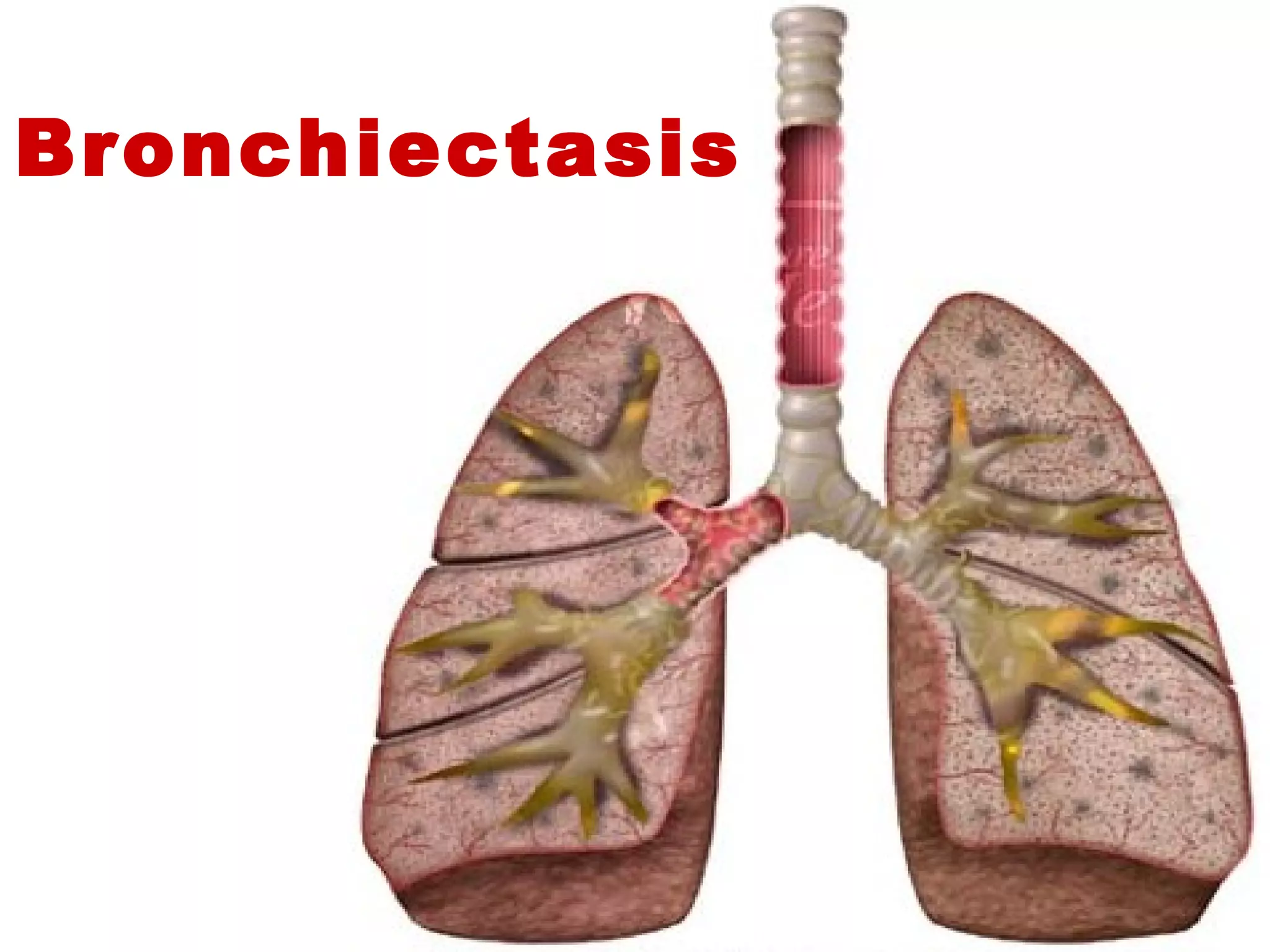
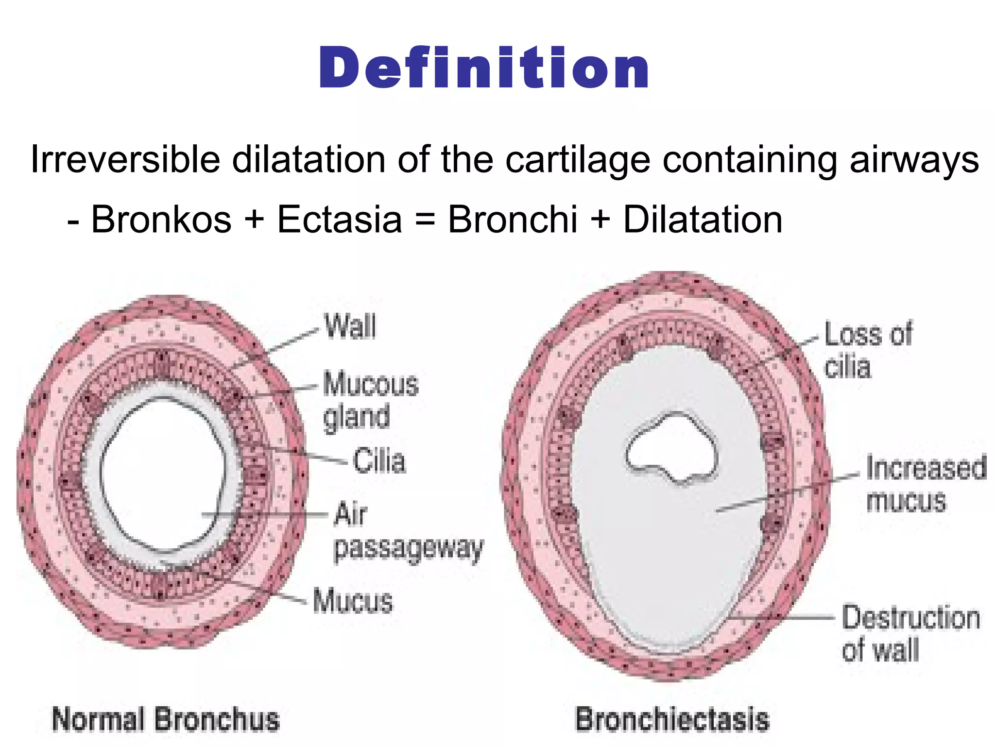
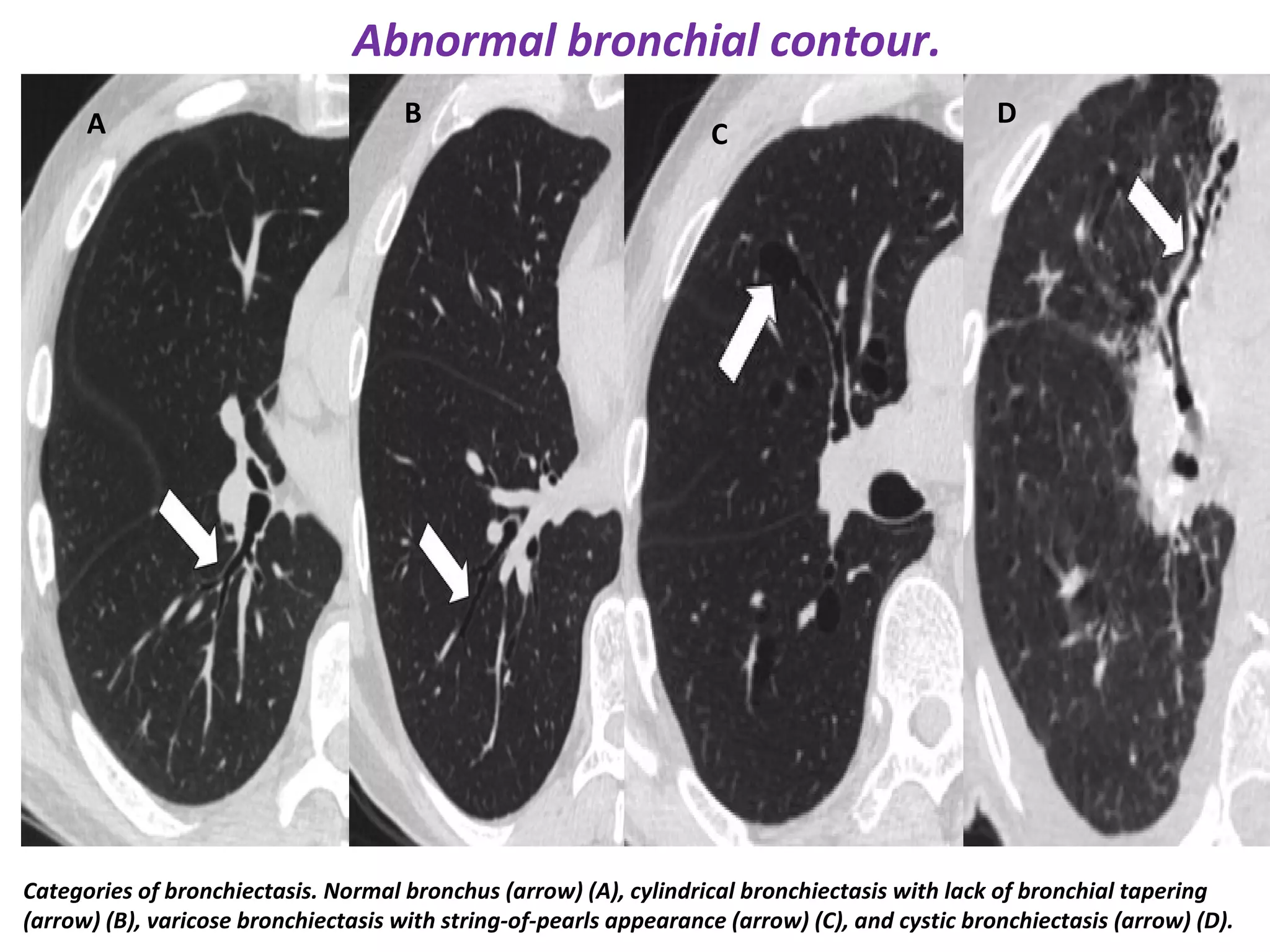
Suppurative LungSuppurative Lung
DiseasesDiseases
Dr/ Riham Hazem Raafat
Lecturer of Chest Diseases
Ainshams University
Bronchiectasis
Definition
Irreversible dilatation of the cartilage containing airways
- Bronkos + Ectasia = Bronchi + Dilatation
colonization
Pathogenesis
Types of bronchiectasis
Causes
- AI
Toxic gas inhalation
-Congenital
(metabolic
gross structure,
ultrastructure)
- Acquired
Diagnostic Approach
Clinical Features:Clinical Features:
• Cough
• Daily sputum production: green/yellow sputum (patients with
bronchiectasis may produce 240ml (8 oz) of sputum daily).
• Dyspnea
• Wheezing
• Hemoptysis
• Bluish skin color
• Recurrent pleurisy
• Dry Bronchiectasis:
• Breath odor
• Clubbing of fingers
• Fatigue
• Paleness
• Weight loss
• Acute exacerbation: purulence, amount of Sputum & Dyspnea
• Late: RF, Corpulmonale
Complications:
Recurrent hemoptysis, pneumonia and pleurisy
Lung abscess and metastatic brain abscess
Empyema
Amyloidosis
Cor pulmonale and respiratory failure
• CXR:
Suspicious but not diagnostic radiographic findings include:
focal pneumonitis, scattered irregular opacities that may represent
mucopurulent plugs, linear or plate-like atelectasis , dilated and thickened
airways that appear as ring-like shadows (of airways that are seen on end)
or tram lines (airways that are perpendicular to the x-ray beam)
• HRCT:
Major features on HRCT include AW dilatation & bronchial wall thickening
• Bronchoscopy:
For diagnosis of tumor, foreign body, localize site of hemoptysis.
• PFTs
• Blood (including serology) & Sputum
Sputum Organisms
Typical offending organisms:
• Klebsiella species,
• Staphylococcus aureus,
• Mycobacterium tuberculosis,
• Mycoplasma pneumoniae,
• Non-tuberculous mycobacteria,
Once a patient develops bronchiectasis:
- Many of these same organisms colonize the damaged bronchi and may
result in ongoing damage and episodic infectious exacerbations.
- The organisms found most typically include Haemophilus species and
Pseudomonas species (Staph Aureus in CF)
•Measles virus,
•Pertussis virus,
•Influenza virus,
•Herpes simplex virus, and
•Certain types of adenovirus.
•Aspergillus fumigatus
Plain radiographic signs
Chest radiography showing a) cystic bronchiectasis with multiple cystic airspaces and
b) cylindrical bronchiectasis and tram track opacities in a cystic fibrosis patient.
(a) (b)
Cystic bronchiectasis with air-fluid levels.
Mucoid Impaction
CT signs
*Cause can be
seen as well
SIGNET RING SIGN. Chest CT shows small bronchiectasis.
High-resolution computed tomography image showing
non tapering bronchi, in keeping with bronchiectasis.
Visibility of peripheral air ways within 1cm from the
costal pleura
Categories of bronchiectasis. Normal bronchus (arrow) (A), cylindrical bronchiectasis with lack of bronchial tapering
(arrow) (B), varicose bronchiectasis with string-of-pearls appearance (arrow) (C), and cystic bronchiectasis (arrow) (D).
BA C
D
Abnormal bronchial contour.
*
High-resolution computed tomography image demonstrating bronchiectasis with bronchial
wall thickening (asterisk) and mucous plugging (arrow) in the right lower lobe.
Bronchial wall thickening
Inspiratory high-resolution computed tomography image showing bronchiectasis
and widespread areas of low attenuation, representing air-trapping.
Air-trapping Sign.
High-resolution computed tomography showing a) proximal bronchiectasis affecting
segmental airways and b) high attenuation mucous plugs in patients with allergic
bronchopulmonary aspergillosis. No intravenous contrast medium was used in (b).
a)
b)
Mucous plugs Impaction.
Cystic Bronchiectasis. CT: Markedly dilated bronchi are seen, some
with air-fluid levels (yellow arrows), mostly in the right lung.
Cystic changes with air-fluid levels.
Tree in budd opacities
Mounier-Kuhn syndrome, also known as tracheobronchomegaly, is a rare
congenital abnormality of the trachea and main bronchi characterized by
cystic dilatation of the tracheobronchial tree and recurrent respiratory infections.
Treatment• Goals:
1. Controlling infections and bronchial secretions
2. Relieving airway obstructions
3. Removal of affected portions of lung by surgical removal or artery 
embolization 
4. Preventing complications.
• Treatment of bronchiectasis include:
1. The prolonged usage of Antibiotics to prevent detrimental infections
2. Eliminating accumulated fluid with Bronchial Hygiene therapy +
Humidification & Mucolytics (dornase, HS, ACC) +/- Anti-oxidants
3. Inhaled Bronchodilators (in obst.) & Steroids (in exac., ABPA)
4. Surgery is used to treat localized bronchiectasis, removing obstructions, 
recurrent hemoptysis & exacerbations  Transplantation
5. Vaccination (S. pn, H. Inf., M., P.), IgG (25mg/kg /wk), O2, Smoke Cess
6. Pulmonary Rehabilitation (add ADEK, Panc enz to CF)
Antibiotic Use
During Exacerbation:
• Mild to Moderate Exacerbation (7-10 ds): Amoxacillin,
Tetracycline, Trimethoprim-Sulfamethoxazole, New Macrolides,
Cephalosporin, Quinolones
• Moderate to Severe Exacerbation (10-14 ds):
Antipseudomonal IV (dual therapy) or MAC treatment if proved
(clarithromycin, ethambutol, rifampicin, sterptomycin 18-14 Ms)
Regular Treatment for Colonization:
• Intermittent courses are used for 7 days and Ab- free periods of 7
days each month can be used (oral previous drugs) or
• Long term antibiotics for 3 to 6 Ms (macrolides)
• Inhaled Antibiotics: Tobramycin, Colistin, Gentamycin, Astreonam
• Postural drainage
• Percussion 
• Directed cough: as Forced expiratory technique (huffing: 
small long (LL) or big short huff (UL) in cycle; 10 mins twice /d)
• Active cycle of breathing (breathing control (hands on abd.), 
deep breathing exercises e’ breath hold (ribs) & huffing +/- 
manual technique)
• Autogenic drainage (self drainage: unstick, collect, evacuate)
• Positive expiratory pressure (behind mucus to push)
• Incentive Spirometry
Bronchial Hygiene Techniques
Can be associated with others
Diaphragmatic
Breathing
Vibratory PEP
Flutter device
Acapella
Lung AbscessLung Abscess
Definition:Definition:
• A lung abscess is a localized area of destruction of lungA lung abscess is a localized area of destruction of lung
parenchyma in which infection by a Pyogenic organism resultsparenchyma in which infection by a Pyogenic organism results
in tissue necrosis and suppuration and cavity formation.in tissue necrosis and suppuration and cavity formation.
Classification
Lung abscesses can be classified based on:
- Duration:
• Acute abscesses are less than 4-6 wks old
• Chronic abscesses are of longer duration
- Etiology:
• Primary Abscess is infectious in origin: caused by aspiration or
pneumonia in the healthy host.
• Secondary Abscess
- Pre-existing lung condition (obstruction, bronchiectasis, cyctic lung,
c).
- Hematogenous: Spread from an extra-pulmonary site (septic emboli)
- Immuno-compromised state.
- Inhalation of infected material
- Necrotizing pneumonia (klebsiella, staph., pseudomonus, anearobic,
- Number: single or multiple
Pathogenesis:Pathogenesis:
• Lung  abscess  begin  as  an  area  of  pneumonia Lung  abscess  begin  as  an  area  of  pneumonia    
necrosis  or  microabscess necrosis  or  microabscess    coalesce  to  form  a   coalesce  to  form  a 
single or sometimes multiple areas of suppuration single or sometimes multiple areas of suppuration 
 reach a size > 1cm in diameter  reach a size > 1cm in diameter  lung abscess.  lung abscess. 
• Inflammation  erodes  adjacent  bronchi Inflammation  erodes  adjacent  bronchi    
suppuration is expectorated suppuration is expectorated  air finds its way to  air finds its way to 
the abscess cavity the abscess cavity  fluid air interface. fluid air interface.
Clinical features:Clinical features:
• Presence of the predisposing factorsPresence of the predisposing factors
• Symptoms of acute lung abscess:Symptoms of acute lung abscess:
• TheThe onsetonset may be abruptmay be abrupt  or gradual.or gradual.
• FeverFever withwith rigorsrigors,, sweatingsweating,, coughcough (dry cough at first(dry cough at first
which is followed by sudden onset ofwhich is followed by sudden onset of expectorationexpectoration ofof
large amounts of sputum after which fever drop).large amounts of sputum after which fever drop).
• Sputum:Sputum: foul-odor, purulent with relation to posture.foul-odor, purulent with relation to posture.
• Chest pain:Chest pain: it may be dull aching or severe pleuritic pain.it may be dull aching or severe pleuritic pain.
• HemoptysisHemoptysis may occur and may be massive.may occur and may be massive.
• Weight lossWeight loss,, anaemiaanaemia andand clubbingclubbing in chronic lungin chronic lung
abscess (8-12 weeks).abscess (8-12 weeks).
Complications:
• Local spread to the same or to the other lung.Local spread to the same or to the other lung.
• Prolonged fever and chronicity.Prolonged fever and chronicity.
• Massive hemoptysis.Massive hemoptysis.
• Residual bronchiectasis or fibrosisResidual bronchiectasis or fibrosis  Trapped lungTrapped lung
• Extension to the pleura or skinExtension to the pleura or skin Empyema, BPFEmpyema, BPF or PCFor PCF
• Metastatic brain abscessMetastatic brain abscess..
• Perinephric abscess.Perinephric abscess.
• Amyloidosis.Amyloidosis.
Investigations:Investigations:
1.1. Chest x-ray PA and lateral viewChest x-ray PA and lateral view
In the acute abscess the wall is thin with surroundingIn the acute abscess the wall is thin with surrounding
consolidation, and the chronic abscess the wall is thick.consolidation, and the chronic abscess the wall is thick.
2. Laboratory:2. Laboratory:
-- Sputum examination:Sputum examination:
o Gram- stain film and culture (85% anaerobes)Gram- stain film and culture (85% anaerobes)
o Ziehl- Neelsen staining to exclude TBZiehl- Neelsen staining to exclude TB
o Sputum cytology for malignant cellsSputum cytology for malignant cells
o Sputum culture for fungi and parasitic ova (if suspected)Sputum culture for fungi and parasitic ova (if suspected)
-- Blood pictureBlood picture: leucocytosis (>30.000/mm3) and anemia.: leucocytosis (>30.000/mm3) and anemia.
Irregularly shaped cavity with an air-fluid level inside
posterior segments of the upper lobes or
the superior segments of the lower lobes
3. Bronchoscopy:3. Bronchoscopy:
- Diagnostic:- Diagnostic: foreign body, tumors, or inspissatedforeign body, tumors, or inspissated
secretions.secretions.
- Therapeutic:- Therapeutic:
• Aspiration of secretions.Aspiration of secretions.
• Instillation of antibiotics.Instillation of antibiotics.
• Foreign body extraction.Foreign body extraction.
• Palliative treatment of bronchial carcinoma with laserPalliative treatment of bronchial carcinoma with laser
or cryotherapy.or cryotherapy.
4. C-T chest:4. C-T chest: for suspected bronchial carcinoma.for suspected bronchial carcinoma.
Treatment:Treatment:
1.1. Prophylactic:Prophylactic: proper attention to the etiologic factors.proper attention to the etiologic factors.
2.2. Acute lung abscess:Acute lung abscess:
• Medical treatment is usually sufficient:Medical treatment is usually sufficient:
a)a) Antibiotics:Antibiotics: 4 -6 wks4 -6 wks  till resolution or small stabletill resolution or small stable
• It is usually given empirically in the form of combinedIt is usually given empirically in the form of combined
antibiotics to cover the spectrum of Gram positive,antibiotics to cover the spectrum of Gram positive,
Gram negative and anaerobic organisms (Clindamycin).Gram negative and anaerobic organisms (Clindamycin).
b)b) Postural drainagePostural drainage is an important item in theis an important item in the
management of lung abscess.management of lung abscess.
c)c) BronchoscopicBronchoscopic aspirationaspiration and instillation of antibiotics.and instillation of antibiotics.
d)d) Symptomatic treatmentSymptomatic treatment::
• Analgesics for pain.Analgesics for pain.
• Expectorants.Expectorants.
e)e) Rest, good nutritionRest, good nutrition withwith high protein diet.high protein diet.
33.. Chronic lung abscessChronic lung abscess::
• ContinueContinue thethe medicalmedical treatment for another 6 wks.treatment for another 6 wks.
• Surgical treatmentSurgical treatment (lobectomy or pneumonectomy)(lobectomy or pneumonectomy)
is indicated in chronic situation with:is indicated in chronic situation with:
• Serious hemoptysis.Serious hemoptysis.
• Failed medical treatment.Failed medical treatment.
• Suspected neoplasmSuspected neoplasm
• Congenital lung malformationCongenital lung malformation
Empyema with
Broncho-Pleural
Fistula
DefinitionDefinition
• Pus in the pleural space or infected pleuralPus in the pleural space or infected pleural
fluid with fistula (opening) between the pleuralfluid with fistula (opening) between the pleural
space and the bronchial tree.space and the bronchial tree.
Causes:Causes:
• Direct spreadDirect spread from adjacent bacterial pneumonia.from adjacent bacterial pneumonia.
• RuptureRupture of a lung abscess into the pleural space.of a lung abscess into the pleural space.
• InvasionInvasion from subphrenic collection either pyogenic orfrom subphrenic collection either pyogenic or
amoebic.amoebic.
• TraumaticTraumatic penetration orpenetration or IatrogenicIatrogenic
-- It may beIt may be acuteacute oror chronic (> 3 months).chronic (> 3 months).
- It may beIt may be loculatedloculated oror free.free.
- It may beIt may be post-operativepost-operative (2/3) or(2/3) or non-operativenon-operative (1/3)(1/3)
Clinical featuresClinical features
• Fever and chest pain.Fever and chest pain.
• Dyspnea.Dyspnea.
• Broncho-pleural fistula is characterized by posturalBroncho-pleural fistula is characterized by postural
cough and big amount of expectorated pus.cough and big amount of expectorated pus.
• Chronicity: pallor, malaise, weakness, easyChronicity: pallor, malaise, weakness, easy
fatigability, fever, anorexia and weight loss.fatigability, fever, anorexia and weight loss.
• Clubbing and pleural rub.Clubbing and pleural rub.
• Bubbling from chest tubeBubbling from chest tube
InvestigationsInvestigations
• Chest x-rayChest x-ray (air-fluid level, tension pnx)(air-fluid level, tension pnx)
• CT of the chest:CT of the chest: the condition of the underlying lung, Pnxthe condition of the underlying lung, Pnx
• ThoracentesisThoracentesis::
o Foul - smelling aspirate (anaerobic infection).Foul - smelling aspirate (anaerobic infection).
o Gram stain and culture and sensitivity: identification of the causativeGram stain and culture and sensitivity: identification of the causative
organisms.organisms.
o Low PH (<7.2).Low PH (<7.2).
o Pleural fluid white cell count > 15.000/ mmPleural fluid white cell count > 15.000/ mm33
..
• Methylene blue test:Methylene blue test: injection of methylene blue 1% in theinjection of methylene blue 1% in the
pleural space, it will be expectorated in the sputum.pleural space, it will be expectorated in the sputum.
• Inhalation of radioactive isotopes:Inhalation of radioactive isotopes: detected in the pleuraldetected in the pleural
space.space.
• FOB: Methylene blue test and visualization of bubbles after bronchial
wash
TreatmentTreatment::
• Appropriate antibiotic therapy.Appropriate antibiotic therapy.
• Intercostal tube drainage under water seal &Intercostal tube drainage under water seal &
pleurodesispleurodesis
• DecorticationDecortication
• Muscle flap closure of fistulaMuscle flap closure of fistula
• Pleuropneumonectomy.Pleuropneumonectomy.
Suppurative lung diseases

Suppurative lung diseases

  • 1.
    Suppurative LungSuppurative Lung DiseasesDiseases Dr/Riham Hazem Raafat Lecturer of Chest Diseases Ainshams University
  • 2.
  • 3.
    Definition Irreversible dilatation ofthe cartilage containing airways - Bronkos + Ectasia = Bronchi + Dilatation
  • 4.
  • 5.
  • 6.
    Causes - AI Toxic gasinhalation -Congenital (metabolic gross structure, ultrastructure) - Acquired
  • 10.
  • 12.
    Clinical Features:Clinical Features: •Cough • Daily sputum production: green/yellow sputum (patients with bronchiectasis may produce 240ml (8 oz) of sputum daily). • Dyspnea • Wheezing • Hemoptysis • Bluish skin color • Recurrent pleurisy • Dry Bronchiectasis: • Breath odor • Clubbing of fingers • Fatigue • Paleness • Weight loss • Acute exacerbation: purulence, amount of Sputum & Dyspnea • Late: RF, Corpulmonale
  • 14.
    Complications: Recurrent hemoptysis, pneumoniaand pleurisy Lung abscess and metastatic brain abscess Empyema Amyloidosis Cor pulmonale and respiratory failure
  • 15.
    • CXR: Suspicious butnot diagnostic radiographic findings include: focal pneumonitis, scattered irregular opacities that may represent mucopurulent plugs, linear or plate-like atelectasis , dilated and thickened airways that appear as ring-like shadows (of airways that are seen on end) or tram lines (airways that are perpendicular to the x-ray beam) • HRCT: Major features on HRCT include AW dilatation & bronchial wall thickening • Bronchoscopy: For diagnosis of tumor, foreign body, localize site of hemoptysis. • PFTs • Blood (including serology) & Sputum
  • 17.
    Sputum Organisms Typical offendingorganisms: • Klebsiella species, • Staphylococcus aureus, • Mycobacterium tuberculosis, • Mycoplasma pneumoniae, • Non-tuberculous mycobacteria, Once a patient develops bronchiectasis: - Many of these same organisms colonize the damaged bronchi and may result in ongoing damage and episodic infectious exacerbations. - The organisms found most typically include Haemophilus species and Pseudomonas species (Staph Aureus in CF) •Measles virus, •Pertussis virus, •Influenza virus, •Herpes simplex virus, and •Certain types of adenovirus. •Aspergillus fumigatus
  • 18.
  • 19.
    Chest radiography showinga) cystic bronchiectasis with multiple cystic airspaces and b) cylindrical bronchiectasis and tram track opacities in a cystic fibrosis patient. (a) (b)
  • 20.
    Cystic bronchiectasis withair-fluid levels.
  • 21.
  • 22.
    CT signs *Cause canbe seen as well
  • 23.
    SIGNET RING SIGN.Chest CT shows small bronchiectasis.
  • 24.
    High-resolution computed tomographyimage showing non tapering bronchi, in keeping with bronchiectasis.
  • 25.
    Visibility of peripheralair ways within 1cm from the costal pleura
  • 26.
    Categories of bronchiectasis.Normal bronchus (arrow) (A), cylindrical bronchiectasis with lack of bronchial tapering (arrow) (B), varicose bronchiectasis with string-of-pearls appearance (arrow) (C), and cystic bronchiectasis (arrow) (D). BA C D Abnormal bronchial contour.
  • 27.
    * High-resolution computed tomographyimage demonstrating bronchiectasis with bronchial wall thickening (asterisk) and mucous plugging (arrow) in the right lower lobe. Bronchial wall thickening
  • 28.
    Inspiratory high-resolution computedtomography image showing bronchiectasis and widespread areas of low attenuation, representing air-trapping. Air-trapping Sign.
  • 29.
    High-resolution computed tomographyshowing a) proximal bronchiectasis affecting segmental airways and b) high attenuation mucous plugs in patients with allergic bronchopulmonary aspergillosis. No intravenous contrast medium was used in (b). a) b) Mucous plugs Impaction.
  • 30.
    Cystic Bronchiectasis. CT:Markedly dilated bronchi are seen, some with air-fluid levels (yellow arrows), mostly in the right lung. Cystic changes with air-fluid levels.
  • 31.
    Tree in buddopacities
  • 32.
    Mounier-Kuhn syndrome, alsoknown as tracheobronchomegaly, is a rare congenital abnormality of the trachea and main bronchi characterized by cystic dilatation of the tracheobronchial tree and recurrent respiratory infections.
  • 33.
    Treatment• Goals: 1. Controlling infections and bronchial secretions 2.Relieving airway obstructions 3. Removal of affected portions of lung by surgical removal or artery  embolization  4. Preventing complications. • Treatment of bronchiectasis include: 1. The prolonged usage of Antibiotics to prevent detrimental infections 2. Eliminating accumulated fluid with Bronchial Hygiene therapy + Humidification & Mucolytics (dornase, HS, ACC) +/- Anti-oxidants 3. Inhaled Bronchodilators (in obst.) & Steroids (in exac., ABPA) 4. Surgery is used to treat localized bronchiectasis, removing obstructions,  recurrent hemoptysis & exacerbations  Transplantation 5. Vaccination (S. pn, H. Inf., M., P.), IgG (25mg/kg /wk), O2, Smoke Cess 6. Pulmonary Rehabilitation (add ADEK, Panc enz to CF)
  • 34.
    Antibiotic Use During Exacerbation: •Mild to Moderate Exacerbation (7-10 ds): Amoxacillin, Tetracycline, Trimethoprim-Sulfamethoxazole, New Macrolides, Cephalosporin, Quinolones • Moderate to Severe Exacerbation (10-14 ds): Antipseudomonal IV (dual therapy) or MAC treatment if proved (clarithromycin, ethambutol, rifampicin, sterptomycin 18-14 Ms) Regular Treatment for Colonization: • Intermittent courses are used for 7 days and Ab- free periods of 7 days each month can be used (oral previous drugs) or • Long term antibiotics for 3 to 6 Ms (macrolides) • Inhaled Antibiotics: Tobramycin, Colistin, Gentamycin, Astreonam
  • 36.
    • Postural drainage • Percussion  •Directed cough: as Forced expiratory technique (huffing:  small long (LL) or big short huff (UL) in cycle; 10 mins twice /d) • Active cycle of breathing (breathing control (hands on abd.),  deep breathing exercises e’ breath hold (ribs) & huffing +/-  manual technique) • Autogenic drainage (self drainage: unstick, collect, evacuate) • Positive expiratory pressure (behind mucus to push) • Incentive Spirometry Bronchial Hygiene Techniques Can be associated with others
  • 37.
  • 38.
  • 40.
  • 41.
    Definition:Definition: • A lungabscess is a localized area of destruction of lungA lung abscess is a localized area of destruction of lung parenchyma in which infection by a Pyogenic organism resultsparenchyma in which infection by a Pyogenic organism results in tissue necrosis and suppuration and cavity formation.in tissue necrosis and suppuration and cavity formation.
  • 42.
    Classification Lung abscesses canbe classified based on: - Duration: • Acute abscesses are less than 4-6 wks old • Chronic abscesses are of longer duration - Etiology: • Primary Abscess is infectious in origin: caused by aspiration or pneumonia in the healthy host. • Secondary Abscess - Pre-existing lung condition (obstruction, bronchiectasis, cyctic lung, c). - Hematogenous: Spread from an extra-pulmonary site (septic emboli) - Immuno-compromised state. - Inhalation of infected material - Necrotizing pneumonia (klebsiella, staph., pseudomonus, anearobic, - Number: single or multiple
  • 43.
    Pathogenesis:Pathogenesis: • Lung  abscess begin  as  an  area  of  pneumonia Lung  abscess  begin  as  an  area  of  pneumonia     necrosis  or  microabscess necrosis  or  microabscess    coalesce  to  form  a   coalesce  to  form  a  single or sometimes multiple areas of suppuration single or sometimes multiple areas of suppuration   reach a size > 1cm in diameter  reach a size > 1cm in diameter  lung abscess.  lung abscess.  • Inflammation  erodes  adjacent  bronchi Inflammation  erodes  adjacent  bronchi     suppuration is expectorated suppuration is expectorated  air finds its way to  air finds its way to  the abscess cavity the abscess cavity  fluid air interface. fluid air interface.
  • 44.
    Clinical features:Clinical features: •Presence of the predisposing factorsPresence of the predisposing factors • Symptoms of acute lung abscess:Symptoms of acute lung abscess: • TheThe onsetonset may be abruptmay be abrupt  or gradual.or gradual. • FeverFever withwith rigorsrigors,, sweatingsweating,, coughcough (dry cough at first(dry cough at first which is followed by sudden onset ofwhich is followed by sudden onset of expectorationexpectoration ofof large amounts of sputum after which fever drop).large amounts of sputum after which fever drop). • Sputum:Sputum: foul-odor, purulent with relation to posture.foul-odor, purulent with relation to posture. • Chest pain:Chest pain: it may be dull aching or severe pleuritic pain.it may be dull aching or severe pleuritic pain. • HemoptysisHemoptysis may occur and may be massive.may occur and may be massive. • Weight lossWeight loss,, anaemiaanaemia andand clubbingclubbing in chronic lungin chronic lung abscess (8-12 weeks).abscess (8-12 weeks).
  • 45.
    Complications: • Local spreadto the same or to the other lung.Local spread to the same or to the other lung. • Prolonged fever and chronicity.Prolonged fever and chronicity. • Massive hemoptysis.Massive hemoptysis. • Residual bronchiectasis or fibrosisResidual bronchiectasis or fibrosis  Trapped lungTrapped lung • Extension to the pleura or skinExtension to the pleura or skin Empyema, BPFEmpyema, BPF or PCFor PCF • Metastatic brain abscessMetastatic brain abscess.. • Perinephric abscess.Perinephric abscess. • Amyloidosis.Amyloidosis.
  • 46.
    Investigations:Investigations: 1.1. Chest x-rayPA and lateral viewChest x-ray PA and lateral view In the acute abscess the wall is thin with surroundingIn the acute abscess the wall is thin with surrounding consolidation, and the chronic abscess the wall is thick.consolidation, and the chronic abscess the wall is thick. 2. Laboratory:2. Laboratory: -- Sputum examination:Sputum examination: o Gram- stain film and culture (85% anaerobes)Gram- stain film and culture (85% anaerobes) o Ziehl- Neelsen staining to exclude TBZiehl- Neelsen staining to exclude TB o Sputum cytology for malignant cellsSputum cytology for malignant cells o Sputum culture for fungi and parasitic ova (if suspected)Sputum culture for fungi and parasitic ova (if suspected) -- Blood pictureBlood picture: leucocytosis (>30.000/mm3) and anemia.: leucocytosis (>30.000/mm3) and anemia.
  • 47.
    Irregularly shaped cavitywith an air-fluid level inside posterior segments of the upper lobes or the superior segments of the lower lobes
  • 48.
    3. Bronchoscopy:3. Bronchoscopy: -Diagnostic:- Diagnostic: foreign body, tumors, or inspissatedforeign body, tumors, or inspissated secretions.secretions. - Therapeutic:- Therapeutic: • Aspiration of secretions.Aspiration of secretions. • Instillation of antibiotics.Instillation of antibiotics. • Foreign body extraction.Foreign body extraction. • Palliative treatment of bronchial carcinoma with laserPalliative treatment of bronchial carcinoma with laser or cryotherapy.or cryotherapy. 4. C-T chest:4. C-T chest: for suspected bronchial carcinoma.for suspected bronchial carcinoma.
  • 49.
    Treatment:Treatment: 1.1. Prophylactic:Prophylactic: properattention to the etiologic factors.proper attention to the etiologic factors. 2.2. Acute lung abscess:Acute lung abscess: • Medical treatment is usually sufficient:Medical treatment is usually sufficient: a)a) Antibiotics:Antibiotics: 4 -6 wks4 -6 wks  till resolution or small stabletill resolution or small stable • It is usually given empirically in the form of combinedIt is usually given empirically in the form of combined antibiotics to cover the spectrum of Gram positive,antibiotics to cover the spectrum of Gram positive, Gram negative and anaerobic organisms (Clindamycin).Gram negative and anaerobic organisms (Clindamycin).
  • 50.
    b)b) Postural drainagePosturaldrainage is an important item in theis an important item in the management of lung abscess.management of lung abscess. c)c) BronchoscopicBronchoscopic aspirationaspiration and instillation of antibiotics.and instillation of antibiotics. d)d) Symptomatic treatmentSymptomatic treatment:: • Analgesics for pain.Analgesics for pain. • Expectorants.Expectorants. e)e) Rest, good nutritionRest, good nutrition withwith high protein diet.high protein diet.
  • 51.
    33.. Chronic lungabscessChronic lung abscess:: • ContinueContinue thethe medicalmedical treatment for another 6 wks.treatment for another 6 wks. • Surgical treatmentSurgical treatment (lobectomy or pneumonectomy)(lobectomy or pneumonectomy) is indicated in chronic situation with:is indicated in chronic situation with: • Serious hemoptysis.Serious hemoptysis. • Failed medical treatment.Failed medical treatment. • Suspected neoplasmSuspected neoplasm • Congenital lung malformationCongenital lung malformation
  • 52.
  • 53.
    DefinitionDefinition • Pus inthe pleural space or infected pleuralPus in the pleural space or infected pleural fluid with fistula (opening) between the pleuralfluid with fistula (opening) between the pleural space and the bronchial tree.space and the bronchial tree.
  • 54.
    Causes:Causes: • Direct spreadDirectspread from adjacent bacterial pneumonia.from adjacent bacterial pneumonia. • RuptureRupture of a lung abscess into the pleural space.of a lung abscess into the pleural space. • InvasionInvasion from subphrenic collection either pyogenic orfrom subphrenic collection either pyogenic or amoebic.amoebic. • TraumaticTraumatic penetration orpenetration or IatrogenicIatrogenic -- It may beIt may be acuteacute oror chronic (> 3 months).chronic (> 3 months). - It may beIt may be loculatedloculated oror free.free. - It may beIt may be post-operativepost-operative (2/3) or(2/3) or non-operativenon-operative (1/3)(1/3)
  • 55.
    Clinical featuresClinical features •Fever and chest pain.Fever and chest pain. • Dyspnea.Dyspnea. • Broncho-pleural fistula is characterized by posturalBroncho-pleural fistula is characterized by postural cough and big amount of expectorated pus.cough and big amount of expectorated pus. • Chronicity: pallor, malaise, weakness, easyChronicity: pallor, malaise, weakness, easy fatigability, fever, anorexia and weight loss.fatigability, fever, anorexia and weight loss. • Clubbing and pleural rub.Clubbing and pleural rub. • Bubbling from chest tubeBubbling from chest tube
  • 57.
    InvestigationsInvestigations • Chest x-rayChestx-ray (air-fluid level, tension pnx)(air-fluid level, tension pnx) • CT of the chest:CT of the chest: the condition of the underlying lung, Pnxthe condition of the underlying lung, Pnx • ThoracentesisThoracentesis:: o Foul - smelling aspirate (anaerobic infection).Foul - smelling aspirate (anaerobic infection). o Gram stain and culture and sensitivity: identification of the causativeGram stain and culture and sensitivity: identification of the causative organisms.organisms. o Low PH (<7.2).Low PH (<7.2). o Pleural fluid white cell count > 15.000/ mmPleural fluid white cell count > 15.000/ mm33 .. • Methylene blue test:Methylene blue test: injection of methylene blue 1% in theinjection of methylene blue 1% in the pleural space, it will be expectorated in the sputum.pleural space, it will be expectorated in the sputum. • Inhalation of radioactive isotopes:Inhalation of radioactive isotopes: detected in the pleuraldetected in the pleural space.space. • FOB: Methylene blue test and visualization of bubbles after bronchial wash
  • 58.
    TreatmentTreatment:: • Appropriate antibiotictherapy.Appropriate antibiotic therapy. • Intercostal tube drainage under water seal &Intercostal tube drainage under water seal & pleurodesispleurodesis • DecorticationDecortication • Muscle flap closure of fistulaMuscle flap closure of fistula • Pleuropneumonectomy.Pleuropneumonectomy.