Downloaded 130 times
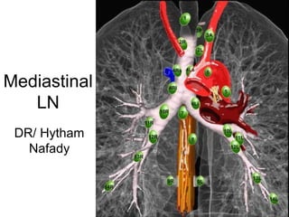
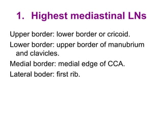
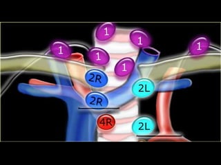
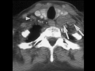
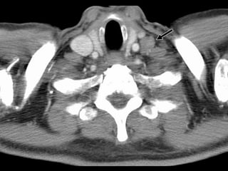
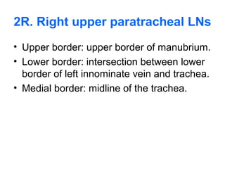
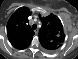

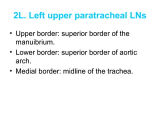
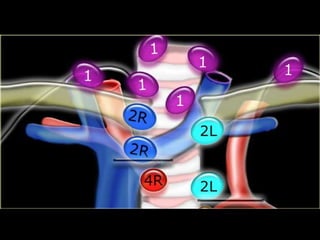
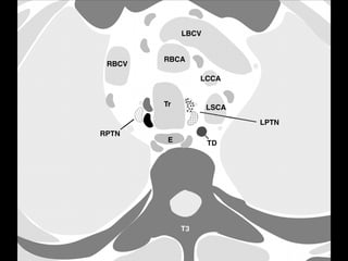
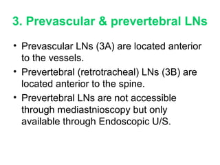
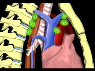
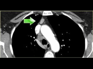
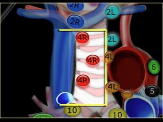

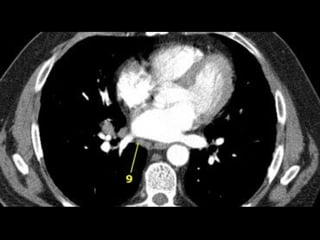
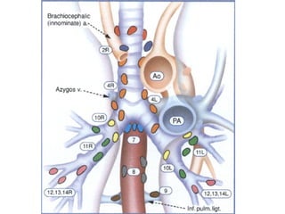
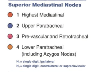
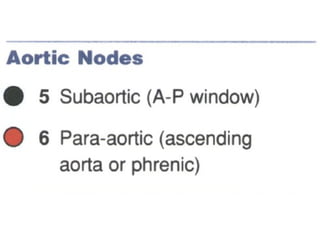
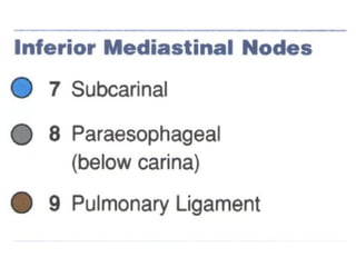
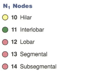
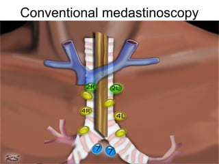
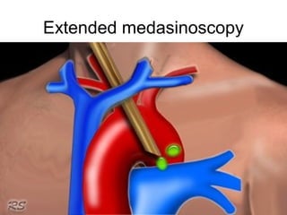
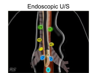
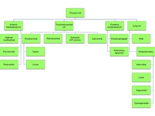
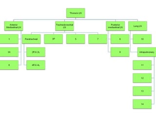

This document describes the 10 different regions of mediastinal lymph nodes, including their anatomical locations and borders. It notes that prevertebral lymph nodes are only accessible through endoscopic ultrasound and not mediastinoscopy. Finally, it mentions conventional mediastinoscopy, extended mediastinoscopy, and endoscopic ultrasound as techniques to access different mediastinal lymph nodes.