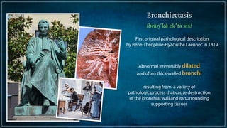
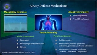
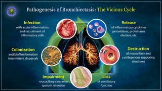
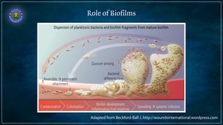
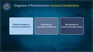
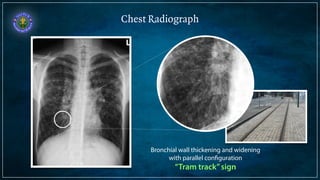
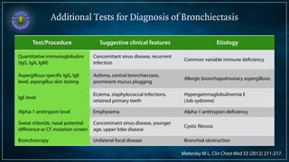
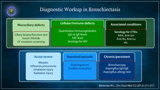
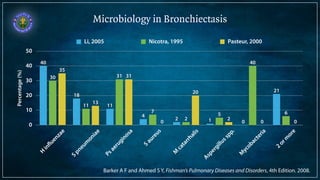
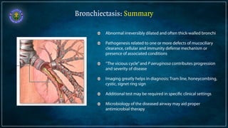
Bronchiectasis is defined as abnormal irreversibly dilated and thick-walled bronchi resulting from destruction of the bronchial wall. Its pathogenesis involves defects in mucociliary clearance, cellular immunity, or associated conditions. High-resolution CT is helpful for diagnosis by showing features like tram lines or honeycombing. Additional tests may be needed to identify underlying causes. Microbiology of infected airways guides antimicrobial therapy for managing the vicious cycle of infection and inflammation that can progress the disease.
History Class XII Ch. 3 Kinship, Caste and Class (1).pptx
Bronchiectasis Causes Diagnosis Guide
1. Bronchiectasis
Causes and Diagnosis
S a n t i S i l a i r a t a n a , M D
Pulmonar y Medicine Uni t , Depar tment of Medicine,
Facul ty of Medicine Vaj i ra Hospi tal
Navamindradhiraj Unive r s i ty
3. Bronchiectasis
/bräŋ′kē ek′tə sis/
First original pathological description
by René-Théophile-Hyacinthe Laennec in 1819
Abnormal irreversibly dilated
and often thick-walled bronchi
!
resulting from a variety of
pathologic process that cause destruction
of the bronchial wall and its surrounding
supporting tissues
4. Airway Defense Mechanisms
Mucociliary clearance
Innate immunity
Neutrophils
Macrophages and dentritic cells
NK cells
Adaptive Immunity
Cellular components Protein components
Immunoglobulins
T and B lymphocytes
Toll-like receptors
Antimicrobial proteins: Lysozymes,
lactoferrins, peroxidase, defensin, cathecidins
Inflammatory cytokines: interferons,
cytokines, chemokines, complements
Waterer G W. Clin Chest Med 33 (2012) 211-217.
6. Reported Etiologies of Bronchiectasis
Others
6%
2%
2%
3%
5%
7%
8%
24%
45% Idiopathic
Rheumatoid arthritis
GERD or aspiration
Young syndrome
Primary ciliary dyskinesia
Allergic
bronchopulmonary
aspergillosis
Immunodeficiency Postinfectious
Quast T M, Self A R, Browning R F et al. Dis Mon 2008; 54: 527-539.
Pasteur M C, Helliwell S M, Hughton S J et al. Am J Respir Crit Care Med 2006; 100: 2183-2189.
7. Pathogenesis of Bronchiectasis: The Vicious Cycle
Release
of inflammatory cytokines
peroxidases, proteinases
elastase, etc.
Destruction
of mucociliary and
cartilagenous supporing
structures
Infection
with acute inflammation
and recruitment of
inflmmatory cells
Colonization
and biofilm formation
intermittent dispersals
Impairment
mucociliary clearance
sputum retention
Loss
of ventilatory
function
8. Pseudomonas aeruginosa in Bronchiectasis
Flagellum
Mobility
Release of the proinflammatory
chemokines on attachment
Pili
Adherence to lipid membrane
Release of toll-like receptors
Recruitment of phagocytic cells
Lipopolysaccharide
Quarum sensing
Alginate
Adherence to the epithelial cells
Pyocyanin
Disruption of the epithelial cell wall
Impairment of ciliary function
Systemic inflammation
Production of lactone
cell-cell signaling
Barker A F, Moulton B C. Clin Chest Med 33 (2012) 211-217.
9. Role of Biofilms
Adapted from Beckford-Ball J. http://woundsinternational.wordpress.com
10. Histologic Changes in Bronchiectasis
Normal bronchus Bronchiectasis
Cartilage
destruction
and fibrosis
Mucosal and
mucous gland
hyperplasia
Inflammatory
cells
infiltration
Increased
mucus and
exudates
11. Diagnosis of Bronchiectasis: General Consideration
Underlying or
associated condition(s)
Microbiology of
the bronchiectatic airway
Definitive diagnosis
Severity of impairment
12. Clinical Manifestations of Bronchiectasis
Clinical Features of
bronchiectasis
Clinical features of the
associated/causative
condition(s)
History
Chronic productive cough*
Sputum production*
Reported bouts of respiratory tract infection
Physical examination
Wheezing, rhonchi, crackles
Clubbing
Cyanosis
CTDs
Arthritis
Sicca syndrome
ABPA
Prominent
wheezing
Bronchial
obstruction
localized
wheezing
PCD, CF, Young Syndrome
Recurrent sinus disease
Infertility
17. Spirometry and Arterial Blood Gas Analysis
Supportive evidence of airway disease (Obstructive defect)
Severity assessment (Severe impairment = poor prognosis)
Bronchodilator therapy (Positive bronchodilator response)
Oxygen therapy (Hypoxemia)
18. Diagnosis of Bronchiectasis: General Consideration
Microbiology of
the bronchiectatic airway
Definitive diagnosis
Severity of impairment
Underlying or
associated condition(s)
19. Additional Tests for Diagnosis of Bronchiectasis
Test/Procedure Suggestive clinical features Etiology
Quantitative immunoglobulins
(IgG, IgA, IgM)
Concomitant sinus disease, recurrent
infection Common variable immune deficiency
Aspergillous-specific IgG, IgE
level, aspergillus skin testing
Asthma, central bronchiectasis,
prominent mucus plugging Allergic bronchopulmonary aspergillosis
IgE level Eczema, staphylococcal infections,
retained primary teeth
Hypergammaglobulinemia E
(Job sydrome)
Alpha-1 antitrypsin level Emphysema Alpha-1 antitrypsin deficiency
Sweat chloride, nasal potential
Concominant sinus disease, younger
difference or CF mutation screen
age, upper lobe disease Cystic fibrosis
Bronchoscopy Unilateral focal disease Bronchial obstruction
Metersky M L. Clin Chest Med 33 (2012) 211-217.
20. Additional Tests for Diagnosis of Bronchiectasis (2)
Test/Procedure Suggestive clinical features Etiology
Bronchoscopy with mycobacterial
cultures
“Tree-in-bud” on HRCT, nodules,
cavity, scoliosis, pectus
Nontuberculous mycobacteria and
tuberculosis
Ciliary biopsy, ciliary functional
testing, nasal nitric oxide
Childhood onset, sinus or ear
infections, situs inversus, infertility Primary ciliary dyskinesia
Swallow evaluation of esophageal pH
measurement M avium disease Gastroesophageal reflux disease or
aspiration
IgG subclass levels, response to
immunization, tests for qualitative
Childhood onset, recurrent infections,
Congenital immunodeficiency
immune defects
dysmorphic features
Serology for autoimmune disease Arthritis, sicca syndrome Rheumatoid arthritis, Sjögren
syndrome
Serology for HIV infection Opportunistic infections, recurrent
infections HIV disease
Metersky M L. Clin Chest Med 33 (2012) 211-217.
21. Diagnostic Workup in Bronchiectasis
Chronic/persistent
!
Endobronchial TB
Bronchial obstruction
ABPA
Repeated/episodic
GERD
Aspirations
Posterior nasal drip
Mucociliary defects
1o ciliary dyskinesia
Cystic fibrosis
Acute/severe
Measles
!
!
Influenza pneumonia
inhalation injury
Radiation injury
Cellular/immune defects
Common variable immune deficiency
X-link agammaglobulinemia
Hyper IgE syndrome
Alpha1-antitrypsin deficiency
HIV infection
Associated conditions
Rheumatoid arthritis
Sjögren syndrome
Relapsing polychondritis
SLE
Ciliary biopsy/function test
Sweat chloride
CF mutation screening
Quantitative immunoglobulins
IgG or IgE levels
AAT level
Serology for HIV
Serology for CTDs:
ANA, Anti-Sm
Anti-Ro, Anti-La
etc.
Esophageal pH
Swallow evaluation
Bronchoscopy
Aspergillus IgG/IgE
Aspergillus allergy test
Metersky M L. Clin Chest Med 33 (2012) 211-217.
22. Diagnosis of Bronchiectasis: General Consideration
Definitive diagnosis
Severity of impairment
Underlying or
associated condition(s)
Microbiology of
the affected airways
23. Microbiology in Bronchiectasis
Percentage (%)
50
40
30
20
10
0
H influenzae
S pneumoniae
Ps aeruginosa
S aureus
M catarrhalis
2 0 0
Aspergillus spp.
Mycobacteria
2 or more
20
0
31
13
35
6
40
5
2
7
31
11
30
21
2 1 0 4
11
18
40
Li, 2005 Nicotra, 1995 Pasteur, 2000
Barker A F and Ahmed S Y, Fishman’s Pulmonary Diseases and Disorders, 4th Edition. 2008.
24. Bronchiectasis: Summary
Abnormal irreversibly dilated and often thick-walled bronchi
Pathogenesis related to one or more defects of mucociliary
clearance, cellular and immunity defense mechanism or
presence of associated conditions
“The vicious cycle” and P aeruginosa contributes progression
and severity of disease
Imaging greatly helps in diagnosis: Tram line, honeycombing,
cystic, signet ring sign
Additional test may be required in specific clinical settings
Microbiology of the diseased airway may aid proper
antimicrobial therapy