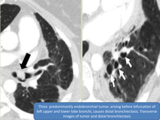
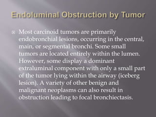
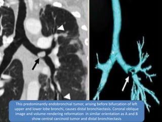
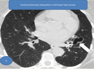
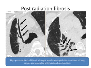
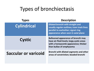
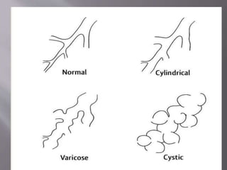
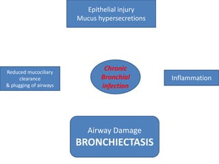
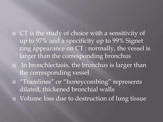
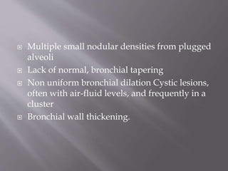
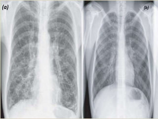
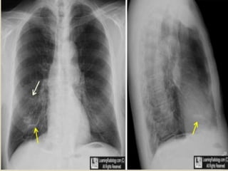
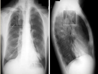
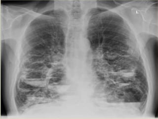
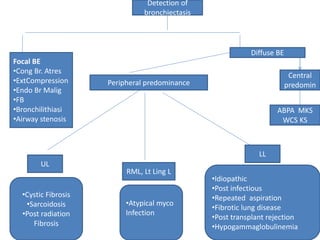
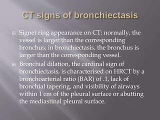
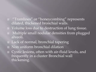
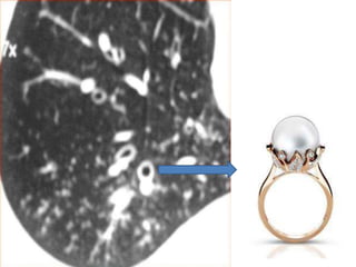
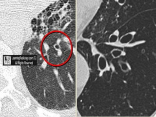
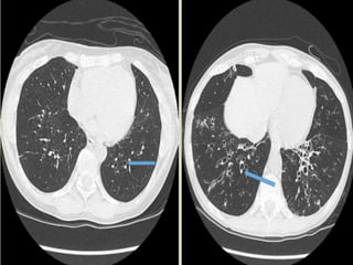
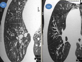
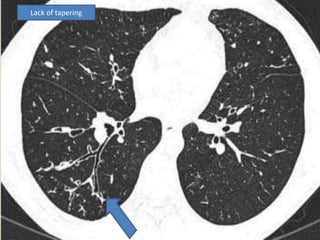
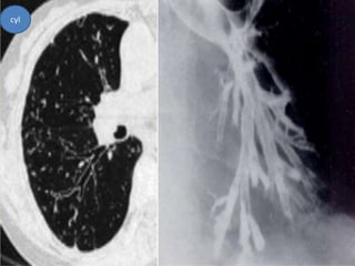
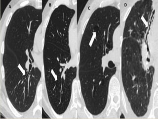
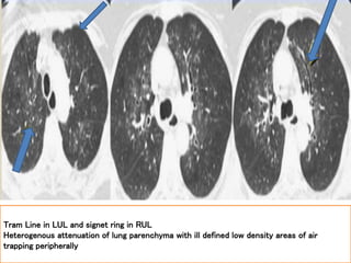
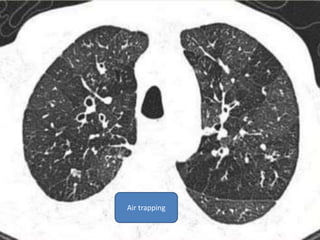
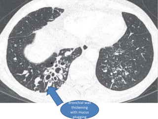
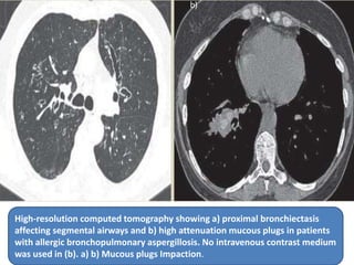
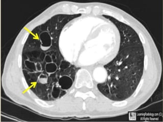
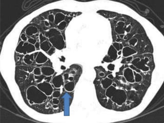
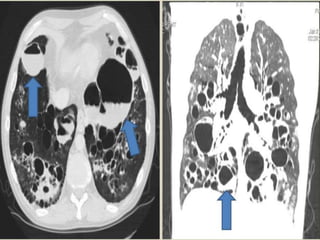
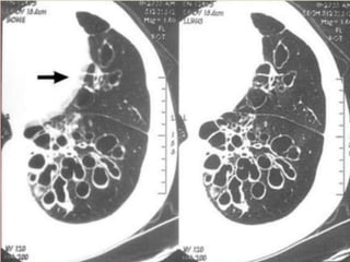
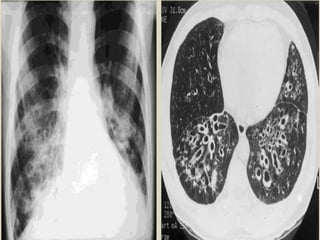
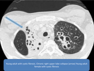
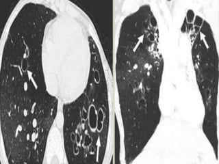
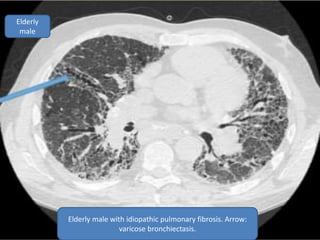
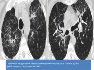
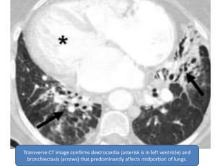
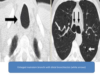
SlideShare a Scribd company logo
Presented by Dr Vrishit Saraswat
IInd Yr Resident
Guided by Prof Dr Dharmraj Meena
Department of Radiodiagnosis
 Bronchiectasis ( Bronchos- airways ; ectasia-
dilatation) is a morphological term used to
describe abnormal irreversibly dilated and
often thick walled bronchi.
 Bronchiectasis represents the end stage of
variety of pathological precesses that cause
destruction of bronchial wall and its
surrounding tissues.
 Clinical Findings:
Classical clinical triad: chronic cough, excess
sputum production and repeated infection.
Hemoptysis.
Shortness of breath.
Wheezing.
Fever is rare even with infection.
Types of bronchiectasis
Types
Cylindrical
Cystic
Saccular or varicoid
Description
Dilated bronchi with straight and
usually regular outlines; tram track lines
parallel to eachother; signet ring
appearance when seen in axial plane
Ballooned appearance of bronchi may
have air-fluid levels; large,cystic areas
with a honeycomb appearance; thicker
than bullae of emphysema
Bronchi with dilated segments and other
areas of constriction; beaded bronchi
 Bronchial dilatation is associated with
destructive and inflammatory changes in the
walls of medium-sized airways, often at the
level of segmental or subsegmental bronchi.
 Airway inflammation is primarily mediated by
neutrophils and results in upregulation of
enzymes, such as elastase and matrix
metalloprotienases.
 This leads to detruction of cartiulage, muscle
and elastic tissue in bronchial wall.
 The dilated airways frequently contain pools of
thick, purulent material, while more peripheral
airways afe often occluded by secretions or
obliterated and replaced by fibrous tissue.
 The parenchyma mormally supplied by
affected airways is abnormal, containing
varying combinations of fibrosis, emphysema,
bronchopneumonia and atelactsis
Epithelial injury
Mucus hypersecretions
Reduced mucociliary
clearance
& plugging of airways
Inflammation
Chronic
Bronchial
infection
Airway Damage
BRONCHIECTASIS
Location
Proximal, frequently upper lobe, bronchiectasis is
characteristic of allergic bronchopulmonary
aspergillosis (ABPA)
Bronchiectasis from viral or pyogenic infections is
usually at the bases
Tuberculous bronchiectasis is usually at the apices
Diffuse bronchiectasis :
Impaired mucus clearing e.g. cystic fibrosis and
Kartagener
Chronic diffuse airways disease (chronic
bronchitis, asthma, BO)
Immune deficiency states
 CT is the study of choice with a sensitivity of
up to 97% and a specificity up to 99% Signet
ring appearance on CT : normally, the vessel is
larger than the corresponding bronchus
 In bronchiectasis, the bronchus is larger than
the corresponding vessel
 “Tramlines” or “honeycombing” represents
dilated, thickened bronchial walls
 Volume loss due to destruction of lung tissue
 Multiple small nodular densities from plugged
alveoli
 Lack of normal, bronchial tapering
 Non uniform bronchial dilation Cystic lesions,
often with air-fluid levels, and frequently in a
cluster
 Bronchial wall thickening.
 Scintigraphy Prior to the advent of HRCT, ventilation
(with or without perfusion) scintigraphy was used to
aid disease evaluation in bronchiectasis. DOLLERY
and HUGH-JONES studied the physiological
implications of bronchiectasis and found reduced
blood flow and impaired ventilation in bronchiectatic
areas. V/Q scintigraphy typically demonstrates
matched ventilation and perfusion defects, reflecting
abnormal ventilation secondary to bronchiectasis and
associated small airways obstruction V/Q scintigraphy
and lung function are additive tools to aid diagnosis
and guide therapeutic management. The ongoing issue
of radiation dose and absence of useful anatomical
information, however, limit the value of V/Q
scintigraphy in routine practice.
 Signs on CXR include the identification of
parallel linear densities, tram-track opacities, or
ring shadows reflecting thickened and
abnormally dilated bronchial walls. These
bronchial abnormalities form a spectrum from
subtle or barely perceptible 5-mm ring
shadows to obvious cysts. Tubular branching
opacities conforming to the expected bronchial
branching pattern may result from fluid or
mucous filling of bronchi
Mucoid impactation
Detection of
bronchiectasis
Focal BE
•Cong Br. Atres
•ExtCompression
•Endo Br Malig
•FB
•Bronchilithiasi
•Airway stenosis
Diffuse BE
Central
predomin
ABPA MKS
WCS KS
Peripheral predominance
UL
LL
RML, Lt Ling L
•Cystic Fibrosis
•Sarcoidosis
•Post radiation
Fibrosis
•Atypical myco
Infection
•Idiopathic
•Post infectious
•Repeated aspiration
•Fibrotic lung disease
•Post transplant rejection
•Hypogammaglobulinemia
 Signet ring appearance on CT: normally, the
vessel is larger than the corresponding
bronchus; in bronchiectasis, the bronchus is
larger than the corresponding vessel.
 Bronchial dilation, the cardinal sign of
bronchiectasis, is characterised on HRCT by a
bronchoarterial ratio (BAR) of .1, lack of
bronchial tapering, and visibility of airways
within 1 cm of the pleural surface or abutting
the mediastinal pleural surface.
 “Tramlines” or “honeycombing” represents
dilated, thickened bronchial walls.
 Volume loss due to destruction of lung tissue.
 Multiple small nodular densities from plugged
alveoli.
 Lack of normal, bronchial tapering
 Non uniform bronchial dilation
 Cystic lesions, often with air-fluid levels, and
frequently in a cluster Bronchial wall
thickening.
Var
Cys
Lack of tapering
cyl
Tram Line in LUL and signet ring in RUL
Heterogenous attenuation of lung parenchyma with ill defined low density areas of air
trapping peripherally
Air trapping
Bronchial wall
thickening
with mucus
plugging
High-resolution computed tomography showing a) proximal bronchiectasis
affecting segmental airways and b) high attenuation mucous plugs in patients
with allergic bronchopulmonary aspergillosis. No intravenous contrast medium
was used in (b). a) b) Mucous plugs Impaction.
• In advance disease of cystic bronchiectasis, it
is often impossible to distinguish between
markedly dilated bronchi and cystic air spaces
in destroyed lung.
• Air fluid levels and history of patients usually
helps differentiating the two conditions
Thisis predominantly endobronchial tumor, arising before bifurcation of
left upper and lower lobe bronchi, causes distal bronchiectasis. Transverse
images of tumor and distal bronchiectasis
 Most carcinoid tumors are primarily
endobronchial lesions, occurring in the central,
main, or segmental bronchi. Some small
tumors are located entirely within the lumen.
However, some display a dominant
extraluminal component with only a small part
of the tumor lying within the airway (iceberg
lesion). A variety of other benign and
malignant neoplasms can also result in
obstruction leading to focal bronchiectasis.
This predominantly endobronchial tumor, arising before bifurcation of left
upper and lower lobe bronchi, causes distal bronchiectasis. Coronal oblique
image and volume-rendering reformation in similar orientation as A and B
show central carcinoid tumor and distal bronchiectasis
 Calcified or ossified material within the bronchial
lumen can cause focal bronchiectasis. By far the
most common cause of broncholithiasis is erosion
by and extrusion of a calcified adjacent lymph
node, usually associated with a long-standing
focus of necrotizing granulomatous lymphadenitis,
especially after tuberculosis. Nevertheless, the
frequency of broncholithiasis complicating
granulomatous infection is quite low. The most
common sites are the proximal right middle lobe
bronchus and the origin of the anterior segmental
bronchus of the upper lobes because of airway
anatomy and lymph node distribution.
Calcified left upper lobe endobronchial broncholithiasis (arrow) from
previous tuberculosis exposure is seen on transverse image (A), minimum-
intensity- projection reformation in coronal oblique plane (B), and volume-
rendering reformation (C) in similar orientation. In C, arrow points to distal
bronchiectasis.
A B
C
 Airway stenosis causing focal bronchiectasis
can result from a broad spectrum of entities
including infection, intubation stricture,
healing of a tracheostomy stoma,
tracheobronchopathia osteochondroplastica,
amyloidosis, relapsing polychondritis,
sarcoidosis, and fibrosing mediastinitis.
Congenital stenosis of left mainstem bronchus. Transverse images show
stenosis (arrow, A), distal bronchiectasis, and mucoid impaction (arrows, B).
A B
 The most common cause of congenital focal
bronchiectasis is bronchial atresia,
characterized by obliteration of a bronchus
with distal bronchiectasis, mucoid impaction,
and air trapping that is most commonly seen in
the left upper lobe. In this rare lesion, the
bronchial tree peripheral to the point of
obliteration is patent and the lung parenchyma
is overinflated because of collateral air drift.
Focal bronchiectasis (idiopathic) in left lower lobe (arrow).
A
B
Post radiation fibrosis
Right para-mediastinal fibrotic changes, which developed after treatment of lung
cancer, are associated with traction bronchiectasis
 The most common cause of congenital upper-
lung-predominant bronchiectasis is cystic
fibrosis, commonly associated with enlarged
lung volumes and interstitial alterations. An
autosomal recessive genetic disorder causing
ineffective clearance of secretions, cystic
fibrosis presents with recurrent pneumonias,
sinusitis, pancreatic insufficiency, and
infertility. Milder forms of cystic fibrosis,
however, can remain unrecognized until
adulthood.
Young adult with cystic fibrosis. Chronic right upper lobe collapse (arrow) Young adult
female with cystic fibrosis
Elderly
male
Elderly male with idiopathic pulmonary fibrosis. Arrow:
varicose bronchiectasis.
 Parenchymal involvement by sarcoidosis can
lead to upper and mid lung fibrosis and
traction bronchiectasis, typically associated
with multiple nodules in a perilymphatic
distribution. Mediastinal and bilateral
symmetric lymphadenopathy is common,
although it can regress as the interstitial disease
worsens.
Transverse images show fibrosis and traction bronchiectasis (arrows, B) that
predominantly involve upper lobes.
 Aka Immobile Cilia Syndrome.
 Recurrent URT infections(rhinitis, sinusitis,
otitis media)
 This rare congenital cause of bronchiectasis,
which primarily involves the middle lung, is
characterized by ineffective clearing of
secretions, causing bronchiectasis, recurrent
pneumonias, sinusitis, and infertility. In 50% of
cases, total situs inversus is present, a condition
known as Kartagener's syndrome.
Transverse CT image confirms dextrocardia (asterisk is in left ventricle) and
bronchiectasis (arrows) that predominantly affects midportion of lungs.
 Also known as tracheobronchomegaly, is a rare
congenital abnormality of the trachea and main
bronchi characterized by cystic dilatation of the
tracheobronchial tree and recurrent respiratory
infections.
 Central Bronchietasis
 Histopathologic evidence of deficiency of
tracheobronchial muscle fibers and absence of the
myenteric plexus, as well as an association with
other congenital or connective tissue disorders,
including ankylosing spondylitis, Marfan
syndrome, CF, Ehlers- Danlos syndrome, and cutis
laxa in children
Enlarged mainstem bronchi with distal bronchiectsis (white arrows)
 Cystic bronchiectasis of 4th to 6th order bronchi
 typically in childhood ,due to defective
cartilage.
 Ballooning of central bronchi on inspiration
and collapse on expiration has also been
reported.
Williams-Campbell syndrome with
bronchiectasis in a patient who had left lung
transplantation. Histologic evaluation of the
resected lung revealed deficient cartilage in
central bronchi. HRCT at three levels shows
marked dilatation of bronchi within central
lung regions. Peripheral lung appears lucent,
particularly when compared with the normal
left lung transplant. This lucency reflects
air trapping and mosaic perfusion.
Thank YOU

More Related Content

What's hot

Bronchiectasis
BronchiectasisBronchiectasis
Bronchiectasis
Denis Katatwire
 
BRONCHIECTASIS
BRONCHIECTASISBRONCHIECTASIS
BRONCHIECTASIS
32KISHOREKUMAR
 
Abpa
AbpaAbpa
Interventions in pulmonary medicine
Interventions in pulmonary medicineInterventions in pulmonary medicine
Interventions in pulmonary medicineDrDon Mascarenhas
 
Bronchiectasis
BronchiectasisBronchiectasis
Bronchiectasis
Amr Eldakroury
 
Cystic lung diseases
Cystic lung diseasesCystic lung diseases
Cystic lung diseasesGamal Agmy
 
Pneumothorax and pneumomediastinum
Pneumothorax and pneumomediastinum Pneumothorax and pneumomediastinum
Pneumothorax and pneumomediastinum
Dr Emad efat
 
lung hrct patterns
lung hrct patterns lung hrct patterns
lung hrct patterns Satish Naga
 
interstitial lung diseases
interstitial lung diseasesinterstitial lung diseases
interstitial lung diseases
DrBasith Lateef
 
Snap Shots in ILDs.ppt
Snap Shots in ILDs.pptSnap Shots in ILDs.ppt
Snap Shots in ILDs.ppt
Gamal Agmy
 
Electromagnetic Navigation Bronchoscopy (ENB): Clinical Review
Electromagnetic Navigation Bronchoscopy (ENB): Clinical ReviewElectromagnetic Navigation Bronchoscopy (ENB): Clinical Review
Electromagnetic Navigation Bronchoscopy (ENB): Clinical Review
Bassel Ericsoussi, MD
 
Pulmonary diseases of vascular origin(pulmonary embolism)
Pulmonary diseases of vascular origin(pulmonary embolism)Pulmonary diseases of vascular origin(pulmonary embolism)
Pulmonary diseases of vascular origin(pulmonary embolism)
imrana tanvir
 
Pneumothorax
PneumothoraxPneumothorax
Pneumothorax
Waqas Ahmed Khan
 
Approach To Interstitial Lung Diseases
Approach To Interstitial Lung DiseasesApproach To Interstitial Lung Diseases
Approach To Interstitial Lung Diseases
Ashraf ElAdawy
 
Know "Solitary Pulmonary Nodule" in a simple way !! (Radiology)
Know "Solitary Pulmonary Nodule" in a simple way !! (Radiology)Know "Solitary Pulmonary Nodule" in a simple way !! (Radiology)
Know "Solitary Pulmonary Nodule" in a simple way !! (Radiology)
Dr.Santosh Atreya
 
Pulmonary aspergilloma
Pulmonary aspergillomaPulmonary aspergilloma
Pulmonary aspergilloma
Deepak Chinagi
 
Bronchogenic carcinoma
Bronchogenic carcinomaBronchogenic carcinoma
Bronchogenic carcinoma
maimusirdan
 
Airway stents
Airway stents Airway stents
Airway stents
Santosh Jha
 
Presentation1.pptx, radiological imaging of bronchiectasis.
Presentation1.pptx, radiological imaging of bronchiectasis.Presentation1.pptx, radiological imaging of bronchiectasis.
Presentation1.pptx, radiological imaging of bronchiectasis.
Abdellah Nazeer
 

What's hot (20)

Bronchiectasis
BronchiectasisBronchiectasis
Bronchiectasis
 
Bronchiectasis
BronchiectasisBronchiectasis
Bronchiectasis
 
BRONCHIECTASIS
BRONCHIECTASISBRONCHIECTASIS
BRONCHIECTASIS
 
Abpa
AbpaAbpa
Abpa
 
Interventions in pulmonary medicine
Interventions in pulmonary medicineInterventions in pulmonary medicine
Interventions in pulmonary medicine
 
Bronchiectasis
BronchiectasisBronchiectasis
Bronchiectasis
 
Cystic lung diseases
Cystic lung diseasesCystic lung diseases
Cystic lung diseases
 
Pneumothorax and pneumomediastinum
Pneumothorax and pneumomediastinum Pneumothorax and pneumomediastinum
Pneumothorax and pneumomediastinum
 
lung hrct patterns
lung hrct patterns lung hrct patterns
lung hrct patterns
 
interstitial lung diseases
interstitial lung diseasesinterstitial lung diseases
interstitial lung diseases
 
Snap Shots in ILDs.ppt
Snap Shots in ILDs.pptSnap Shots in ILDs.ppt
Snap Shots in ILDs.ppt
 
Electromagnetic Navigation Bronchoscopy (ENB): Clinical Review
Electromagnetic Navigation Bronchoscopy (ENB): Clinical ReviewElectromagnetic Navigation Bronchoscopy (ENB): Clinical Review
Electromagnetic Navigation Bronchoscopy (ENB): Clinical Review
 
Pulmonary diseases of vascular origin(pulmonary embolism)
Pulmonary diseases of vascular origin(pulmonary embolism)Pulmonary diseases of vascular origin(pulmonary embolism)
Pulmonary diseases of vascular origin(pulmonary embolism)
 
Pneumothorax
PneumothoraxPneumothorax
Pneumothorax
 
Approach To Interstitial Lung Diseases
Approach To Interstitial Lung DiseasesApproach To Interstitial Lung Diseases
Approach To Interstitial Lung Diseases
 
Know "Solitary Pulmonary Nodule" in a simple way !! (Radiology)
Know "Solitary Pulmonary Nodule" in a simple way !! (Radiology)Know "Solitary Pulmonary Nodule" in a simple way !! (Radiology)
Know "Solitary Pulmonary Nodule" in a simple way !! (Radiology)
 
Pulmonary aspergilloma
Pulmonary aspergillomaPulmonary aspergilloma
Pulmonary aspergilloma
 
Bronchogenic carcinoma
Bronchogenic carcinomaBronchogenic carcinoma
Bronchogenic carcinoma
 
Airway stents
Airway stents Airway stents
Airway stents
 
Presentation1.pptx, radiological imaging of bronchiectasis.
Presentation1.pptx, radiological imaging of bronchiectasis.Presentation1.pptx, radiological imaging of bronchiectasis.
Presentation1.pptx, radiological imaging of bronchiectasis.
 

Similar to Bronchiectasis airway disease

HRCT Chest
HRCT ChestHRCT Chest
HRCT Chest
Manjit Tendolkar
 
Air way disease
Air way diseaseAir way disease
Air way disease
Abhishek Mishra
 
Radiological signs in chest medicine Part 1
Radiological signs in chest medicine Part 1Radiological signs in chest medicine Part 1
Radiological signs in chest medicine Part 1Gamal Agmy
 
Thoracic imaging terminology
Thoracic imaging terminology   Thoracic imaging terminology
Thoracic imaging terminology
Ahmed Bahnassy
 
Interstitial lung-diseases
Interstitial lung-diseases Interstitial lung-diseases
Interstitial lung-diseases
mohamedrafi112
 
HRCT Reticular pattern
HRCT Reticular pattern HRCT Reticular pattern
HRCT Reticular pattern
Sakher Alkhaderi
 
congenital lung.pptx
congenital lung.pptxcongenital lung.pptx
congenital lung.pptx
ranjitharadhakrishna3
 
Hrct
HrctHrct
PATHOGENESIS OF BRONCHIECTASIS BY DR BASHIR AHMED DAR ASSOCIATE PROFESSOR MED...
PATHOGENESIS OF BRONCHIECTASIS BY DR BASHIR AHMED DAR ASSOCIATE PROFESSOR MED...PATHOGENESIS OF BRONCHIECTASIS BY DR BASHIR AHMED DAR ASSOCIATE PROFESSOR MED...
PATHOGENESIS OF BRONCHIECTASIS BY DR BASHIR AHMED DAR ASSOCIATE PROFESSOR MED...
Prof Dr Bashir Ahmed Dar
 
BRONCHIECTASIS BY DR BASHIR AHMED DAR ASSOCIATE PROFESSOR MEDICINE SOPORE KAS...
BRONCHIECTASIS BY DR BASHIR AHMED DAR ASSOCIATE PROFESSOR MEDICINE SOPORE KAS...BRONCHIECTASIS BY DR BASHIR AHMED DAR ASSOCIATE PROFESSOR MEDICINE SOPORE KAS...
BRONCHIECTASIS BY DR BASHIR AHMED DAR ASSOCIATE PROFESSOR MEDICINE SOPORE KAS...Prof Dr Bashir Ahmed Dar
 
The road to HRCT evaluation of pediatric diffuse lung diseases .part 1
The road to HRCT evaluation of pediatric diffuse lung diseases .part 1The road to HRCT evaluation of pediatric diffuse lung diseases .part 1
The road to HRCT evaluation of pediatric diffuse lung diseases .part 1
Ahmed Bahnassy
 
Radiological signs of chest diseases
Radiological signs of chest diseasesRadiological signs of chest diseases
Radiological signs of chest diseases
Gamal Agmy
 
Thoracic imaging terms 2
Thoracic imaging terms 2Thoracic imaging terms 2
Thoracic imaging terms 2
Ahmed Bahnassy
 
tuberculosis - CAVERNOUS TUBERCULOSIS
tuberculosis - CAVERNOUS TUBERCULOSIStuberculosis - CAVERNOUS TUBERCULOSIS
tuberculosis - CAVERNOUS TUBERCULOSIS
Dr. Hament Sharma
 
Common suppurative diseases of lung- Bronchiectasis...!
Common suppurative diseases of lung- Bronchiectasis...!Common suppurative diseases of lung- Bronchiectasis...!
Common suppurative diseases of lung- Bronchiectasis...!
Sharmin Susiwala
 
presentation1-140523194630-phpapp01.pptx
presentation1-140523194630-phpapp01.pptxpresentation1-140523194630-phpapp01.pptx
presentation1-140523194630-phpapp01.pptx
Oluseyi7
 
Interstitial lung diseases- HRCT
Interstitial lung diseases- HRCTInterstitial lung diseases- HRCT
Interstitial lung diseases- HRCTNavdeep Shah
 
Interactive radiology case presentation
Interactive radiology case presentationInteractive radiology case presentation
Interactive radiology case presentationGamal Agmy
 
Presentation1.pptx, radiological imaging of copd.
Presentation1.pptx, radiological imaging of copd.Presentation1.pptx, radiological imaging of copd.
Presentation1.pptx, radiological imaging of copd.Abdellah Nazeer
 
Patterns in HRCT chest
Patterns in HRCT chest Patterns in HRCT chest
Patterns in HRCT chest
Ranjith Kumar
 

Similar to Bronchiectasis airway disease (20)

HRCT Chest
HRCT ChestHRCT Chest
HRCT Chest
 
Air way disease
Air way diseaseAir way disease
Air way disease
 
Radiological signs in chest medicine Part 1
Radiological signs in chest medicine Part 1Radiological signs in chest medicine Part 1
Radiological signs in chest medicine Part 1
 
Thoracic imaging terminology
Thoracic imaging terminology   Thoracic imaging terminology
Thoracic imaging terminology
 
Interstitial lung-diseases
Interstitial lung-diseases Interstitial lung-diseases
Interstitial lung-diseases
 
HRCT Reticular pattern
HRCT Reticular pattern HRCT Reticular pattern
HRCT Reticular pattern
 
congenital lung.pptx
congenital lung.pptxcongenital lung.pptx
congenital lung.pptx
 
Hrct
HrctHrct
Hrct
 
PATHOGENESIS OF BRONCHIECTASIS BY DR BASHIR AHMED DAR ASSOCIATE PROFESSOR MED...
PATHOGENESIS OF BRONCHIECTASIS BY DR BASHIR AHMED DAR ASSOCIATE PROFESSOR MED...PATHOGENESIS OF BRONCHIECTASIS BY DR BASHIR AHMED DAR ASSOCIATE PROFESSOR MED...
PATHOGENESIS OF BRONCHIECTASIS BY DR BASHIR AHMED DAR ASSOCIATE PROFESSOR MED...
 
BRONCHIECTASIS BY DR BASHIR AHMED DAR ASSOCIATE PROFESSOR MEDICINE SOPORE KAS...
BRONCHIECTASIS BY DR BASHIR AHMED DAR ASSOCIATE PROFESSOR MEDICINE SOPORE KAS...BRONCHIECTASIS BY DR BASHIR AHMED DAR ASSOCIATE PROFESSOR MEDICINE SOPORE KAS...
BRONCHIECTASIS BY DR BASHIR AHMED DAR ASSOCIATE PROFESSOR MEDICINE SOPORE KAS...
 
The road to HRCT evaluation of pediatric diffuse lung diseases .part 1
The road to HRCT evaluation of pediatric diffuse lung diseases .part 1The road to HRCT evaluation of pediatric diffuse lung diseases .part 1
The road to HRCT evaluation of pediatric diffuse lung diseases .part 1
 
Radiological signs of chest diseases
Radiological signs of chest diseasesRadiological signs of chest diseases
Radiological signs of chest diseases
 
Thoracic imaging terms 2
Thoracic imaging terms 2Thoracic imaging terms 2
Thoracic imaging terms 2
 
tuberculosis - CAVERNOUS TUBERCULOSIS
tuberculosis - CAVERNOUS TUBERCULOSIStuberculosis - CAVERNOUS TUBERCULOSIS
tuberculosis - CAVERNOUS TUBERCULOSIS
 
Common suppurative diseases of lung- Bronchiectasis...!
Common suppurative diseases of lung- Bronchiectasis...!Common suppurative diseases of lung- Bronchiectasis...!
Common suppurative diseases of lung- Bronchiectasis...!
 
presentation1-140523194630-phpapp01.pptx
presentation1-140523194630-phpapp01.pptxpresentation1-140523194630-phpapp01.pptx
presentation1-140523194630-phpapp01.pptx
 
Interstitial lung diseases- HRCT
Interstitial lung diseases- HRCTInterstitial lung diseases- HRCT
Interstitial lung diseases- HRCT
 
Interactive radiology case presentation
Interactive radiology case presentationInteractive radiology case presentation
Interactive radiology case presentation
 
Presentation1.pptx, radiological imaging of copd.
Presentation1.pptx, radiological imaging of copd.Presentation1.pptx, radiological imaging of copd.
Presentation1.pptx, radiological imaging of copd.
 
Patterns in HRCT chest
Patterns in HRCT chest Patterns in HRCT chest
Patterns in HRCT chest
 

More from Vrishit Saraswat

Introduction to Data Science.pptx
Introduction to Data Science.pptxIntroduction to Data Science.pptx
Introduction to Data Science.pptx
Vrishit Saraswat
 
Artificial intelligence-in-radiology
Artificial intelligence-in-radiologyArtificial intelligence-in-radiology
Artificial intelligence-in-radiology
Vrishit Saraswat
 
The thyroid gland
The thyroid gland The thyroid gland
The thyroid gland
Vrishit Saraswat
 
The scrotum
The scrotumThe scrotum
The scrotum
Vrishit Saraswat
 
The peripheral veins
The peripheral veinsThe peripheral veins
The peripheral veins
Vrishit Saraswat
 
The peripheral arteries
The peripheral arteries   The peripheral arteries
The peripheral arteries
Vrishit Saraswat
 
Nuchal translucency
Nuchal translucencyNuchal translucency
Nuchal translucency
Vrishit Saraswat
 
Ist trimester ultrasound
Ist trimester ultrasoundIst trimester ultrasound
Ist trimester ultrasound
Vrishit Saraswat
 
Head trauma
Head traumaHead trauma
Head trauma
Vrishit Saraswat
 
Follicular study
Follicular studyFollicular study
Follicular study
Vrishit Saraswat
 
Fetal urogenital usg
Fetal urogenital usgFetal urogenital usg
Fetal urogenital usg
Vrishit Saraswat
 
Fetal head & neck usg
Fetal head & neck usgFetal head & neck usg
Fetal head & neck usg
Vrishit Saraswat
 
Fetal brain usg 3
Fetal brain usg  3Fetal brain usg  3
Fetal brain usg 3
Vrishit Saraswat
 
Fetal brain usg 2
Fetal brain usg 2Fetal brain usg 2
Fetal brain usg 2
Vrishit Saraswat
 
Fetal brain usg 1
Fetal brain usg   1Fetal brain usg   1
Fetal brain usg 1
Vrishit Saraswat
 
The fetal musculoskeletal system
The fetal musculoskeletal systemThe fetal musculoskeletal system
The fetal musculoskeletal system
Vrishit Saraswat
 
The fetal heart
The fetal heartThe fetal heart
The fetal heart
Vrishit Saraswat
 
Role of color doppler and mr breast in
Role of color doppler and mr breast inRole of color doppler and mr breast in
Role of color doppler and mr breast in
Vrishit Saraswat
 
Patttern of lung disease on chest x ray
Patttern of lung disease on   chest x rayPatttern of lung disease on   chest x ray
Patttern of lung disease on chest x ray
Vrishit Saraswat
 
NSIP
NSIPNSIP

More from Vrishit Saraswat (20)

Introduction to Data Science.pptx
Introduction to Data Science.pptxIntroduction to Data Science.pptx
Introduction to Data Science.pptx
 
Artificial intelligence-in-radiology
Artificial intelligence-in-radiologyArtificial intelligence-in-radiology
Artificial intelligence-in-radiology
 
The thyroid gland
The thyroid gland The thyroid gland
The thyroid gland
 
The scrotum
The scrotumThe scrotum
The scrotum
 
The peripheral veins
The peripheral veinsThe peripheral veins
The peripheral veins
 
The peripheral arteries
The peripheral arteries   The peripheral arteries
The peripheral arteries
 
Nuchal translucency
Nuchal translucencyNuchal translucency
Nuchal translucency
 
Ist trimester ultrasound
Ist trimester ultrasoundIst trimester ultrasound
Ist trimester ultrasound
 
Head trauma
Head traumaHead trauma
Head trauma
 
Follicular study
Follicular studyFollicular study
Follicular study
 
Fetal urogenital usg
Fetal urogenital usgFetal urogenital usg
Fetal urogenital usg
 
Fetal head & neck usg
Fetal head & neck usgFetal head & neck usg
Fetal head & neck usg
 
Fetal brain usg 3
Fetal brain usg  3Fetal brain usg  3
Fetal brain usg 3
 
Fetal brain usg 2
Fetal brain usg 2Fetal brain usg 2
Fetal brain usg 2
 
Fetal brain usg 1
Fetal brain usg   1Fetal brain usg   1
Fetal brain usg 1
 
The fetal musculoskeletal system
The fetal musculoskeletal systemThe fetal musculoskeletal system
The fetal musculoskeletal system
 
The fetal heart
The fetal heartThe fetal heart
The fetal heart
 
Role of color doppler and mr breast in
Role of color doppler and mr breast inRole of color doppler and mr breast in
Role of color doppler and mr breast in
 
Patttern of lung disease on chest x ray
Patttern of lung disease on   chest x rayPatttern of lung disease on   chest x ray
Patttern of lung disease on chest x ray
 
NSIP
NSIPNSIP
NSIP
 

Recently uploaded

Novas diretrizes da OMS para os cuidados perinatais de mais qualidade
Novas diretrizes da OMS para os cuidados perinatais de mais qualidadeNovas diretrizes da OMS para os cuidados perinatais de mais qualidade
Novas diretrizes da OMS para os cuidados perinatais de mais qualidade
Prof. Marcus Renato de Carvalho
 
POST OPERATIVE OLIGURIA and its management
POST OPERATIVE OLIGURIA and its managementPOST OPERATIVE OLIGURIA and its management
POST OPERATIVE OLIGURIA and its management
touseefaziz1
 
The Normal Electrocardiogram - Part I of II
The Normal Electrocardiogram - Part I of IIThe Normal Electrocardiogram - Part I of II
The Normal Electrocardiogram - Part I of II
MedicoseAcademics
 
New Drug Discovery and Development .....
New Drug Discovery and Development .....New Drug Discovery and Development .....
New Drug Discovery and Development .....
NEHA GUPTA
 
263778731218 Abortion Clinic /Pills In Harare ,
263778731218 Abortion Clinic /Pills In Harare ,263778731218 Abortion Clinic /Pills In Harare ,
263778731218 Abortion Clinic /Pills In Harare ,
sisternakatoto
 
NVBDCP.pptx Nation vector borne disease control program
NVBDCP.pptx Nation vector borne disease control programNVBDCP.pptx Nation vector borne disease control program
NVBDCP.pptx Nation vector borne disease control program
Sapna Thakur
 
HOT NEW PRODUCT! BIG SALES FAST SHIPPING NOW FROM CHINA!! EU KU DB BK substit...
HOT NEW PRODUCT! BIG SALES FAST SHIPPING NOW FROM CHINA!! EU KU DB BK substit...HOT NEW PRODUCT! BIG SALES FAST SHIPPING NOW FROM CHINA!! EU KU DB BK substit...
HOT NEW PRODUCT! BIG SALES FAST SHIPPING NOW FROM CHINA!! EU KU DB BK substit...
GL Anaacs
 
Pulmonary Thromboembolism - etilogy, types, medical- Surgical and nursing man...
Pulmonary Thromboembolism - etilogy, types, medical- Surgical and nursing man...Pulmonary Thromboembolism - etilogy, types, medical- Surgical and nursing man...
Pulmonary Thromboembolism - etilogy, types, medical- Surgical and nursing man...
VarunMahajani
 
Cervical & Brachial Plexus By Dr. RIG.pptx
Cervical & Brachial Plexus By Dr. RIG.pptxCervical & Brachial Plexus By Dr. RIG.pptx
Cervical & Brachial Plexus By Dr. RIG.pptx
Dr. Rabia Inam Gandapore
 
Phone Us ❤85270-49040❤ #ℂall #gIRLS In Surat By Surat @ℂall @Girls Hotel With...
Phone Us ❤85270-49040❤ #ℂall #gIRLS In Surat By Surat @ℂall @Girls Hotel With...Phone Us ❤85270-49040❤ #ℂall #gIRLS In Surat By Surat @ℂall @Girls Hotel With...
Phone Us ❤85270-49040❤ #ℂall #gIRLS In Surat By Surat @ℂall @Girls Hotel With...
Savita Shen $i11
 
Triangles of Neck and Clinical Correlation by Dr. RIG.pptx
Triangles of Neck and Clinical Correlation by Dr. RIG.pptxTriangles of Neck and Clinical Correlation by Dr. RIG.pptx
Triangles of Neck and Clinical Correlation by Dr. RIG.pptx
Dr. Rabia Inam Gandapore
 
Ocular injury ppt Upendra pal optometrist upums saifai etawah
Ocular injury  ppt  Upendra pal  optometrist upums saifai etawahOcular injury  ppt  Upendra pal  optometrist upums saifai etawah
Ocular injury ppt Upendra pal optometrist upums saifai etawah
pal078100
 
Knee anatomy and clinical tests 2024.pdf
Knee anatomy and clinical tests 2024.pdfKnee anatomy and clinical tests 2024.pdf
Knee anatomy and clinical tests 2024.pdf
vimalpl1234
 
Non-respiratory Functions of the Lungs.pdf
Non-respiratory Functions of the Lungs.pdfNon-respiratory Functions of the Lungs.pdf
Non-respiratory Functions of the Lungs.pdf
MedicoseAcademics
 
basicmodesofventilation2022-220313203758.pdf
basicmodesofventilation2022-220313203758.pdfbasicmodesofventilation2022-220313203758.pdf
basicmodesofventilation2022-220313203758.pdf
aljamhori teaching hospital
 
BENIGN PROSTATIC HYPERPLASIA.BPH. BPHpdf
BENIGN PROSTATIC HYPERPLASIA.BPH. BPHpdfBENIGN PROSTATIC HYPERPLASIA.BPH. BPHpdf
BENIGN PROSTATIC HYPERPLASIA.BPH. BPHpdf
DR SETH JOTHAM
 
How to Give Better Lectures: Some Tips for Doctors
How to Give Better Lectures: Some Tips for DoctorsHow to Give Better Lectures: Some Tips for Doctors
How to Give Better Lectures: Some Tips for Doctors
LanceCatedral
 
micro teaching on communication m.sc nursing.pdf
micro teaching on communication m.sc nursing.pdfmicro teaching on communication m.sc nursing.pdf
micro teaching on communication m.sc nursing.pdf
Anurag Sharma
 
24 Upakrama.pptx class ppt useful in all
24 Upakrama.pptx class ppt useful in all24 Upakrama.pptx class ppt useful in all
24 Upakrama.pptx class ppt useful in all
DrSathishMS1
 
Hemodialysis: Chapter 3, Dialysis Water Unit - Dr.Gawad
Hemodialysis: Chapter 3, Dialysis Water Unit - Dr.GawadHemodialysis: Chapter 3, Dialysis Water Unit - Dr.Gawad
Hemodialysis: Chapter 3, Dialysis Water Unit - Dr.Gawad
NephroTube - Dr.Gawad
 

Recently uploaded (20)

Novas diretrizes da OMS para os cuidados perinatais de mais qualidade
Novas diretrizes da OMS para os cuidados perinatais de mais qualidadeNovas diretrizes da OMS para os cuidados perinatais de mais qualidade
Novas diretrizes da OMS para os cuidados perinatais de mais qualidade
 
POST OPERATIVE OLIGURIA and its management
POST OPERATIVE OLIGURIA and its managementPOST OPERATIVE OLIGURIA and its management
POST OPERATIVE OLIGURIA and its management
 
The Normal Electrocardiogram - Part I of II
The Normal Electrocardiogram - Part I of IIThe Normal Electrocardiogram - Part I of II
The Normal Electrocardiogram - Part I of II
 
New Drug Discovery and Development .....
New Drug Discovery and Development .....New Drug Discovery and Development .....
New Drug Discovery and Development .....
 
263778731218 Abortion Clinic /Pills In Harare ,
263778731218 Abortion Clinic /Pills In Harare ,263778731218 Abortion Clinic /Pills In Harare ,
263778731218 Abortion Clinic /Pills In Harare ,
 
NVBDCP.pptx Nation vector borne disease control program
NVBDCP.pptx Nation vector borne disease control programNVBDCP.pptx Nation vector borne disease control program
NVBDCP.pptx Nation vector borne disease control program
 
HOT NEW PRODUCT! BIG SALES FAST SHIPPING NOW FROM CHINA!! EU KU DB BK substit...
HOT NEW PRODUCT! BIG SALES FAST SHIPPING NOW FROM CHINA!! EU KU DB BK substit...HOT NEW PRODUCT! BIG SALES FAST SHIPPING NOW FROM CHINA!! EU KU DB BK substit...
HOT NEW PRODUCT! BIG SALES FAST SHIPPING NOW FROM CHINA!! EU KU DB BK substit...
 
Pulmonary Thromboembolism - etilogy, types, medical- Surgical and nursing man...
Pulmonary Thromboembolism - etilogy, types, medical- Surgical and nursing man...Pulmonary Thromboembolism - etilogy, types, medical- Surgical and nursing man...
Pulmonary Thromboembolism - etilogy, types, medical- Surgical and nursing man...
 
Cervical & Brachial Plexus By Dr. RIG.pptx
Cervical & Brachial Plexus By Dr. RIG.pptxCervical & Brachial Plexus By Dr. RIG.pptx
Cervical & Brachial Plexus By Dr. RIG.pptx
 
Phone Us ❤85270-49040❤ #ℂall #gIRLS In Surat By Surat @ℂall @Girls Hotel With...
Phone Us ❤85270-49040❤ #ℂall #gIRLS In Surat By Surat @ℂall @Girls Hotel With...Phone Us ❤85270-49040❤ #ℂall #gIRLS In Surat By Surat @ℂall @Girls Hotel With...
Phone Us ❤85270-49040❤ #ℂall #gIRLS In Surat By Surat @ℂall @Girls Hotel With...
 
Triangles of Neck and Clinical Correlation by Dr. RIG.pptx
Triangles of Neck and Clinical Correlation by Dr. RIG.pptxTriangles of Neck and Clinical Correlation by Dr. RIG.pptx
Triangles of Neck and Clinical Correlation by Dr. RIG.pptx
 
Ocular injury ppt Upendra pal optometrist upums saifai etawah
Ocular injury  ppt  Upendra pal  optometrist upums saifai etawahOcular injury  ppt  Upendra pal  optometrist upums saifai etawah
Ocular injury ppt Upendra pal optometrist upums saifai etawah
 
Knee anatomy and clinical tests 2024.pdf
Knee anatomy and clinical tests 2024.pdfKnee anatomy and clinical tests 2024.pdf
Knee anatomy and clinical tests 2024.pdf
 
Non-respiratory Functions of the Lungs.pdf
Non-respiratory Functions of the Lungs.pdfNon-respiratory Functions of the Lungs.pdf
Non-respiratory Functions of the Lungs.pdf
 
basicmodesofventilation2022-220313203758.pdf
basicmodesofventilation2022-220313203758.pdfbasicmodesofventilation2022-220313203758.pdf
basicmodesofventilation2022-220313203758.pdf
 
BENIGN PROSTATIC HYPERPLASIA.BPH. BPHpdf
BENIGN PROSTATIC HYPERPLASIA.BPH. BPHpdfBENIGN PROSTATIC HYPERPLASIA.BPH. BPHpdf
BENIGN PROSTATIC HYPERPLASIA.BPH. BPHpdf
 
How to Give Better Lectures: Some Tips for Doctors
How to Give Better Lectures: Some Tips for DoctorsHow to Give Better Lectures: Some Tips for Doctors
How to Give Better Lectures: Some Tips for Doctors
 
micro teaching on communication m.sc nursing.pdf
micro teaching on communication m.sc nursing.pdfmicro teaching on communication m.sc nursing.pdf
micro teaching on communication m.sc nursing.pdf
 
24 Upakrama.pptx class ppt useful in all
24 Upakrama.pptx class ppt useful in all24 Upakrama.pptx class ppt useful in all
24 Upakrama.pptx class ppt useful in all
 
Hemodialysis: Chapter 3, Dialysis Water Unit - Dr.Gawad
Hemodialysis: Chapter 3, Dialysis Water Unit - Dr.GawadHemodialysis: Chapter 3, Dialysis Water Unit - Dr.Gawad
Hemodialysis: Chapter 3, Dialysis Water Unit - Dr.Gawad
 

Bronchiectasis airway disease

  • 1. Presented by Dr Vrishit Saraswat IInd Yr Resident Guided by Prof Dr Dharmraj Meena Department of Radiodiagnosis
  • 2.  Bronchiectasis ( Bronchos- airways ; ectasia- dilatation) is a morphological term used to describe abnormal irreversibly dilated and often thick walled bronchi.  Bronchiectasis represents the end stage of variety of pathological precesses that cause destruction of bronchial wall and its surrounding tissues.
  • 3.  Clinical Findings: Classical clinical triad: chronic cough, excess sputum production and repeated infection. Hemoptysis. Shortness of breath. Wheezing. Fever is rare even with infection.
  • 4. Types of bronchiectasis Types Cylindrical Cystic Saccular or varicoid Description Dilated bronchi with straight and usually regular outlines; tram track lines parallel to eachother; signet ring appearance when seen in axial plane Ballooned appearance of bronchi may have air-fluid levels; large,cystic areas with a honeycomb appearance; thicker than bullae of emphysema Bronchi with dilated segments and other areas of constriction; beaded bronchi
  • 5.
  • 6.  Bronchial dilatation is associated with destructive and inflammatory changes in the walls of medium-sized airways, often at the level of segmental or subsegmental bronchi.  Airway inflammation is primarily mediated by neutrophils and results in upregulation of enzymes, such as elastase and matrix metalloprotienases.  This leads to detruction of cartiulage, muscle and elastic tissue in bronchial wall.
  • 7.  The dilated airways frequently contain pools of thick, purulent material, while more peripheral airways afe often occluded by secretions or obliterated and replaced by fibrous tissue.  The parenchyma mormally supplied by affected airways is abnormal, containing varying combinations of fibrosis, emphysema, bronchopneumonia and atelactsis
  • 8. Epithelial injury Mucus hypersecretions Reduced mucociliary clearance & plugging of airways Inflammation Chronic Bronchial infection Airway Damage BRONCHIECTASIS
  • 9. Location Proximal, frequently upper lobe, bronchiectasis is characteristic of allergic bronchopulmonary aspergillosis (ABPA) Bronchiectasis from viral or pyogenic infections is usually at the bases Tuberculous bronchiectasis is usually at the apices Diffuse bronchiectasis : Impaired mucus clearing e.g. cystic fibrosis and Kartagener Chronic diffuse airways disease (chronic bronchitis, asthma, BO) Immune deficiency states
  • 10.  CT is the study of choice with a sensitivity of up to 97% and a specificity up to 99% Signet ring appearance on CT : normally, the vessel is larger than the corresponding bronchus  In bronchiectasis, the bronchus is larger than the corresponding vessel  “Tramlines” or “honeycombing” represents dilated, thickened bronchial walls  Volume loss due to destruction of lung tissue
  • 11.  Multiple small nodular densities from plugged alveoli  Lack of normal, bronchial tapering  Non uniform bronchial dilation Cystic lesions, often with air-fluid levels, and frequently in a cluster  Bronchial wall thickening.
  • 12.  Scintigraphy Prior to the advent of HRCT, ventilation (with or without perfusion) scintigraphy was used to aid disease evaluation in bronchiectasis. DOLLERY and HUGH-JONES studied the physiological implications of bronchiectasis and found reduced blood flow and impaired ventilation in bronchiectatic areas. V/Q scintigraphy typically demonstrates matched ventilation and perfusion defects, reflecting abnormal ventilation secondary to bronchiectasis and associated small airways obstruction V/Q scintigraphy and lung function are additive tools to aid diagnosis and guide therapeutic management. The ongoing issue of radiation dose and absence of useful anatomical information, however, limit the value of V/Q scintigraphy in routine practice.
  • 13.  Signs on CXR include the identification of parallel linear densities, tram-track opacities, or ring shadows reflecting thickened and abnormally dilated bronchial walls. These bronchial abnormalities form a spectrum from subtle or barely perceptible 5-mm ring shadows to obvious cysts. Tubular branching opacities conforming to the expected bronchial branching pattern may result from fluid or mucous filling of bronchi
  • 14.
  • 15.
  • 17.
  • 18. Detection of bronchiectasis Focal BE •Cong Br. Atres •ExtCompression •Endo Br Malig •FB •Bronchilithiasi •Airway stenosis Diffuse BE Central predomin ABPA MKS WCS KS Peripheral predominance UL LL RML, Lt Ling L •Cystic Fibrosis •Sarcoidosis •Post radiation Fibrosis •Atypical myco Infection •Idiopathic •Post infectious •Repeated aspiration •Fibrotic lung disease •Post transplant rejection •Hypogammaglobulinemia
  • 19.  Signet ring appearance on CT: normally, the vessel is larger than the corresponding bronchus; in bronchiectasis, the bronchus is larger than the corresponding vessel.  Bronchial dilation, the cardinal sign of bronchiectasis, is characterised on HRCT by a bronchoarterial ratio (BAR) of .1, lack of bronchial tapering, and visibility of airways within 1 cm of the pleural surface or abutting the mediastinal pleural surface.
  • 20.  “Tramlines” or “honeycombing” represents dilated, thickened bronchial walls.  Volume loss due to destruction of lung tissue.  Multiple small nodular densities from plugged alveoli.  Lack of normal, bronchial tapering  Non uniform bronchial dilation  Cystic lesions, often with air-fluid levels, and frequently in a cluster Bronchial wall thickening.
  • 21.
  • 22.
  • 23.
  • 26. cyl
  • 27.
  • 28. Tram Line in LUL and signet ring in RUL Heterogenous attenuation of lung parenchyma with ill defined low density areas of air trapping peripherally
  • 31. High-resolution computed tomography showing a) proximal bronchiectasis affecting segmental airways and b) high attenuation mucous plugs in patients with allergic bronchopulmonary aspergillosis. No intravenous contrast medium was used in (b). a) b) Mucous plugs Impaction.
  • 32.
  • 33.
  • 34.
  • 35.
  • 36.
  • 37. • In advance disease of cystic bronchiectasis, it is often impossible to distinguish between markedly dilated bronchi and cystic air spaces in destroyed lung. • Air fluid levels and history of patients usually helps differentiating the two conditions
  • 38. Thisis predominantly endobronchial tumor, arising before bifurcation of left upper and lower lobe bronchi, causes distal bronchiectasis. Transverse images of tumor and distal bronchiectasis
  • 39.  Most carcinoid tumors are primarily endobronchial lesions, occurring in the central, main, or segmental bronchi. Some small tumors are located entirely within the lumen. However, some display a dominant extraluminal component with only a small part of the tumor lying within the airway (iceberg lesion). A variety of other benign and malignant neoplasms can also result in obstruction leading to focal bronchiectasis.
  • 40. This predominantly endobronchial tumor, arising before bifurcation of left upper and lower lobe bronchi, causes distal bronchiectasis. Coronal oblique image and volume-rendering reformation in similar orientation as A and B show central carcinoid tumor and distal bronchiectasis
  • 41.  Calcified or ossified material within the bronchial lumen can cause focal bronchiectasis. By far the most common cause of broncholithiasis is erosion by and extrusion of a calcified adjacent lymph node, usually associated with a long-standing focus of necrotizing granulomatous lymphadenitis, especially after tuberculosis. Nevertheless, the frequency of broncholithiasis complicating granulomatous infection is quite low. The most common sites are the proximal right middle lobe bronchus and the origin of the anterior segmental bronchus of the upper lobes because of airway anatomy and lymph node distribution.
  • 42. Calcified left upper lobe endobronchial broncholithiasis (arrow) from previous tuberculosis exposure is seen on transverse image (A), minimum- intensity- projection reformation in coronal oblique plane (B), and volume- rendering reformation (C) in similar orientation. In C, arrow points to distal bronchiectasis. A B C
  • 43.  Airway stenosis causing focal bronchiectasis can result from a broad spectrum of entities including infection, intubation stricture, healing of a tracheostomy stoma, tracheobronchopathia osteochondroplastica, amyloidosis, relapsing polychondritis, sarcoidosis, and fibrosing mediastinitis.
  • 44. Congenital stenosis of left mainstem bronchus. Transverse images show stenosis (arrow, A), distal bronchiectasis, and mucoid impaction (arrows, B). A B
  • 45.  The most common cause of congenital focal bronchiectasis is bronchial atresia, characterized by obliteration of a bronchus with distal bronchiectasis, mucoid impaction, and air trapping that is most commonly seen in the left upper lobe. In this rare lesion, the bronchial tree peripheral to the point of obliteration is patent and the lung parenchyma is overinflated because of collateral air drift.
  • 46. Focal bronchiectasis (idiopathic) in left lower lobe (arrow). A
  • 47. B
  • 48. Post radiation fibrosis Right para-mediastinal fibrotic changes, which developed after treatment of lung cancer, are associated with traction bronchiectasis
  • 49.  The most common cause of congenital upper- lung-predominant bronchiectasis is cystic fibrosis, commonly associated with enlarged lung volumes and interstitial alterations. An autosomal recessive genetic disorder causing ineffective clearance of secretions, cystic fibrosis presents with recurrent pneumonias, sinusitis, pancreatic insufficiency, and infertility. Milder forms of cystic fibrosis, however, can remain unrecognized until adulthood.
  • 50. Young adult with cystic fibrosis. Chronic right upper lobe collapse (arrow) Young adult female with cystic fibrosis
  • 51.
  • 52. Elderly male Elderly male with idiopathic pulmonary fibrosis. Arrow: varicose bronchiectasis.
  • 53.  Parenchymal involvement by sarcoidosis can lead to upper and mid lung fibrosis and traction bronchiectasis, typically associated with multiple nodules in a perilymphatic distribution. Mediastinal and bilateral symmetric lymphadenopathy is common, although it can regress as the interstitial disease worsens.
  • 54. Transverse images show fibrosis and traction bronchiectasis (arrows, B) that predominantly involve upper lobes.
  • 55.  Aka Immobile Cilia Syndrome.  Recurrent URT infections(rhinitis, sinusitis, otitis media)  This rare congenital cause of bronchiectasis, which primarily involves the middle lung, is characterized by ineffective clearing of secretions, causing bronchiectasis, recurrent pneumonias, sinusitis, and infertility. In 50% of cases, total situs inversus is present, a condition known as Kartagener's syndrome.
  • 56. Transverse CT image confirms dextrocardia (asterisk is in left ventricle) and bronchiectasis (arrows) that predominantly affects midportion of lungs.
  • 57.  Also known as tracheobronchomegaly, is a rare congenital abnormality of the trachea and main bronchi characterized by cystic dilatation of the tracheobronchial tree and recurrent respiratory infections.  Central Bronchietasis  Histopathologic evidence of deficiency of tracheobronchial muscle fibers and absence of the myenteric plexus, as well as an association with other congenital or connective tissue disorders, including ankylosing spondylitis, Marfan syndrome, CF, Ehlers- Danlos syndrome, and cutis laxa in children
  • 58. Enlarged mainstem bronchi with distal bronchiectsis (white arrows)
  • 59.
  • 60.  Cystic bronchiectasis of 4th to 6th order bronchi  typically in childhood ,due to defective cartilage.  Ballooning of central bronchi on inspiration and collapse on expiration has also been reported.
  • 61. Williams-Campbell syndrome with bronchiectasis in a patient who had left lung transplantation. Histologic evaluation of the resected lung revealed deficient cartilage in central bronchi. HRCT at three levels shows marked dilatation of bronchi within central lung regions. Peripheral lung appears lucent, particularly when compared with the normal left lung transplant. This lucency reflects air trapping and mosaic perfusion.